Lysosomal accumulation of SCMAS (subunit c of mitochondrial ATP synthase) in neurons of the mouse model of mucopolysaccharidosis III B - PubMed (original) (raw)
Lysosomal accumulation of SCMAS (subunit c of mitochondrial ATP synthase) in neurons of the mouse model of mucopolysaccharidosis III B
Sergey Ryazantsev et al. Mol Genet Metab. 2007 Apr.
Abstract
The neurodegenerative disease MPS III B (Sanfilippo syndrome type B) is caused by mutations in the gene encoding the lysosomal enzyme alpha-N-acetylglucosaminidase, with a resulting block in heparan sulfate degradation. A mouse model with disruption of the Naglu gene allows detailed study of brain pathology. In contrast to somatic cells, which accumulate primarily heparan sulfate, neurons accumulate a number of apparently unrelated metabolites, including subunit c of mitochondrial ATP synthase (SCMAS). SCMAS accumulated from 1 month of age, primarily in the medial entorhinal cortex and layer V of the somatosensory cortex. Its accumulation was not due to the absence of specific proteases. Light microscopy of brain sections of 6-months-old mice showed SCMAS to accumulate in the same areas as glycosaminoglycan and unesterified cholesterol, in the same cells as ubiquitin and GM3 ganglioside, and in the same organelles as Lamp 1 and Lamp 2. Cryo-immuno electron microscopy showed SCMAS to be present in Lamp positive vesicles bounded by a single membrane (lysosomes), in fingerprint-like layered arrays. GM3 ganglioside was found in the same lysosomes, but was not associated with the SCMAS arrays. GM3 ganglioside was also seen in lysosomes of microglia, suggesting phagocytosis of neuronal membranes. Samples used for cryo-EM and further processed by standard EM procedures (osmium tetroxide fixation and plastic embedding) showed the disappearance of the SCMAS fingerprint arrays and appearance in the same location of "zebra bodies", well known but little understood inclusions in the brain of patients with mucopolysaccharidoses.
Figures
Fig. 1
Age-dependent increase of neurons staining positively for SCMAS in the medial entorhinal cortex (left panel) and the somatosensory cortex (right panel). The barrel field of the somatosensory cortex (S1BF) was used for counting stained cells after confirmation by the mouse brain atlas [47]. The age in months and the genotype (− for MPS III B, + for wild type control) of the mice are indicated on the horizontal axis. The data represent the mean and standard deviation for the number of SCMAS-positive neurons in a brain hemisphere from each of three mice.
Fig. 2
Storage products revealed by staining and immunostaining in selected areas of the brain of 6 months-old mice. All panels show sections from the medial entorhinal cortex, except for Panels C and D which show sections from the somatosensory cortex. Panels A - D: SCMAS immunostaining and hematoxylin counterstain; A and C from MPS III B mouse, B and D, from wild type control. Panels E and F: glycosaminoglycan stained with colloidal iron in sections from MPS III B (E) and wild type control (F) mice. Panels G and H: unesterified cholesterol fluorescence upon reaction with filipin in sections from MPS III B (G) and wild type control (H) mice. Panels I -L: double immunofluorescence of SCMAS with Lamp 1, ubiquitin and GM3, as indicated on the figure; Panel M: double immunofluorescence of Tom 20 with Lamp1. The scale bars represent 100 μm for panels A-K, while the insets show cells at 10 times higher magnification for panels A-D and 4 times higher magnification for panels E and G. The scale bars for panels L and M represent 10 μm.
Fig. 3
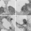
Cryoimmuno EM showing SCMAS in lysosomes of a pyramidal neuron in layer V of somatosensory cortex. Panel A: gold-labeled antibody identifies SCMAS (asterisks); Panel B: higher magnification shows SCMAS in layered finger-print-like array; Panel C: gold-labeled antibodies (arrowheads) identify Lamp at the surface of a single membrane of a vacuole containing a multilayered array (asterisk); Panel D: higher magnification of the array shows the characteristic fingerprint-like structure of SCMAS. Scale bars are 0.2 μm; gold particles, 10 nm.
Fig. 4
Cryo-immuno electron microscopic identification of GM3 ganglioside. Panel A: GM3 ganglioside is identified by 10 nm gold particles (arrowheads) whereas SCMAS is identified by 5 nm gold particles (arrows) and by the characteristic fingerprint-like array (asterisk) in lysosome of a pyramidal neuron. Panel B: GM3 is identified by 10 nm gold particles (arrowheads) in lysosome of a microglial cell. Scale bars are 0.2 μm.
Fig. 5
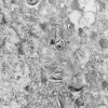
Zebra bodies in neuron after processing for standard electron microscopy. No fingerprint-like arrays are visible, but the arrowhead indicates a zebra body with relatively well organized striations. Scale bar is 0.2 μm.
Similar articles
- Improved behavior and neuropathology in the mouse model of Sanfilippo type IIIB disease after adeno-associated virus-mediated gene transfer in the striatum.
Cressant A, Desmaris N, Verot L, Bréjot T, Froissart R, Vanier MT, Maire I, Heard JM. Cressant A, et al. J Neurosci. 2004 Nov 10;24(45):10229-39. doi: 10.1523/JNEUROSCI.3558-04.2004. J Neurosci. 2004. PMID: 15537895 Free PMC article. - Retrovirally transduced bone marrow has a therapeutic effect on brain in the mouse model of mucopolysaccharidosis IIIB.
Zheng Y, Ryazantsev S, Ohmi K, Zhao HZ, Rozengurt N, Kohn DB, Neufeld EF. Zheng Y, et al. Mol Genet Metab. 2004 Aug;82(4):286-95. doi: 10.1016/j.ymgme.2004.06.004. Mol Genet Metab. 2004. PMID: 15308126 - Neuropathology of murine Sanfilippo D syndrome.
Takahashi K, Le SQ, Kan SH, Jansen MJ, Dickson PI, Cooper JD. Takahashi K, et al. Mol Genet Metab. 2021 Dec;134(4):323-329. doi: 10.1016/j.ymgme.2021.11.010. Epub 2021 Nov 24. Mol Genet Metab. 2021. PMID: 34844863 - Glycosaminoglycans and mucopolysaccharidosis type III.
Jakobkiewicz-Banecka J, Gabig-Ciminska M, Kloska A, Malinowska M, Piotrowska E, Banecka-Majkutewicz Z, Banecki B, Wegrzyn A, Wegrzyn G. Jakobkiewicz-Banecka J, et al. Front Biosci (Landmark Ed). 2016 Jun 1;21(7):1393-409. doi: 10.2741/4463. Front Biosci (Landmark Ed). 2016. PMID: 27100513 Review. - Roles of LAMP-1 and LAMP-2 in lysosome biogenesis and autophagy.
Eskelinen EL. Eskelinen EL. Mol Aspects Med. 2006 Oct-Dec;27(5-6):495-502. doi: 10.1016/j.mam.2006.08.005. Epub 2006 Sep 14. Mol Aspects Med. 2006. PMID: 16973206 Review.
Cited by
- Molecular Bases of Neurodegeneration and Cognitive Decline, the Major Burden of Sanfilippo Disease.
Heon-Roberts R, Nguyen ALA, Pshezhetsky AV. Heon-Roberts R, et al. J Clin Med. 2020 Jan 27;9(2):344. doi: 10.3390/jcm9020344. J Clin Med. 2020. PMID: 32012694 Free PMC article. Review. - Mutation update: Review of TPP1 gene variants associated with neuronal ceroid lipofuscinosis CLN2 disease.
Gardner E, Bailey M, Schulz A, Aristorena M, Miller N, Mole SE. Gardner E, et al. Hum Mutat. 2019 Nov;40(11):1924-1938. doi: 10.1002/humu.23860. Epub 2019 Jul 26. Hum Mutat. 2019. PMID: 31283065 Free PMC article. - Pentosan Polysulfate Treatment of Mucopolysaccharidosis Type IIIA Mice.
Guo N, DeAngelis V, Zhu C, Schuchman EH, Simonaro CM. Guo N, et al. JIMD Rep. 2019;43:37-52. doi: 10.1007/8904_2018_96. Epub 2018 Apr 14. JIMD Rep. 2019. PMID: 29654542 Free PMC article. - Feasibility and safety of systemic rAAV9-hNAGLU delivery for treating mucopolysaccharidosis IIIB: toxicology, biodistribution, and immunological assessments in primates.
Murrey DA, Naughton BJ, Duncan FJ, Meadows AS, Ware TA, Campbell KJ, Bremer WG, Walker CM, Goodchild L, Bolon B, La Perle K, Flanigan KM, McBride KL, McCarty DM, Fu H. Murrey DA, et al. Hum Gene Ther Clin Dev. 2014 Jun;25(2):72-84. doi: 10.1089/humc.2013.208. Epub 2014 Apr 10. Hum Gene Ther Clin Dev. 2014. PMID: 24720466 Free PMC article. - Neuromelanin organelles are specialized autolysosomes that accumulate undegraded proteins and lipids in aging human brain and are likely involved in Parkinson's disease.
Zucca FA, Vanna R, Cupaioli FA, Bellei C, De Palma A, Di Silvestre D, Mauri P, Grassi S, Prinetti A, Casella L, Sulzer D, Zecca L. Zucca FA, et al. NPJ Parkinsons Dis. 2018 Jun 5;4:17. doi: 10.1038/s41531-018-0050-8. eCollection 2018. NPJ Parkinsons Dis. 2018. PMID: 29900402 Free PMC article.
References
- Neufeld EF, Muenzer J. The mucopolysaccharidoses. In: Scriver CR, Beaudet AL, Sly WS, Valle D, editors. The Metabolic and Molecular Bases of Inherited Disease. McGraw-Hill; New York: 2001. pp. 3421–3452.
- Yogalingam G, Hopwood JJ. Molecular genetics of mucopolysaccharidosis type IIIA and IIIB: Diagnostic, clinical, and biological implications. Hum Mutat. 2001;18:264–281. - PubMed
- Aronovich EL, Johnston JM, Wang P, Giger U, Whitley CB. Molecular basis of mucopolysaccharidosis type IIIB in emu (Dromaius novaehollandiae): an avian model of Sanfilippo syndrome type B. Genomics. 2001;74:299–305. - PubMed
- Ellinwood NM, Wang P, Skeen T, Sharp NJ, Cesta M, Decker S, Edwards NJ, Bublot I, Thompson JN, Bush W, Hardam E, Haskins ME, Giger U. A model of mucopolysaccharidosis IIIB (Sanfilippo syndrome type IIIB): N-acetyl-alpha-D-glucosaminidase deficiency in Schipperke dogs. J Inherit Metab Dis. 2003;26:489–504. - PubMed
Publication types
MeSH terms
Substances
LinkOut - more resources
Full Text Sources
Other Literature Sources
Molecular Biology Databases
Miscellaneous


