Immunosenescence of ageing - PubMed (original) (raw)
Review
Immunosenescence of ageing
A L Gruver et al. J Pathol. 2007 Jan.
Abstract
Ageing is a complex process that negatively impacts the development of the immune system and its ability to function. The mechanisms that underlie these age-related defects are broad and range from defects in the haematopoietic bone marrow to defects in peripheral lymphocyte migration, maturation and function. The thymus is a central lymphoid organ responsible for production of naïve T cells, which play a vital role in mediating both cellular and humoral immunity. Chronic involution of the thymus gland is thought to be one of the major contributing factors to loss of immune function with increasing age. It has recently been demonstrated that thymic atrophy is mediated by a shift from a stimulatory to a suppressive cytokine microenvironment. In this review we present an overview of the morphological, cellular and biochemical changes that have been implicated in the decline of thymic and peripheral immune function with ageing. We conclude with the clinical implications of age-associated immunosenescence to vaccine development for tumours and infectious disease. A fundamental understanding of the complex mechanisms by which ageing attenuates immune function will enable translational research teams to develop new therapies and vaccines specifically aimed at overcoming these defects in immunological function in the aged.
Copyright (c) 2007 Pathological Society of Great Britain and Ireland.
Conflict of interest statement
No conflicts of interest were declared.
Figures
Figure 1
The human thymus across the lifespan. [A] Representative views of human thymus morphology throughout ageing. All tissue was formalin-fixed, paraffin-embedded and sections stained with haematoxylin and eosin and anti-keratin antibody [brown] to determine the percentage thymic epithelial space [each panel, ×25]. C, cortex; M, medulla; P, perivascular space. [B] Graphical depiction of the impact of age on human thymus morphology. Thymic epithelial space, pink; perivascular space, white. Reprinted with permission from reference [45]
Figure 2
Impact of ageing on human thymic output and peripheral naïve T cell pool. [A] Molecules of sjTREC/mg whole human thymus tissue. [B] Molecules of sjTREC/μg isolated peripheral blood mononuclear cell DNA [n = 100 donors, aged 6 months–80 years]
Figure 3
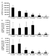
Impact of ageing on BALB/c mouse thymic output and splenic naïve T cell pools. [A] Molecules of mTREC/mg thymus tissue during ageing. Molecules of mTREC in naïve splenic CD4 [B] and CD8 [C] T cells in mice the age range 6–90 weeks [n = 3/group]. Data are mean ± SEM of three mice/group. *p < 0.05 compared to 6 week-old mice [44,45]
Figure 4
Schematic of cytokine stimuli that impact thymopoiesis
Similar articles
- Involution of the mammalian thymus, one of the leading regulators of aging.
Bodey B, Bodey B Jr, Siegel SE, Kaiser HE. Bodey B, et al. In Vivo. 1997 Sep-Oct;11(5):421-40. In Vivo. 1997. PMID: 9427047 - Is thymus redundant after adulthood?
Shanker A. Shanker A. Immunol Lett. 2004 Feb 15;91(2-3):79-86. doi: 10.1016/j.imlet.2003.12.012. Immunol Lett. 2004. PMID: 15019273 Review. - Maturation of B cell precursors is impaired in thymic-deprived nude and old mice.
Szabo P, Zhao K, Kirman I, Le Maoult J, Dyall R, Cruikshank W, Weksler ME. Szabo P, et al. J Immunol. 1998 Sep 1;161(5):2248-53. J Immunol. 1998. PMID: 9725218 - [The ageing immune system].
Djukic M, Nau R, Sieber C. Djukic M, et al. Dtsch Med Wochenschr. 2014 Oct;139(40):1987-90. doi: 10.1055/s-0034-1370283. Epub 2014 Sep 25. Dtsch Med Wochenschr. 2014. PMID: 25254392 Review. German.
Cited by
- Brain inflammaging in the pathogenesis of late-life depression.
Ishizuka T, Nagata W, Nakagawa K, Takahashi S. Ishizuka T, et al. Hum Cell. 2024 Oct 26;38(1):7. doi: 10.1007/s13577-024-01132-4. Hum Cell. 2024. PMID: 39460876 Review. - METTL3 governs thymocyte development and thymic involution by regulating ferroptosis.
Jing H, Song J, Sun J, Su S, Hu J, Zhang H, Bi Y, Wu B. Jing H, et al. Nat Aging. 2024 Oct 23. doi: 10.1038/s43587-024-00724-x. Online ahead of print. Nat Aging. 2024. PMID: 39443728 - Inflammaging: Expansion of Molecular Phenotype and Role in Age-Associated Female Infertility.
Ivanov D, Drobintseva A, Rodichkina V, Mironova E, Zubareva T, Krylova Y, Morozkina S, Marasco MGP, Mazzoccoli G, Nasyrov R, Kvetnoy I. Ivanov D, et al. Biomedicines. 2024 Sep 2;12(9):1987. doi: 10.3390/biomedicines12091987. Biomedicines. 2024. PMID: 39335502 Free PMC article. - Regional lymph node changes on breast MRI in patients with early-stage breast cancer receiving neoadjuvant chemo-immunotherapy.
Jacob S, Christofferson A, Fisch S, Norwood P, Castillo P, Yu H, Hirst G, Soliman H, Nanda R, Mukhtar RA, Ewing C, Majure M, Melisko M, Rugo HS, Esserman L, Price E, Chien AJ. Jacob S, et al. Breast Cancer Res Treat. 2024 Sep 21. doi: 10.1007/s10549-024-07481-w. Online ahead of print. Breast Cancer Res Treat. 2024. PMID: 39305392 - The embodiment of the neighborhood socioeconomic environment in the architecture of the immune system.
Noppert GA, Clarke P, Stebbins RC, Duchowny KA, Melendez R, Rollings K, Aiello AE. Noppert GA, et al. PNAS Nexus. 2024 Jun 27;3(7):pgae253. doi: 10.1093/pnasnexus/pgae253. eCollection 2024 Jul. PNAS Nexus. 2024. PMID: 39006475 Free PMC article.
References
- Miller RA. Aging and immune function. Int Rev Cytol. 1991;124:187–215. - PubMed
- Haynes BF, Markert ML, Sempowski GD, Patel DD, Hale LP. The role of the thymus in immune reconstitution in aging, bone marrow transplantation, and HIV-1 infection. Ann Rev Immunol. 2000;18:529–560. - PubMed
- Compston JE. Bone marrow and bone: a functional unit. J Endocrinol. 2002;173(3):387–394. - PubMed
- Lamberts SW, van den Beld AW, van der Lely AJ. The endocrinology of aging. Science. 1997;278(5337):419–424. - PubMed
- French RA, Broussard SR, Meier WA, Minshall C, Arkins S, Zachary JF, et al. Age-associated loss of bone marrow hematopoietic cells is reversed by GH and accompanies thymic reconstitution. Endocrinology. 2002;143(2):690–699. - PubMed
Publication types
MeSH terms
Substances
LinkOut - more resources
Full Text Sources
Other Literature Sources
Medical


