Features discriminating SARS from other severe viral respiratory tract infections - PubMed (original) (raw)
doi: 10.1007/s10096-006-0246-4.
N Lee, M Ip, A P Galvani, G E Antonio, K T Wong, D P N Chan, A W H Ng, K K Shing, S S L Chau, P Mak, P K S Chan, A T Ahuja, D S Hui, J J Y Sung
Affiliations
- PMID: 17219094
- PMCID: PMC7088160
- DOI: 10.1007/s10096-006-0246-4
Features discriminating SARS from other severe viral respiratory tract infections
T H Rainer et al. Eur J Clin Microbiol Infect Dis. 2007 Feb.
Abstract
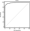
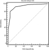
This study investigated the discriminatory features of severe acute respiratory syndrome (SARS) and severe non-SARS community-acquired viral respiratory infection (requiring hospitalization) in an emergency department in Hong Kong. In a case-control study, clinical, laboratory and radiological data from 322 patients with laboratory-confirmed SARS from the 2003 SARS outbreak were compared with the data of 253 non-SARS adult patients with confirmed viral respiratory tract infection from 2004 in order to identify discriminatory features. Among the non-SARS patients, 235 (93%) were diagnosed as having influenza infections (primarily H3N2 subtype) and 77 (30%) had radiological evidence of pneumonia. In the early phase of the illness and after adjusting for baseline characteristics, SARS patients were less likely to have lower respiratory symptoms (e.g. sputum production, shortness of breath, chest pain) and more likely to have myalgia (p < 0.001). SARS patients had lower mean leukocyte and neutrophil counts (p < 0.0001) and more commonly had "ground-glass" radiological changes with no pleural effusion. Despite having a younger average age, SARS patients had a more aggressive respiratory course requiring admission to the ICU and a higher mortality rate. The area under the receiver operator characteristic curve for predicting SARS when all variables were considered was 0.983. Using a cutoff score of >99, the sensitivity was 89.1% (95%CI 82.0-94.0) and the specificity was 98.0% (95%CI 95.4-99.3). The area under the receiver operator characteristic curve for predicting SARS when all variables except radiological change were considered was 0.933. Using a cutoff score of >8, the sensitivity was 80.7% (95%CI 72.4-87.3) and the specificity was 94.5% (95%CI 90.9-96.9). Certain clinical manifestations and laboratory changes may help to distinguish SARS from other influenza-like illnesses. Scoring systems may help identify patients who should receive more specific tests for influenza or SARS.
Figures
Fig. 1
Receiver operating characteristic curve for predicting SARS, including all significant variables
Fig. 2
Receiver operating characteristic curve for predicting SARS, including all significant variables except radiographic appearance
Similar articles
- Role of laboratory variables in differentiating SARS-coronavirus from other causes of community-acquired pneumonia within the first 72 h of hospitalization.
Lee N, Rainer TH, Ip M, Zee B, Ng MH, Antonio GE, Chan E, Lui G, Cockram CS, Sung JJ, Hui DS. Lee N, et al. Eur J Clin Microbiol Infect Dis. 2006 Dec;25(12):765-72. doi: 10.1007/s10096-006-0222-z. Eur J Clin Microbiol Infect Dis. 2006. PMID: 17077967 Free PMC article. - SARS and common viral infections.
Louie JK, Hacker JK, Mark J, Gavali SS, Yagi S, Espinosa A, Schnurr DP, Cossen CK, Isaacson ER, Glaser CA, Fischer M, Reingold AL, Vugia DJ; Unexplained Deaths and Critical Illnesses Working Group. Louie JK, et al. Emerg Infect Dis. 2004 Jun;10(6):1143-6. doi: 10.3201/eid1006.030863. Emerg Infect Dis. 2004. PMID: 15207072 Free PMC article. - [Viral infections of the respiratory tract].
Toczyska I, Targowski T. Toczyska I, et al. Pol Merkur Lekarski. 2012 Nov;33(197):261-4. Pol Merkur Lekarski. 2012. PMID: 23394036 Review. Polish. - Multiplex PCR theranostics of severe respiratory infections.
Pozzetto B, Grattard F, Pillet S. Pozzetto B, et al. Expert Rev Anti Infect Ther. 2010 Mar;8(3):251-3. doi: 10.1586/eri.09.131. Expert Rev Anti Infect Ther. 2010. PMID: 20192677 Free PMC article. No abstract available. - Severe acute respiratory syndrome in children.
Stockman LJ, Massoudi MS, Helfand R, Erdman D, Siwek AM, Anderson LJ, Parashar UD. Stockman LJ, et al. Pediatr Infect Dis J. 2007 Jan;26(1):68-74. doi: 10.1097/01.inf.0000247136.28950.41. Pediatr Infect Dis J. 2007. PMID: 17195709 Review.
Cited by
- Viral Load Dynamics in Sputum and Nasopharyngeal Swab in Patients with COVID-19.
Liu R, Yi S, Zhang J, Lv Z, Zhu C, Zhang Y. Liu R, et al. J Dent Res. 2020 Oct;99(11):1239-1244. doi: 10.1177/0022034520946251. Epub 2020 Aug 3. J Dent Res. 2020. PMID: 32744907 Free PMC article. - Clinical characteristics of COVID-19 complicated with pleural effusion.
Zhan N, Guo Y, Tian S, Huang B, Tian X, Zou J, Xiong Q, Tang D, Zhang L, Dong W. Zhan N, et al. BMC Infect Dis. 2021 Feb 15;21(1):176. doi: 10.1186/s12879-021-05856-8. BMC Infect Dis. 2021. PMID: 33588779 Free PMC article. - The costs of an expanded screening criteria for COVID-19: A modelling study.
Lim JT, Dickens BL, Cook AR, Khoo AL, Dan YY, Fisher DA, Tambyah PA, Chai LYA. Lim JT, et al. Int J Infect Dis. 2020 Nov;100:490-496. doi: 10.1016/j.ijid.2020.08.025. Epub 2020 Aug 12. Int J Infect Dis. 2020. PMID: 32800857 Free PMC article. - Guillain-Barre syndrome during COVID-19 pandemic: an overview of the reports.
Rahimi K. Rahimi K. Neurol Sci. 2020 Nov;41(11):3149-3156. doi: 10.1007/s10072-020-04693-y. Epub 2020 Sep 2. Neurol Sci. 2020. PMID: 32876777 Free PMC article. Review. - Comparison of Pneumonia Severity Indices, qCSI, 4C-Mortality Score and qSOFA in Predicting Mortality in Hospitalized Patients with COVID-19 Pneumonia.
Kibar Akilli I, Bilge M, Uslu Guz A, Korkusuz R, Canbolat Unlu E, Kart Yasar K. Kibar Akilli I, et al. J Pers Med. 2022 May 16;12(5):801. doi: 10.3390/jpm12050801. J Pers Med. 2022. PMID: 35629223 Free PMC article.
References
- Ksiazek TG, Erdman D, Goldsmith CS, Zaki SR, Peret T, Emery S, Tong S, Urbani C, Comer JA, Lim W, Rollin PE, Dowell SF, Ling AE, Humphrey CD, Shieh WJ, Guarner J, Paddock CD, Rota P, Fields B, DeRisi J, Yang JY, Cox N, Hughes JM, LeDuc JW, Bellini WJ, Anderson LJ, SARS Working Group A novel coronavirus associated with severe acute respiratory syndrome. N Engl J Med. 2003;348:1953–1966. doi: 10.1056/NEJMoa030781. - DOI - PubMed
- Peiris JS, Lai ST, Poon LL, Guan Y, Yam LY, Lim W, Nicholls J, Yee WK, Yan WW, Cheung MT, Cheng VC, Chan KH, Tsang DN, Yung RW, Ng TK, Yuen KY, SARS study group Coronavirus as a possible cause of severe acute respiratory syndrome. Lancet. 2003;361:1319–1325. doi: 10.1016/S0140-6736(03)13077-2. - DOI - PMC - PubMed
- Drosten C, Gunther S, Preiser W, van der Werf S, Brodt HR, Becker S, Rabenau H, Panning M, Kolesnikova L, Fouchier RA, Berger A, Burguiere AM, Cinatl J, Eickmann M, Escriou N, Grywna K, Kramme S, Manuguerra JC, Muller S, Rickerts V, Sturmer M, Vieth S, Klenk HD, Osterhaus AD, Schmitz H, Doerr HW. Identification of a novel coronavirus in patients with severe acute respiratory syndrome. N Engl J Med. 2003;348:1967–1976. doi: 10.1056/NEJMoa030747. - DOI - PubMed
- World Health Organization. Case definitions for surveillance of severe acute respiratory syndrome (SARS). http://www.who.int/csr/sars/casedefinition/en/. Cited 4 December 2003
- Rainer TH, Chan PKS, Ip M, Lee N, Hui DS, Wu A, Ahuja AT, Tam JS, Sung JY, Cameron P. Preliminary analysis of the spectrum of severe acute respiratory syndrome-associated coronavirus (SARS-CoV) infection in a large outbreak. Ann Intern Med. 2004;140:614–619. - PubMed
MeSH terms
LinkOut - more resources
Full Text Sources
Medical
Miscellaneous

