Degradation of fibrillar forms of Alzheimer's amyloid beta-peptide by macrophages - PubMed (original) (raw)
Degradation of fibrillar forms of Alzheimer's amyloid beta-peptide by macrophages
Amitabha Majumdar et al. Neurobiol Aging. 2008 May.
Abstract
Cultured microglia internalize fibrillar amyloid Abeta (fAbeta) and deliver it to lysosomes. Degradation of fAbeta by microglia is incomplete, but macrophages degrade fAbeta efficiently. When mannose-6 phosphorylated lysosomal enzymes were added to the culture medium of microglia, degradation of fAbeta was increased, and the increased degradation was inhibited by excess mannose-6-phosphate, which competes for binding and endocytic uptake. This suggests that low activity of one or more lysosomal enzymes in the microglia was responsible for the poor degradation of fAbeta. To further characterize the degradation of fAbeta in late endosomes and lysosomes, we analyzed fAbeta-derived intracellular degradation products in macrophages and microglia by mass spectrometry. Fragments with truncations in the first 12 N-terminal residues were observed in extracts from both cell types. We also analyzed material released by the cells. Microglia released mainly intact Abeta1-42, whereas macrophages released a variety of N-terminal truncated fragments. These results indicate that initial proteolysis near the N-terminus is similar in both cell types, but microglia are limited in their ability to make further cuts in the fAbeta.
Conflict of interest statement
Disclosure Statement: The authors declare that there is no actual or potential conflict of interest with any person or organization.
Figures
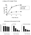
Figure 1. Degradation of labeled fAβ in macrophages and microglia
(A) 125_I-labeled fAβ_. Microglia, J774 cells and primary mice peritoneal macrophages were incubated for 1 h with 125I-labeled fAβ 1 µg/ml) after which the cells were washed and chased for varying times. In parallel samples, excess Ac-LDL (200 µg/ml) or fucoidan (500 µg/ml) was added to the radiolabeled fAβ to determine the extent of nonspecific cell-associated radiolabeled fAβ, for which all measurements were corrected. At each chase time, the chase medium was collected, and cells were solubilized. The chase medium was subjected to TCA precipitation to monitor degradation. The radioactivity of TCA-soluble fractions in peritoneal macrophages (Δ), J774 cells (●), and microglia (■) are shown as the % of total radioactivity. The data presented are averages of the radioactive counts from three dishes per condition from six different experiments using fAβ42. Error bars, S.E. (B) Cy3-fAβ. Cells were incubated for 45 min with Cy3-fAβ42 (1 µg/ml) and chased for various times. After each chase time, the cells were rinsed extensively, fixed and imaged by digital fluorescence microscopy. The Cy3 fluorescence intensity remaining in each cell over the course of the chase was quantified. The integrated cell-associated fluorescence power (arbitrary units) normalized to the fluorescence power at day 0 over the course of 3days is shown in the figure. Data for each time point shows the average of the normalized fluorescence power values from 3 different experiments done on 3 different days. Error bars, represent S.E.
Figure 2. fAβ degradation by microglia in the presence of Man6P tagged enzymes
Cells were incubated for 45 min with Cy3-fAβ42 (1 µg/ml). After incubation the cells were rinsed extensively with complete media and were chased in (A) complete media (B) complete media+30nM mannose-6 phosphate phosphorylated lysosomal enzyme (C) complete media +30nM mannose-6 phosphate phosphorylated lysosomal enzyme + 10mM mannose-6 phosphate. After 3 days, the cells were rinsed extensively, fixed, and imaged by digital fluorescence microscopy. The integrated Cy3 fluorescence power per cell before and after the chase was quantified. The integrated cell-associated fluorescence power (arbitrary units) after 3 days normalized to the fluorescence power at day 0 is shown in the figure. The P value (unpaired Student’s t test) for fAβ degradation by microglia after add-back of 30nM mannose-6 phosphate phosphorylated lysosomal enzyme was less than 0.005 compared to either control microglia or enzyme addition in the presence of 10mM mannose-6 phosphate. Data for each condition show the average of the fluorescence power values from 3 different experiments done on 3 different days. Error bars, represent S.E.
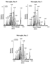
Figure 3. MALDI-TOF-MS spectrum of fAβ fragments in cells after uptake
A). Microglia [(i) chase day 0 (ii) chase day 1 and (iii) chase day 2] B) J774 macrophages [(i) chase day 0 (ii) chase day 1 and (iii) chase day 2] were pulsed with fAβ peptides overnight. The cells were then rinsed and chased for indicated times. At the end of each chase time, the cells were rinsed and lysed in lysis buffer. The cells were sonicated and centrifuged at 100,000 × g for 1 h at 4°C. The resulting pellet was solubilized in 70% formic acid, followed by immunoprecipitation using monoclonal Aβ antibody 4G8. The molecular masses of eluted samples were measured using mass spectrometry. The identities of the observed peaks are indicated using human Aβ sequence numbers. Representative spectra from microglia and J774 cells from ten experiments of each cell type are shown. * indicates the protonated ion peak; ** indicates the mono-sodium adduct peak; *** indicates the disodium adduct peak.
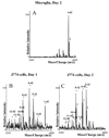
Figure 4. MALDI-TOF-MS of fAβ fragments released into the chase medium
The chase media from microglia (A; chase day 2), J774 cells (B; chase day 1), and J774 cells (C; chase day 2) that were pulsed with fAβ peptides overnight were collected and subjected to TCA precipitation. The TCA insoluble material was solubilized in 70% formic acid, and immunoprecipitated using monoclonal Aβ antibody 4G8. The eluted samples were measured using mass spectrometry. The identities of the observed peaks are indicated using human Aβ sequence numbers. Representative spectra from ten experiments are shown. * indicates the protonated ion peak; ** is the mono-sodium adduct peak.
Similar articles
- Uptake, degradation, and release of fibrillar and soluble forms of Alzheimer's amyloid beta-peptide by microglial cells.
Chung H, Brazil MI, Soe TT, Maxfield FR. Chung H, et al. J Biol Chem. 1999 Nov 5;274(45):32301-8. doi: 10.1074/jbc.274.45.32301. J Biol Chem. 1999. PMID: 10542270 - Activation of microglia acidifies lysosomes and leads to degradation of Alzheimer amyloid fibrils.
Majumdar A, Cruz D, Asamoah N, Buxbaum A, Sohar I, Lobel P, Maxfield FR. Majumdar A, et al. Mol Biol Cell. 2007 Apr;18(4):1490-6. doi: 10.1091/mbc.e06-10-0975. Epub 2007 Feb 21. Mol Biol Cell. 2007. PMID: 17314396 Free PMC article. - Deacetylation of TFEB promotes fibrillar Aβ degradation by upregulating lysosomal biogenesis in microglia.
Bao J, Zheng L, Zhang Q, Li X, Zhang X, Li Z, Bai X, Zhang Z, Huo W, Zhao X, Shang S, Wang Q, Zhang C, Ji J. Bao J, et al. Protein Cell. 2016 Jun;7(6):417-33. doi: 10.1007/s13238-016-0269-2. Epub 2016 May 21. Protein Cell. 2016. PMID: 27209302 Free PMC article. - Fibrillar beta-amyloid induces microglial phagocytosis, expression of inducible nitric oxide synthase, and loss of a select population of neurons in the rat CNS in vivo.
Weldon DT, Rogers SD, Ghilardi JR, Finke MP, Cleary JP, O'Hare E, Esler WP, Maggio JE, Mantyh PW. Weldon DT, et al. J Neurosci. 1998 Mar 15;18(6):2161-73. doi: 10.1523/JNEUROSCI.18-06-02161.1998. J Neurosci. 1998. PMID: 9482801 Free PMC article. Review.
Cited by
- Pathology of Amyloid-β (Aβ) Peptide Peripheral Clearance in Alzheimer's Disease.
Tsoy A, Umbayev B, Kassenova A, Kaupbayeva B, Askarova S. Tsoy A, et al. Int J Mol Sci. 2024 Oct 11;25(20):10964. doi: 10.3390/ijms252010964. Int J Mol Sci. 2024. PMID: 39456746 Free PMC article. Review. - Fast-spiking parvalbumin-positive interneurons in brain physiology and Alzheimer's disease.
Hijazi S, Smit AB, van Kesteren RE. Hijazi S, et al. Mol Psychiatry. 2023 Dec;28(12):4954-4967. doi: 10.1038/s41380-023-02168-y. Epub 2023 Jul 7. Mol Psychiatry. 2023. PMID: 37419975 Free PMC article. Review. - Naturally-aged microglia exhibit phagocytic dysfunction accompanied by gene expression changes reflective of underlying neurologic disease.
Thomas AL, Lehn MA, Janssen EM, Hildeman DA, Chougnet CA. Thomas AL, et al. Sci Rep. 2022 Nov 14;12(1):19471. doi: 10.1038/s41598-022-21920-y. Sci Rep. 2022. PMID: 36376530 Free PMC article. - Polyunsaturated Fatty Acids Mend Macrophage Transcriptome, Glycome, and Phenotype in the Patients with Neurodegenerative Diseases, Including Alzheimer's Disease.
Dover M, Moseley T, Biskaduros A, Paulchakrabarti M, Hwang SH, Hammock B, Choudhury B, Kaczor-Urbanowicz KE, Urbanowicz A, Morselli M, Dang J, Pellegrini M, Paul K, Bentolila LA, Fiala M. Dover M, et al. J Alzheimers Dis. 2023;91(1):245-261. doi: 10.3233/JAD-220764. J Alzheimers Dis. 2023. PMID: 36373322 Free PMC article. - Elevated levels of tripeptidyl peptidase 1 do not ameliorate pathogenesis in a mouse model of Alzheimer disease.
Sleat DE, Maita I, Banach-Petrosky W, Larrimore KE, Liu T, Cruz D, Baker L, Maxfield FR, Samuels B, Lobel P. Sleat DE, et al. Neurobiol Aging. 2022 Oct;118:106-107. doi: 10.1016/j.neurobiolaging.2022.06.012. Epub 2022 Jul 7. Neurobiol Aging. 2022. PMID: 35914472 Free PMC article.
References
- Akiyama H, Schwab C, Kondo H, Mori H, Kametani F, Ikeda K, McGeer PL. Granules in glial cells of patients with Alzheimer's disease are immunopositive for C-terminal sequences of beta-amyloid protein. Neurosci Lett. 1996;206(2–3):169–172. - PubMed
- Antzutkin ON, Leapman RD, Balbach JJ, Tycko R. Supramolecular structural constraints on Alzheimer's beta-amyloid fibrils from electron microscopy and solid-state nuclear magnetic resonance. Biochemistry. 2002;41(51):15436–15450. - PubMed
- Bacskai BJ, Kajdasz ST, Christie RH, Carter C, Games D, Seubert P, Schenk D, Hyman BT. Imaging of amyloid-beta deposits in brains of living mice permits direct observation of clearance of plaques with immunotherapy. Nat Med. 2001;7(3):369–372. - PubMed
- Bard F, Cannon C, Barbour R, Burke RL, Games D, Grajeda H, Guido T, Hu K, Huang J, Johnson-Wood K, Khan K, Kholodenko D, Lee M, Lieberburg I, Motter R, Nguyen M, Soriano F, Vasquez N, Weiss K, Welch B, Seubert P, Schenk D, Yednock T. Peripherally administered antibodies against amyloid beta-peptide enter the central nervous system and reduce pathology in a mouse model of Alzheimer disease. Nat Med. 2000;6(8):916–919. - PubMed
Publication types
MeSH terms
Substances
Grants and funding
- R01 DK027083-26/DK/NIDDK NIH HHS/United States
- R01 DK054317-08/DK/NIDDK NIH HHS/United States
- R01 NS034761-08/NS/NINDS NIH HHS/United States
- R01 DK054317/DK/NIDDK NIH HHS/United States
- DK27083/DK/NIDDK NIH HHS/United States
- R01 DK027083/DK/NIDDK NIH HHS/United States
- R37 DK027083-28/DK/NIDDK NIH HHS/United States
- DK54317/DK/NIDDK NIH HHS/United States
- R01 NS034761-07/NS/NINDS NIH HHS/United States
- NS 34761/NS/NINDS NIH HHS/United States
- R01 NS034761/NS/NINDS NIH HHS/United States
- R37 DK027083-27/DK/NIDDK NIH HHS/United States
- R01 DK054317-07/DK/NIDDK NIH HHS/United States
- R37 DK027083/DK/NIDDK NIH HHS/United States
- R01 NS034761-06/NS/NINDS NIH HHS/United States
LinkOut - more resources
Full Text Sources
Other Literature Sources


