The inflammatory cytokine tumor necrosis factor-alpha generates an autocrine tumor-promoting network in epithelial ovarian cancer cells - PubMed (original) (raw)
The inflammatory cytokine tumor necrosis factor-alpha generates an autocrine tumor-promoting network in epithelial ovarian cancer cells
Hagen Kulbe et al. Cancer Res. 2007.
Abstract
Constitutive expression of the inflammatory cytokine tumor necrosis factor-alpha (TNF-alpha) is characteristic of malignant ovarian surface epithelium. We investigated the hypothesis that this autocrine action of TNF-alpha generates and sustains a network of other mediators that promote peritoneal cancer growth and spread. When compared with two ovarian cancer cell lines that did not make TNF-alpha, constitutive production of TNF-alpha was associated with greater release of the chemokines CCL2 and CXCL12, the cytokines interleukin-6 (IL-6) and macrophage migration-inhibitory factor (MIF), and the angiogenic factor vascular endothelial growth factor (VEGF). TNF-alpha production was associated also with increased peritoneal dissemination when the ovarian cancer cells were xenografted. We next used RNA interference to generate stable knockdown of TNF-alpha in ovarian cancer cells. Production of CCL2, CXCL12, VEGF, IL-6, and MIF was decreased significantly in these cells compared with wild-type or mock-transfected cells, but in vitro growth rates were unaltered. Tumor growth and dissemination in vivo were significantly reduced when stable knockdown of TNF-alpha was achieved. Tumors derived from TNF-alpha knockdown cells were noninvasive and well circumscribed and showed high levels of apoptosis, even in the smallest deposits. This was reflected in reduced vascularization of TNF-alpha knockdown tumors. Furthermore, culture supernatants from such cells failed to stimulate endothelial cell growth in vitro. We conclude that autocrine production of TNF-alpha by ovarian cancer cells stimulates a constitutive network of other cytokines, angiogenic factors, and chemokines that may act in an autocrine/paracrine manner to promote colonization of the peritoneum and neovascularization of developing tumor deposits.
Figures
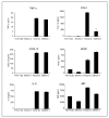
Figure 1
TNF-α release by ovarian cancer cell lines is associated with expression of other protein mediators. Proteins secreted by ovarian cancer cell lines were measured by ELISA after 48 h. Data are representative of three independent experiments done. Columns, mean of triplicate wells for TNF-α, CCL2, CXCL12, VEGF, IL-6, and MIF; bars, SD.
Figure 2
Appearance and location of tumor deposits in nude mice injected with ovarian cancer xenografts. A, gross patterns of peritoneal tumor spread of TOV112D, SKOV-3, TOV21G, and IGROV-1. B, location of tumor deposits in various organs as determined by histologic examination postmortem for TOV112D, SKOV-3, TOV21G, and IGROV-1.
Figure 3
In vitro effects of stable expression of shRNAto TNF-α. In all experiments, IGROV-Mock cells are compared with two independently isolated TNF-α shRNA clones RNAi TNF-α (I) and RNAi TNF-α (II). A, TNF-α protein secretion as measured by ELISA after 72 h. Columns, mean of triplicate wells; bars, SD. Silencing TNF-α expression in IGROV-1 cells also results in decreased protein secretion of CCL2, CXCL12, VEGF, IL-6, and MIF compared with IGROV-Mock as measured by ELISA after 48 h. Columns, mean of triplicate wells; bars, SD. B, proliferation of IGROV-1, IGROV-Mock, RNAi TNF-α (I), and RNAi TNF-α (II) cells in vitro as determined by WST-1 assay over a period of 4 d. Data are representative of three independent experiments. Points, mean for absorbance at 450 nm in triplicates; bars, SD. C, migration of IGROV-1 cells to CXCL12. IGROV-1 cells have low basal migration (black columns) but migrate toward CXCL12 (white columns). Results are representative of three experiments done Columns, mean of 10 determinations; bars, SD.
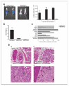
Figure 4
Effects of TNF-α knockdown on growth of cells in vivo. A, a, representative bioluminescence imaging in vivo of IGROV-Mock and RNAi TNF-α IGROV 42 d after i.p. injection. Bioluminescence is presented as a pseudocolor scale: red, highest photon flux; blue, lowest photon flux. b, quantification of bioluminescence from primary tumors (n = 6 each group) of images obtained on days 14, 28, and 42. B, tumor weight of dissectable tumors from each group (n = 5) are shown 6 wks after i.p. injection Columns, mean; bars, SD. C, dissemination of tumor deposits at 6 wks are presented as the percentage of mice with tumors at various anatomic sites. Because no differences between the RNAi TNF-α clones were seen, data from all mice injected with RNAi TNF-α cells were combined (n = 8). D, representative pictures of sections are shown from (a) IGROV-Mock and (b) RNAi TNF-α tumors (magnification, ×20). For detailed histologic analysis, images with higher magnification (magnification, ×40) from (c) IGROV-Mock and (d) RNAi TNF-α tumors are shown. Arrows, areas of apoptotic events.
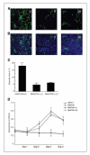
Figure 5
TNF-α knockdown influences tumor angiogenesis. In vivo angiogenesis was evaluated in mice 42 d after tumor cell injection. A, confocal images (magnification, ×20) of representative sections from (I) IGROV-Mock, (II) RNAi TNF-α I, and (III) RNAi TNF-α II tumors, after injection of FITC-conjugated lectin and in (B)4′,6-diamidino-2-phenylindole–stained cell nuclei are shown as control. C, columns, mean vascular area in each group quantified in 10 randomly selected areas of tumor sections (n = 5); bars, SD. D, proliferation of primary mouse lung endothelial cells in vitro with conditioned medium of IGROV-1, IGROV-Mock, and RNAi TNF-α cells. Representative of two independent experiments. Points, mean values for absorbance at 450 nm in triplicates; bars, SD.
Similar articles
- Impact of residual disease as a prognostic factor for survival in women with advanced epithelial ovarian cancer after primary surgery.
Bryant A, Hiu S, Kunonga PT, Gajjar K, Craig D, Vale L, Winter-Roach BA, Elattar A, Naik R. Bryant A, et al. Cochrane Database Syst Rev. 2022 Sep 26;9(9):CD015048. doi: 10.1002/14651858.CD015048.pub2. Cochrane Database Syst Rev. 2022. PMID: 36161421 Free PMC article. Review. - A Blog-Based Study of Autistic Adults' Experiences of Aloneness and Connection and the Interplay with Well-Being: Corpus-Based and Thematic Analyses.
Petty S, Allen S, Pickup H, Woodier B. Petty S, et al. Autism Adulthood. 2023 Dec 1;5(4):437-449. doi: 10.1089/aut.2022.0073. Epub 2023 Dec 12. Autism Adulthood. 2023. PMID: 38116056 Free PMC article. - Depressing time: Waiting, melancholia, and the psychoanalytic practice of care.
Salisbury L, Baraitser L. Salisbury L, et al. In: Kirtsoglou E, Simpson B, editors. The Time of Anthropology: Studies of Contemporary Chronopolitics. Abingdon: Routledge; 2020. Chapter 5. In: Kirtsoglou E, Simpson B, editors. The Time of Anthropology: Studies of Contemporary Chronopolitics. Abingdon: Routledge; 2020. Chapter 5. PMID: 36137063 Free Books & Documents. Review. - Defining the optimum strategy for identifying adults and children with coeliac disease: systematic review and economic modelling.
Elwenspoek MM, Thom H, Sheppard AL, Keeney E, O'Donnell R, Jackson J, Roadevin C, Dawson S, Lane D, Stubbs J, Everitt H, Watson JC, Hay AD, Gillett P, Robins G, Jones HE, Mallett S, Whiting PF. Elwenspoek MM, et al. Health Technol Assess. 2022 Oct;26(44):1-310. doi: 10.3310/ZUCE8371. Health Technol Assess. 2022. PMID: 36321689 Free PMC article. - Anti-angiogenic therapy for high-grade glioma.
Ameratunga M, Pavlakis N, Wheeler H, Grant R, Simes J, Khasraw M. Ameratunga M, et al. Cochrane Database Syst Rev. 2018 Nov 22;11(11):CD008218. doi: 10.1002/14651858.CD008218.pub4. Cochrane Database Syst Rev. 2018. PMID: 30480778 Free PMC article.
Cited by
- Expression of tumor necrosis factor-alpha-mediated genes predicts recurrence-free survival in lung cancer.
Wang B, Song N, Yu T, Zhou L, Zhang H, Duan L, He W, Zhu Y, Bai Y, Zhu M. Wang B, et al. PLoS One. 2014 Dec 30;9(12):e115945. doi: 10.1371/journal.pone.0115945. eCollection 2014. PLoS One. 2014. PMID: 25548907 Free PMC article. - Elucidating tumor heterogeneity from spatially resolved transcriptomics data by multi-view graph collaborative learning.
Zuo C, Zhang Y, Cao C, Feng J, Jiao M, Chen L. Zuo C, et al. Nat Commun. 2022 Oct 10;13(1):5962. doi: 10.1038/s41467-022-33619-9. Nat Commun. 2022. PMID: 36216831 Free PMC article. - Anti-tumour effects of a specific anti-ADAM17 antibody in an ovarian cancer model in vivo.
Richards FM, Tape CJ, Jodrell DI, Murphy G. Richards FM, et al. PLoS One. 2012;7(7):e40597. doi: 10.1371/journal.pone.0040597. Epub 2012 Jul 11. PLoS One. 2012. PMID: 22792380 Free PMC article. - Skewed Signaling through the Receptor for Advanced Glycation End-Products Alters the Proinflammatory Profile of Tumor-Associated Macrophages.
Rojas A, Araya P, Romero J, Delgado-López F, Gonzalez I, Añazco C, Perez-Castro R. Rojas A, et al. Cancer Microenviron. 2018 Dec;11(2-3):97-105. doi: 10.1007/s12307-018-0214-4. Epub 2018 Aug 8. Cancer Microenviron. 2018. PMID: 30091031 Free PMC article. Review. - NF-κB Signaling in Ovarian Cancer.
Harrington BS, Annunziata CM. Harrington BS, et al. Cancers (Basel). 2019 Aug 15;11(8):1182. doi: 10.3390/cancers11081182. Cancers (Basel). 2019. PMID: 31443240 Free PMC article. Review.
References
- Moore R, Owens D, Stamp G, et al. Tumour necrosis factor-α deficient mice are resistant to skin carcinogenesis. Nat Med. 1999;5:828–31. - PubMed
- Arnott CH, Scott KA, Moore RJ, et al. Tumour necrosis factor-α mediates tumour promotion via a PKCα- AP-1-dependent pathway. Oncogene. 2002;21:4728–38. - PubMed
- Pikarsky E, Porat RM, Stein I, et al. NF-κB functions as a tumour promoter in inflammation-associated cancer. Nature. 2004;431:4461–6. - PubMed
- Szlosarek PW, Grimshaw MJ, Kulbe H, et al. Expression and regulation of tumor necrosis factor-α in normal and malignant ovarian epithelium. Mol Cancer Ther. 2006;5:382–90. - PubMed
- Szlosarek P, Balkwill F. Tumour necrosis factor-α: a potential target in the therapy of solid tumors. Lancet Oncol. 2003;4:565–73. - PubMed
Publication types
MeSH terms
Substances
LinkOut - more resources
Full Text Sources
Other Literature Sources
Medical
Miscellaneous


