Wandering minds: the default network and stimulus-independent thought - PubMed (original) (raw)
Wandering minds: the default network and stimulus-independent thought
Malia F Mason et al. Science. 2007.
Abstract
Despite evidence pointing to a ubiquitous tendency of human minds to wander, little is known about the neural operations that support this core component of human cognition. Using both thought sampling and brain imaging, the current investigation demonstrated that mind-wandering is associated with activity in a default network of cortical regions that are active when the brain is "at rest." In addition, individuals' reports of the tendency of their minds to wander were correlated with activity in this network.
Figures
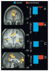
Fig. 1
Graphs depict regions of the default network exhibiting significantly greater activity during practiced blocks (red) relative to novel blocks (blue) at a threshold of P < 0.001, number of voxels (k) = 10. Mean activity was computed for each participant by averaging the signal in regions within 10 mm of the peak, across the duration of the entire block. Graphs depict the mean signal change across all participants. (A) Left (L.) mPFC (BA 9; –6, 54, 22); (B) BXXX (B.) cingulate (BA 24; 0, –7, 36); (C) Right (R.) insula (45, –26, 4); and (D) L. posterior cingulate (BA 23/31; –9, –39, 27). Activity is plotted on the average high-resolution anatomical image and displayed in neurological convention (left hemisphere is depicted on the left).
Fig. 2
Graphs depict regions that exhibited a significant positive relation, r(14) > 0.50, P < 0.05, between the frequency of mind-wandering and the change in BOLD signal observed when people performed practiced relative to novel blocks. Participants’ BOLD difference scores (practiced – novel) are plotted against their standardized IPI daydreaming score. BOLD signal values for the two blocks were computed for each participant by averaging the signal in regions within 10 mm of the peak, from 4 TRs (10 s) until 10 TRs (22.5 s) after the block onset. (A) B. mPFC (BA 10; –6, 51, –9; k = 25); (B) B. precuneus and p. cingulate (BA 31, 7; –3, –45, 37; k = 72); (C) R. cingulate (BA 31; 7, –21, 51; k = 73); (D) L. insula (BA 13; –36, –16, 17; k = 10); (E) R. insula (BA 13; 47, 0, 4; k = 13). Activity is plotted on the average high-resolution anatomical image and displayed in neurological convention (left hemisphere is depicted on the left).
Comment in
- Comment on "Wandering minds: the default network and stimulus-independent thought".
Gilbert SJ, Dumontheil I, Simons JS, Frith CD, Burgess PW. Gilbert SJ, et al. Science. 2007 Jul 6;317(5834):43; author reply 43. doi: 10.1126/science.317.5834.43. Science. 2007. PMID: 17615325
Similar articles
- Comment on "Wandering minds: the default network and stimulus-independent thought".
Gilbert SJ, Dumontheil I, Simons JS, Frith CD, Burgess PW. Gilbert SJ, et al. Science. 2007 Jul 6;317(5834):43; author reply 43. doi: 10.1126/science.317.5834.43. Science. 2007. PMID: 17615325 - Shaped by our thoughts--a new task to assess spontaneous cognition and its associated neural correlates in the default network.
O'Callaghan C, Shine JM, Lewis SJ, Andrews-Hanna JR, Irish M. O'Callaghan C, et al. Brain Cogn. 2015 Feb;93:1-10. doi: 10.1016/j.bandc.2014.11.001. Epub 2014 Nov 18. Brain Cogn. 2015. PMID: 25463243 - Mind wandering away from pain dynamically engages antinociceptive and default mode brain networks.
Kucyi A, Salomons TV, Davis KD. Kucyi A, et al. Proc Natl Acad Sci U S A. 2013 Nov 12;110(46):18692-7. doi: 10.1073/pnas.1312902110. Epub 2013 Oct 28. Proc Natl Acad Sci U S A. 2013. PMID: 24167282 Free PMC article. - A Neural Model of Mind Wandering.
Mittner M, Hawkins GE, Boekel W, Forstmann BU. Mittner M, et al. Trends Cogn Sci. 2016 Aug;20(8):570-578. doi: 10.1016/j.tics.2016.06.004. Epub 2016 Jun 25. Trends Cogn Sci. 2016. PMID: 27353574 Review. - Mind-wandering as spontaneous thought: a dynamic framework.
Christoff K, Irving ZC, Fox KC, Spreng RN, Andrews-Hanna JR. Christoff K, et al. Nat Rev Neurosci. 2016 Nov;17(11):718-731. doi: 10.1038/nrn.2016.113. Epub 2016 Sep 22. Nat Rev Neurosci. 2016. PMID: 27654862 Review.
Cited by
- Neural activity changes associated with impulsive responding in the sustained attention to response task.
Sakai H, Uchiyama Y, Shin D, Hayashi MJ, Sadato N. Sakai H, et al. PLoS One. 2013 Jun 25;8(6):e67391. doi: 10.1371/journal.pone.0067391. Print 2013. PLoS One. 2013. PMID: 23825657 Free PMC article. - Brain activity and connectivity during poetry composition: Toward a multidimensional model of the creative process.
Liu S, Erkkinen MG, Healey ML, Xu Y, Swett KE, Chow HM, Braun AR. Liu S, et al. Hum Brain Mapp. 2015 Sep;36(9):3351-72. doi: 10.1002/hbm.22849. Epub 2015 May 26. Hum Brain Mapp. 2015. PMID: 26015271 Free PMC article. - Culture-related differences in default network activity during visuo-spatial judgments.
Goh JO, Hebrank AC, Sutton BP, Chee MW, Sim SK, Park DC. Goh JO, et al. Soc Cogn Affect Neurosci. 2013 Feb;8(2):134-42. doi: 10.1093/scan/nsr077. Epub 2011 Nov 22. Soc Cogn Affect Neurosci. 2013. PMID: 22114080 Free PMC article. - A shift in perspective: Decentering through mindful attention to imagined stressful events.
Lebois LA, Papies EK, Gopinath K, Cabanban R, Quigley KS, Krishnamurthy V, Barrett LF, Barsalou LW. Lebois LA, et al. Neuropsychologia. 2015 Aug;75:505-24. doi: 10.1016/j.neuropsychologia.2015.05.030. Epub 2015 Jun 22. Neuropsychologia. 2015. PMID: 26111487 Free PMC article. - Mindfulness in the focus of the neurosciences - The contribution of neuroimaging to the understanding of mindfulness.
Weder BJ. Weder BJ. Front Behav Neurosci. 2022 Oct 17;16:928522. doi: 10.3389/fnbeh.2022.928522. eCollection 2022. Front Behav Neurosci. 2022. PMID: 36325155 Free PMC article.
References
- Singer JL. Daydreaming. Plenum Press; New York: 1966.
- Antrobus JS, Singer JL, Greenberg S. Percept Mot Skills. 1966;23:399.
- Singer JL, McRaven V. Int J Soc Psychiatry. 1962;8:272. - PubMed
- Klinger E. Structure and Functions of Fantasy. New York: Wiley; 1971.
- Smallwood J, Schooler JW. Psychol Bull. 2006;132:946. - PubMed
Publication types
MeSH terms
LinkOut - more resources
Full Text Sources
Other Literature Sources

