Nitric oxide and peroxynitrite in health and disease - PubMed (original) (raw)
Review
Nitric oxide and peroxynitrite in health and disease
Pál Pacher et al. Physiol Rev. 2007 Jan.
Abstract
The discovery that mammalian cells have the ability to synthesize the free radical nitric oxide (NO) has stimulated an extraordinary impetus for scientific research in all the fields of biology and medicine. Since its early description as an endothelial-derived relaxing factor, NO has emerged as a fundamental signaling device regulating virtually every critical cellular function, as well as a potent mediator of cellular damage in a wide range of conditions. Recent evidence indicates that most of the cytotoxicity attributed to NO is rather due to peroxynitrite, produced from the diffusion-controlled reaction between NO and another free radical, the superoxide anion. Peroxynitrite interacts with lipids, DNA, and proteins via direct oxidative reactions or via indirect, radical-mediated mechanisms. These reactions trigger cellular responses ranging from subtle modulations of cell signaling to overwhelming oxidative injury, committing cells to necrosis or apoptosis. In vivo, peroxynitrite generation represents a crucial pathogenic mechanism in conditions such as stroke, myocardial infarction, chronic heart failure, diabetes, circulatory shock, chronic inflammatory diseases, cancer, and neurodegenerative disorders. Hence, novel pharmacological strategies aimed at removing peroxynitrite might represent powerful therapeutic tools in the future. Evidence supporting these novel roles of NO and peroxynitrite is presented in detail in this review.
Figures
FIG. 1
Cellular diffusion of superoxide, peroxynitrite, and hydroxyl radical within their estimated first half-lives. These circles indicate the extent to where the concentration of each species from a point source would decrease by 50%. The diffusion of peroxynitrite accounts for its rapid reaction with carbon dioxide and with intracellular thiols. The diffusion distance for nitric oxide is calculated based on its half-life of 1 s in vivo, which results mostly from its diffusion into red blood cells. The diffusion distance for hydroxyl radical is about the same diameter as a small protein, or 10,000 times smaller than peroxynitrite. All of these estimates involve many approximations, but varying the estimated half-lives by 10-fold would only alter the diameters by the square root of 10 or by 3.2-fold.
FIG. 2
Comparison of oxidant production by the reaction of nitric oxide with superoxide versus oxygen. Both reactions are generally given equal weight, but this obscures the vast difference in oxidant productions because of the vast difference in rates. Because the formation of peroxynitrite depends on the product of the concentration of nitric oxide and superoxide, the rate of formation is proportional to the area. Left: estimate of peroxynitrite formation in the cytosol if a cell produces 10 nM nitric oxide, sufficient to activate guanylate cyclase enough to cause at least 10% relaxation of vessels, using 0.1 nM superoxide as an estimate of the basal steady-state concentration of superoxide (777). Right: increase in peroxynitrite formation if the formation of superoxide production increased either 100-fold (yellow) or 1,000-fold (yellow-orange), increases that can reasonably occur with the activation of NADPH oxidase. Nitric oxide is shown to increase only 10-fold and could rise to ~1 _μ_M in highly inflamed states. Far right (orange square): proportional area of nitrogen dioxide formation from 100 nM nitric oxide reacting with oxygen (estimated to be 50 _μ_M in cells), which is magnified 100-fold. This rate is the faster rate occurring in hydrophobic membranes and would be 300-fold smaller in solution (784). Pathways that stimulate the synthesis of superoxide vastly increase oxidant production compared with the reaction of nitric oxide with oxygen.
FIG. 3
The chemical structure of nitric oxide is intermediate between molecular oxygen and nitrogen. The dot illustrates the unpaired electron on nitric oxide and two unpaired electrons on oxygen. These unpaired electrons are in antibonding orbitals, counteracting the three bonding orbitals characteristic of nitrogen gas. Thus nitric oxide has effectively 2.5 bonds and a slightly longer distance separating the nuclei. Oxygen has only two bonds and an even longer intranuclear distance.
FIG. 4
Diffusion of nitric oxide into the phagolysosome and the recycling of peroxynitrite-derived nitrite. Only a miniscule volume of extracellular fluid is engulfed into phagocytic vacuoles, which provides a limited amount of chloride as a substrate for myeloperoxidase. In contrast, nitric oxide can readily diffuse into the phagolysosome. Neutrophils produce superoxide by NADPH oxidases, but superoxide is unlikely to penetrate cell membranes or cell walls of pathogens, and can reversibly inactivate myeloperoxidase. Peroxynitrite is a substrate for myeloperoxidase and can reverse this inhibition. In addition, nitrite formed from peroxynitrite decomposition is entrapped within the phagolysosome and serves as an additional substrate for myeloperoxidase. Myeloperoxidase is not a predominant protein in macrophages, where formation of peroxynitrite from superoxide and nitric oxide appears to be a major mechanism of cytotoxicity.
FIG. 5
Alveolar macrophages produce peroxynitrite. When alveolar macrophages are stimulated to produce both superoxide and nitric oxide, peroxynitrite is quantitatively produced (611) as evidenced by the amount of nitric oxide and superoxide produced and the amount of oxygen consumed. Extracellular addition of superoxide dismutase (SOD) in high concentrations does not significantly reduce the amount of peroxynitrite formed and instead serves as a catalyst of tyrosine nitration. This suggests that superoxide produced at the membrane surface and nitric oxide diffusing through the membrane react at the membrane interface so quickly that SOD in the bulk phase cannot compete.
FIG. 6
The interplay of nitric oxide, superoxide, peroxynitrite, and nitrogen dioxide. When nitric oxide and superoxide are both present, they may also react with nitrogen dioxide to form N2O3 and peroxynitrate. Peroxynitrate decomposes to give nitrite and oxygen, while N2O3 can react with thiols to give nitrosothiols or with hydroxide anion to give nitrite. Goldstein et al. (452) showed that it also reacts at a diffusion-limited rate with peroxynitrite to yield two molecules of nitrogen dioxide and one of nitrite. This creates a cycle to generate more nitrogen dioxide when bolus additions of peroxynitrite are added at neutral pH and substantially increases the number of potential reactions occurring. These same reactions will also occur in vivo, particularly when nitric oxide is produced faster than superoxide.
FIG. 7
Molecular mechanisms of peroxynitrite-mediated cell death. A number of pathological conditions are associated with the simultaneous generation of nitric oxide (NO) and O2•−. NO sources are restricted to the activity of the various NO synthases, whereas O2•− arises from multiple sources, including electron leak from the mitochondria, NADPH oxidase, xanthine oxidase, and uncoupling of NO synthases. Once a flux of NO and O2•− is produced simultaeously in close proximity, the generation of peroxynitrite is considerably enhanced. Peroxynitrite-dependent cytotoxicity is then mediated by a myriad of effects including lipid peroxidation, protein nitration and oxidation, DNA oxidative damage, activation of matrix metalloproteinases (MMP), and inactivation of a series of enzymes. Mitochondrial enzymes are particularly vulnerable to attacks by peroxynitrite, leading to reduced ATP formation and induction of mitochondrial permeability transition by opening of the permeability transition pore, which dissipates the mitochondrial membrane potential (Δ_ψ_m). These events result in cessation of electron transport and ATP formation, mitochondrial swelling, and permeabilization of the outer mitochondrial membrane, allowing the efflux of several proapoptotic molecules, including cytochrome c and apoptosis-inducing factor (AIF). In turn, cytochrome c and AIF activate a series of downstream effectors that eventually lead to the fragmentation of nuclear DNA. In addition to its damaging effects on mitochondria, peroxynitrite inflicts more or less severe oxidative injury to DNA, resulting in DNA strand breakage which in turn activates the nuclear enzyme poly(ADP-ribose) polymerase (PARP). Activated PARP consumes NAD to build-up poly(ADP-ribose) polymers (PAR), which are themselves rapidly metabolized by the activity of poly(ADP-ribose) glycohydrolase (PARG). Some free PAR may exit the nucleus and travel to the mitochondria, where they amplify the mitochondrial efflux of AIF (nuclear to mitochondria cross-talk). Mild damage of DNA activates the DNA repair machinery. On the contrary, once excessive oxidative and nitrosative stress-induced DNA damage occurs, like in various forms of reperfusion injury and other pathophysiological conditions, the cell may be executed by apoptosis in case of moderate PTP opening and PARP activation with preservation of cellular ATP, or by necrosis in the case of widespread PTP opening and PARP overactivation, leading to massive NAD consumption and collapse of cellular ATP. [Derived from Pacher et al. (995) with permission from Elsevier.]
FIG. 8
Schematic diagram of mitogen-activated protein kinase (MAPK) signaling and stimulating effects of peroxynitrite. MAPKs are activated by a dual phosphorylation at a specific tripeptide motif, as indicated on the left, mediated by a conserved protein kinase cascade, involving MAPK kinases (MAPKK or MKK) and MAPK kinase kinases (MAPKKK or MKKK). The activation of the upstream MKKK is mediated by various cell surface receptors, including G protein-coupled receptors (GPCRs) and receptor tyrosine kinases (RTK), such as the receptor for epidermal growth factor (EGFR), which activate several small G proteins, such as Ras, Rho, Rac, and Cdc42. Three groups of MAPKs exist in mammalian cells, including extracellular signal-regulated protein kinase (ERK), p38 MAPK, and the c-Jun NH2-terminal kinase (JNK), whose upstream signaling intermediates include raf-1 and MKK1–2 (ERK pathway), MLK/Ask-1 (mixed lineage kinase/apoptosis-signal regulating kinase-1) and MEK 3–6 (p38), and MLK-1/Ask-1 and MKK 4–7 (JNK). Downstream targets of MAPKs are transcription factors, enzymes, and various proteins, which regulate cell growth, apoptosis, as well as inflammation. The Western blots at the bottom show the activation pattern of the three MAPKs, evidenced by their phosphorylation, induced in the cardiomyoblast cell line H9C2 by treatment with increasing concentrations of peroxynitrite. [Adapted from Pesse et al. (1024).]
FIG. 9
Role of nitric oxide (NO) and peroxynitrite in cardiovascular pathophysiology. On the one hand, NO by activating soluble guanylate cyclase (sGC)-cGMP signal transduction pathway mediates various physiological/beneficial effects in the cardiovascular system including vasodilation, inhibition of platelet aggregation, anti-inflammatory, antiremodelling, and antiapoptotic effects. On the other hand, under pathological conditions associated with increased oxidative stress and inflammation (myocardial infarction, ischemic heart disease, myocarditis, cardiomyopathy, hypertension, etc.), NO and superoxide (O2•−) react to form peroxynitrite (ONOO−) which induces cell damage via lipid peroxidation, inactivation of enzymes and other proteins by oxidation and nitration, and also activation of stress signaling, matrix metalloproteinases (MMPs) among others (see also Table 2). Peroxynitrite also triggers the release of proapoptotic factors such as cytochrome c and apoptosis-inducing factor (AIF) from the mitochondria, which mediate caspase-dependent and -independent apoptotic death pathways. Moreover, peroxynitrite, in concert with other oxidants, causes stand breaks in DNA, activating the nuclear enzyme poly(ADP-ribose) polymerase-1 (PARP-1). Mild damage of DNA activates the DNA repair machinery. In contrast, once excessive oxidative and nitrosative stress-induced DNA damage occurs, like in various forms of myocardial reperfusion injury and heart failure, overactivated PARP initiates an energy-consuming cycle by transferring ADP-ribose units from nicotinamide adenine dinucleotide (NAD+) to nuclear proteins, resulting in rapid depletion of the intracellular NAD+ and ATP pools, slowing the rate of glycolysis and mitochondrial respiration, eventually leading to cellular dysfunction and death. Poly(ADP-ribose) glycohydrolase (PARG) degrades poly(ADP-ribose) (PAR) polymers, generating free PAR polymer and ADP-ribose. Overactivated PARP also facilitates the expression of a variety of inflammatory genes leading to increased inflammation and associated oxidative stress, thus facilitating the progression of cardiovascular dysfunction and heart failure.
FIG. 10
Role of peroxynitrite in myocardial infarction. Peroxynitrite scavenger MnTBAP reduces infart size (A) and suppresses myocardial 3-nitrotyrosine formation (B) in rat. [From Levrand et al. (750) with permission from Elsevier.]
FIG. 11
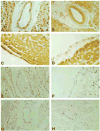
Evidence for nitrotyrosine formation in human myocardial inflammation. Representative examples are shown of nitrotyrosine immunoreactivity in cardiac tissue samples from patients with myocarditis, sepsis, or no cardiac disease (control patients). A: low-power (×20) photomicrograph of nitrotyrosine immunoreactivity (brown staining) in the myocardium, vascular endothelium, and vascular smooth muscle of myocarditis patient. B: low-power (×20) photomicrograph of nitrotyrosine immunoreactivity in the myocardium, vascular endothelium, and vascular smooth muscle of sepsis patient. Note the relative absence of staining of the connective tissue elements. C: higher-power (×40) photomicrograph of intense nitrotyrosine immunoreactivity in the endocardium of myocarditis patient. D: higher-power (×40) photomicrograph of nitrotyrosine immunoreactivity in the endocardium of sepsis patient. E: low-power (×20) photomicrograph of minimal nitrotyrosine immunoreactivity in the myocardium and the virtual absence of nitrotyrosine immunoreactivity in the vascular endothelium and vascular smooth muscle of control patient 1C. F: higher-power (×40) photomicrograph of nitrotyrosine immunoreactivity in the endocardium of control patient. G: low-power (×20) photomicrograph demonstrating the inhibition of nitrotyrosine immunoreactivity by the preincubation of the primary antibody with 10 mM nitrotyrosine before tissue staining in myocarditis patient. H: low-power (×20) photomicrograph demonstrating the inhibition of nitrotyrosine immunoreactivity by the preincubation of the primary antibody with 10 mM nitrotyrosine before tissue staining in sepsis patient. [From Kooy et al. (707) with permission from Lippincott Williams & Wilkins.]
FIG. 12
Progression of heart failure and the role of oxidative stress and peroxynitrite. The mechanisms leading to heart failure are of multiple origins and include acute and chronic ischemic heart disease, cardiomyopathies, myocarditis, and pressure overload just to mention a few. These diseases result in mismatch between the load applied to the heart and the energy needed for contraction, leading to mechanoenergic uncoupling. After initial insult, secondary mediators such as angiotensin II (AII), norepinephrine (NE), endothelin (ET), proinflammatory cytokines [e.g., tumor necrosis factor-α (TNF-α) and interleukin 6 (IL-6), in concert with oxidative stress and peroxynitrite, activate downstream effectors (e.g., PARP-1 or MMPs)], act directly on the myocardium or indirectly via changes in hemodynamic loading conditions to cause endothelial and myocardial dysfunction, cardiac and vascular remodeling with hypertrophy, fibrosis, cardiac dilation, and myocardial necrosis, leading eventually to heart failure. The adverse remodeling and increased peripheral resistance further aggravate heart failure. MMPs, matrix metalloproteinases; PARP-1, poly(ADP-ribose) polymerase. [Derived from Pacher et al. (995) with permission from Elsevier.]
FIG. 13
Role of peroxynitrite in doxorubicin (DOX)-induced heart failure. A: evidence of severe cardiac dysfunction 5 days after DOX injection in mice. Representative PV loops (top) and left ventricular pressure signal (bottom) from control and DOX and DOX + INO-1001-treated mice. Please note that the rightward shift of PV loops in DOX-treated animals, the decrease of maximal left ventricular pressure, and +dP/d_t_ indicate depressed cardiac contractility. B: peroxynitrite scavenger FP15 (black bars) attenuates DOX-induced (hatched bars) acute (aDOX; single dose of 25 mg/kg ip) and chronic (cDOX; 3 doses of 9 mg/kg ip every 10th day for 25 days) cardiac dysfunction. Hemodynamic parameters were measured 5 (aDOX) or 25 (cDOX) days after DOX administration. Results are means ± SE of 10–14 experiments in each group. *P < 0.05 vs. CO. #P < 0.05 vs. aDOX or cDOX. C: evidence of increased myocardial nitrotyrosine formation (widespread dark brown staining) 5 days after DOX injection in mice and reduction by FP15. [From Pacher and co-workers (985, 988) with permission from Lippincott Williams & Wilkins and Prof. Demetrios A. Spandidos.]
FIG. 14
Mechanisms of amplification of inflammation by peroxynitrite. Inflammation is triggered by the activation of multiple signaling cascades culminating in the upregulated production of an array of proinflammatory cytokines and chemokines. Those initiate a more complex inflammatory reaction characterized by the activation of inflammatory cells and the stimulated activity of enzymes, including inducible NO synthase (iNOS), which produces high amounts of NO, and the superoxide (O2•−)-producing enzymes NADPH oxidase (NADPHox) and xanthine oxidase (XO). The simultaneous production of NO and O2•− results in the generation of peroxynitrite (ONOO−), which in turn damages target molecules including proteins, glutathione (GSH), mitochondria, and DNA. DNA damage can initiate apoptotic cell death and is also the obligatory trigger for the activation of poly(ADP-ribose) polymerase (PARP), which may induce cell necrosis by ATP depletion. Both ONOO− and PARP further participate to the upregulation of proinflammatory signal transduction pathways, thereby producing a self-amplifying cycle of inflammatory cell injury, as indicated by the black arrow.
FIG. 15
Roles of NO and peroxynitrite in the pathophysiology of stroke. Brain ischemia and reperfusion leads to transient stimulation of the activity of endothelial NO synthase (eNOS), resulting in brief increases in endothelial NO generation, associated with neuroprotective actions in stroke. In parallel, ischemic energy depletion and oxidant (ROS) production triggers the release of glutamate, which results in neuronal calcium overload from extracellular (activation of calcium channels) and intracellular (phosphoinositol-3-kinase-endoplasmic reticulum signaling) sources. Calcium overload results in prolonged synthesis of NO, due to stimulated activity of the neuronal isoform of NO synthase (nNOS). Enhanced NO generation also depends on the induced expression of inducible NOS (iNOS) in various types of reactive inflammatory cells, upon the activation of several cell signaling pathways (HIF-1, STAT-3, and NF_κ_B) in response to hypoxia, cytokines, oxidants, and glutamate. During the same period of time, superoxide production is enhanced due to uncoupling of eNOS, mitochondrial dysfunction, and the stimulated activity of NADPH oxidase, xanthine oxidase, and cyclooxygnease-2 (COX-2). Formation of peroxynitrite is then markedly favored, damaging lipids, proteins, DNA, and triggering the activation of poly(ADP-ribose) polymerase (PARP), which all contribute significantly to neurotoxicity in stroke.
FIG. 16
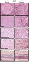
Evidence for nitrotyrosine formation from various tissues of diabetic mice and rats. Immunohistochemical staining for nitrotyrosine (dark brown staining) from control (left) and diabetic (right) murine tissues. [Derived from Pacher et al. (994) and Szabo et al. (1234), with permissions from The Feinstein Institute for Medical Research and Bentham Science Publishers.]
FIG. 17
Mechanisms of cardiovascular dysfunction in diabetes: role of superoxide and peroxynitrite. Hyperglycemia induces increased superoxide anion (O2•−) production via activation of multiple pathways including xanthine and NAD(P)H oxidases, cyclooxygenase, uncoupled nitric oxide synthase (NOS), glucose autoxidation, mitochondrial respiratory chain, polyol pathway, and formation of advanced glycation end products (AGE). Superoxide activates AGE, protein kinase C (PKC), polyol (sorbitol), hexosamine, and stress-signaling pathways leading to increased expression of inflammatory cytokines, angiotensin II (Ang II), endothelin-1 (ET-1), and NAD(P)H oxidases, which in turn generate more superoxide via multiple mechanisms. Hyperglycemia-induced increased superoxide generation may also favor an increased expression of nitric oxide synthases (NOS) through the activation of NF_κ_B, which may increase the generation of nitric oxide (NO). Superoxide anion may quench NO, thereby reducing the efficacy of a potent endothelium-derived vasodilator system. Superoxide can also be converted to hydrogen peroxide (H2O2) by superoxide dismutase (SOD) and interact with NO to form a reactive oxidant peroxynitrite (ONOO−), which induces cell damage via lipid peroxidation, inactivation of enzymes and other proteins by oxidation and nitration, and activation of matrix metalloproteinases (MMPs) among others. Peroxynitrite also acts on mitochondria [decreasing the membrane potential (Ψ)], triggering the release of proapoptotic factors such as cytochrome c (Cyt c) and apoptosis-inducing factor (AIF). These factors mediate caspase-dependent and caspase-independent apoptotic death pathways. Peroxynitrite, in concert with other oxidants (e.g., H2O2), causes strand breaks in DNA, activating the nuclear enzyme poly(ADP-ribose) polymerase-1 (PARP-1). Mild damage to DNA activates the DNA repair machinery. In contrast, once excessive oxidative and nitrosative stress-induced DNA damage occurs, overactivated PARP-1 initiates an energy-consuming cycle by transferring ADP-ribose units (small red spheres) from NAD+ to nuclear proteins, resulting in rapid depletion of the intracellular NAD+ and ATP pools, slowing the rate of glycolysis and mitochondrial respiration, and eventually leading to cellular dysfunction and death. Poly(ADP-ribose) glycohydrolase (PARG) degrades poly(ADP-ribose) (PAR) polymers, generating free PAR polymer and ADP-ribose, which may signal to the mitochondria to induce AIF release. PARP-1 activation also leads to the inhibition of cellular glyceraldehyde-3-phosphate dehydrogenase (GAPDH) activity, which in turn favors the activation of PKC, AGE, and hexosamine pathway leading to increased superoxide generation. PARP-1 also regulates the expression of a variety of inflammatory mediators, which might facilitate the progression of diabetic cardiovascular complications. [From Pacher and Szabo (996), with permission from Elsevier.]
FIG. 18
Evidence for nitrotyrosine formation and PARP activation in diabetic vasculature. Immunohistochemical staining for nitrotyrosine, terminal deoxyribonucleotidyl transferase-mediated dUTP nick-end labeling (indicator of DNA breaks), and poly-(ADP-ribose) (index of PARP activity) in control rings (top row) and in rings from diabetic mice (bottom row). [Derived from Garcia Soriano et al. (430) with permission from Nature Publishing Group.]
Similar articles
- Nitric oxide-derived oxidants with a focus on peroxynitrite: molecular targets, cellular responses and therapeutic implications.
Calcerrada P, Peluffo G, Radi R. Calcerrada P, et al. Curr Pharm Des. 2011 Dec;17(35):3905-32. doi: 10.2174/138161211798357719. Curr Pharm Des. 2011. PMID: 21933142 Review. - Cellular mechanisms of peroxynitrite-induced neuronal death.
Ramdial K, Franco MC, Estevez AG. Ramdial K, et al. Brain Res Bull. 2017 Jul;133:4-11. doi: 10.1016/j.brainresbull.2017.05.008. Epub 2017 Jun 24. Brain Res Bull. 2017. PMID: 28655600 Review. - Role of redox signaling and poly (adenosine diphosphate-ribose) polymerase activation in vascular smooth muscle cell growth inhibition by nitric oxide and peroxynitrite.
Huang J, Lin SC, Nadershahi A, Watts SW, Sarkar R. Huang J, et al. J Vasc Surg. 2008 Mar;47(3):599-607. doi: 10.1016/j.jvs.2007.11.006. J Vasc Surg. 2008. PMID: 18295111 - The superoxide radical switch in the biology of nitric oxide and peroxynitrite.
Piacenza L, Zeida A, Trujillo M, Radi R. Piacenza L, et al. Physiol Rev. 2022 Oct 1;102(4):1881-1906. doi: 10.1152/physrev.00005.2022. Epub 2022 May 23. Physiol Rev. 2022. PMID: 35605280 Review. - Pathophysiological roles of peroxynitrite in circulatory shock.
Szabó C, Módis K. Szabó C, et al. Shock. 2010 Sep;34 Suppl 1(0 1):4-14. doi: 10.1097/SHK.0b013e3181e7e9ba. Shock. 2010. PMID: 20523270 Free PMC article. Review.
Cited by
- Nitrosative stress plays an important role in Wnt pathway activation in diabetic retinopathy.
Liu Q, Li J, Cheng R, Chen Y, Lee K, Hu Y, Yi J, Liu Z, Ma JX. Liu Q, et al. Antioxid Redox Signal. 2013 Apr 1;18(10):1141-53. doi: 10.1089/ars.2012.4583. Epub 2012 Oct 15. Antioxid Redox Signal. 2013. PMID: 23066786 Free PMC article. - Immunological characterization of recombinant Salmonella enterica serovar Typhi FliC protein expressed in Escherichia coli.
Jindal G, Tewari R, Gautam A, Pandey SK, Rishi P. Jindal G, et al. AMB Express. 2012 Oct 15;2(1):55. doi: 10.1186/2191-0855-2-55. AMB Express. 2012. PMID: 23067582 Free PMC article. - Antimicrobial and anti-inflammatory activity of Cystatin C on human gingival fibroblast incubated with Porphyromonas gingivalis.
Blancas-Luciano BE, Becker-Fauser I, Zamora-Chimal J, Delgado-Domínguez J, Ruíz-Remigio A, Leyva-Huerta ER, Portilla-Robertson J, Fernández-Presas AM. Blancas-Luciano BE, et al. PeerJ. 2022 Oct 25;10:e14232. doi: 10.7717/peerj.14232. eCollection 2022. PeerJ. 2022. PMID: 36312752 Free PMC article. - Convergence of biological nitration and nitrosation via symmetrical nitrous anhydride.
Vitturi DA, Minarrieta L, Salvatore SR, Postlethwait EM, Fazzari M, Ferrer-Sueta G, Lancaster JR Jr, Freeman BA, Schopfer FJ. Vitturi DA, et al. Nat Chem Biol. 2015 Jul;11(7):504-10. doi: 10.1038/nchembio.1814. Epub 2015 May 25. Nat Chem Biol. 2015. PMID: 26006011 Free PMC article.
References
- Abdelkarim GE, Gertz K, Harms C, Katchanov J, Dirnagl U, Szabo C, Endres M. Protective effects of PJ34, a novel, potent inhibitor of poly(ADP-ribose) polymerase (PARP) in in vitro and in vivo models of stroke. Int J Mol Med. 2001;7:255–260. - PubMed
- Abe K, Aoki M, Kawagoe J, Yoshida T, Hattori A, Kogure K, Itoyama Y. Ischemic delayed neuronal death. A mitochondrial hypothesis. Stroke. 1995;26:1478–1489. - PubMed
- Abe K, Pan LH, Watanabe M, Kato T, Itoyama Y. Induction of nitrotyrosine-like immunoreactivity in the lower motor neuron of amyotrophic lateral sclerosis. Neurosci Lett. 1995;199:152–154. - PubMed
- Abe K, Pan LH, Watanabe M, Konno H, Kato T, Itoyama Y. Upregulation of protein-tyrosine nitration in the anterior horn cells of amyotrophic lateral sclerosis. Neurol Res. 1997;19:124–128. - PubMed
- Abou-Mohamed G, Johnson JA, Jin L, El-Remessy AB, Do K, Kaesemeyer WH, Caldwell RB, Caldwell RW. Roles of superoxide, peroxynitrite, and protein kinase C in the development of tolerance to nitroglycerin. J Pharmacol Exp Ther. 2004;308:289–299. - PubMed
Publication types
MeSH terms
Substances
Grants and funding
- AT-02034/AT/NCCIH NIH HHS/United States
- P01 ES000040-390003/ES/NIEHS NIH HHS/United States
- P01 ES000040/ES/NIEHS NIH HHS/United States
- ES-00240/ES/NIEHS NIH HHS/United States
- Z01 AA000375-02/Intramural NIH HHS/United States
- P01 ES000040-436360/ES/NIEHS NIH HHS/United States
- P01 AT002034/AT/NCCIH NIH HHS/United States
- ES-00040/ES/NIEHS NIH HHS/United States
LinkOut - more resources
Full Text Sources
Other Literature Sources


