Thymic stromal lymphopoietin is released by human epithelial cells in response to microbes, trauma, or inflammation and potently activates mast cells - PubMed (original) (raw)
Comparative Study
. 2007 Feb 19;204(2):253-8.
doi: 10.1084/jem.20062211. Epub 2007 Jan 22.
Affiliations
- PMID: 17242164
- PMCID: PMC2118732
- DOI: 10.1084/jem.20062211
Comparative Study
Thymic stromal lymphopoietin is released by human epithelial cells in response to microbes, trauma, or inflammation and potently activates mast cells
Zoulfia Allakhverdi et al. J Exp Med. 2007.
Abstract
Compelling evidence suggests that the epithelial cell-derived cytokine thymic stromal lymphopoietin (TSLP) may initiate asthma or atopic dermatitis through a dendritic cell-mediated T helper (Th)2 response. Here, we describe how TSLP might initiate and aggravate allergic inflammation in the absence of T lymphocytes and immunoglobulin E antibodies via the innate immune system. We show that TSLP, synergistically with interleukin 1 and tumor necrosis factor, stimulates the production of high levels of Th2 cytokines by human mast cells (MCs). We next report that TSLP is released by primary epithelial cells in response to certain microbial products, physical injury, or inflammatory cytokines. Direct epithelial cell-mediated, TSLP-dependent activation of MCs may play a central role in "intrinsic" forms of atopic diseases and explain the aggravating role of infection and scratching in these diseases.
Figures
Figure 1.
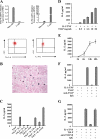
Human MCs expressed functional receptor for TSLP. (A) TSLP-R and IL-7Rα chain expression was determined at mRNA on MCs and peripheral blood T cells (mean ± SEM of eight experiments on different MC lines) and protein levels. (B) Tissue sections from the bronchial mucosa of asthmatic patients were stained with TSLP-R mAb using an HRP system (brown) and Astra Blue (blue) for the identification of MCs. (C) MCs were stimulated with TSLP alone or together with different inflammatory cytokines. The 24-h culture supernatants were tested for their content in IL-5. One representative of three experiments is shown; mean ± SD of triplicates. (D) MCs were stimulated with varying concentrations of TSLP in the presence of 10 ng/ml IL-1 and 25 ng/ml TNF. One representative of three experiments is shown; mean ± SD of triplicates. (E) Time course of cytokine production by MCs stimulated with 10 ng/ml IL-1β/TNF and TSLP. Mean ± SEM (n = 5). (F and G) MCs were stimulated in the presence of neutralizing mAb to TSLP (F) or TSLP-R (G) and isotype control IgG (each at 10 μg/ml). One representative of three experiments is shown; mean ± SD of triplicates.
Figure 2.
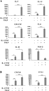
TSLP-stimulated secretion of cytokines and chemokines by MCs. Cytokine (A) and chemokine (B) secretion by MCs (105 cells/ml) stimulated for 24 h with 10 ng/ml IL-1β/TNF or/and TSLP was assessed by ELISA. Mean ± SEM (n = 11).
Figure 3.
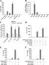
Induction of TSLP production by primary human airway epithelial cells. (A) SAECs were stimulated as indicated, and the 48-h culture supernatants were tested for their content in TSLP by ELISA. Mean ± SEM (n = 5). (B) Expression of the indicated TLR mRNA in SAECs was determined by real-time PCR. (C) BAF cells (104 cells/well) expressing the human TSLP-R and IL-7Rα chains were cultured in the presence of SAEC supernatants and in the presence or absence of neutralizing anti-TSLP mAb, and their proliferation was assessed after 3 d. One representative of three experiments is shown; mean ± SD of triplicates. (D–F) MCs were cultured in the presence or absence of supernatants of SAECs (50% vol/vol) described in A that were obtained upon stimulation with IL-1α/TNF (D), PGN (E), or polyI:C (F). IL-13 and IL-5 (not depicted) were measured after 24 h of MC stimulation. Mean ± SEM of four to five experiments.
Figure 4.
MC activation by skin-derived TSLP. (A) MCs were cultured with or without lesional or nonlesional skin fragments from AD patients in the presence of IL-1β/TNF with or without neutralizing mAb to TSLP. IL-13 and IL-5 (not depicted) were measured in the supernatants after 24 h of culture. (B) TSLP mRNA was assessed in the lesional and nonlesional skin of AD patients by real-time PCR. (C) Skin explants from nonallergic patients undergoing plastic surgery were minced and cultured for 24 h. Their cell-free culture supernatants (50% vol/vol) were used to stimulate MCs in the presence of IL-1β/TNF with or without mAb to TSLP and TSLP-R or isotype control. IL-13 was measured after 24 h of culture. One representative of three experiments is shown; mean ± SD of triplicates. (D) TSLP mRNA was assessed on freshly isolated or cultured for 24-h skin explants. (E) TSLP protein was measured in the supernatant fluids of these cultures. One representative of three experiments is shown; mean ± SD of triplicates.
Similar articles
- Expression and Regulation of Thymic Stromal Lymphopoietin and Thymic Stromal Lymphopoietin Receptor Heterocomplex in the Innate-Adaptive Immunity of Pediatric Asthma.
Lin SC, Cheng FY, Liu JJ, Ye YL. Lin SC, et al. Int J Mol Sci. 2018 Apr 18;19(4):1231. doi: 10.3390/ijms19041231. Int J Mol Sci. 2018. PMID: 29670037 Free PMC article. Review. - Thymic stromal lymphopoietin, OX40-ligand, and interleukin-25 in allergic responses.
Wang YH, Liu YJ. Wang YH, et al. Clin Exp Allergy. 2009 Jun;39(6):798-806. doi: 10.1111/j.1365-2222.2009.03241.x. Epub 2009 Apr 7. Clin Exp Allergy. 2009. PMID: 19400908 Free PMC article. Review. - Thymic stromal lymphopoietin and OX40 ligand pathway in the initiation of dendritic cell-mediated allergic inflammation.
Liu YJ. Liu YJ. J Allergy Clin Immunol. 2007 Aug;120(2):238-44; quiz 245-6. doi: 10.1016/j.jaci.2007.06.004. J Allergy Clin Immunol. 2007. PMID: 17666213 Review. - Thymic stromal lymphopoietin.
He R, Geha RS. He R, et al. Ann N Y Acad Sci. 2010 Jan;1183:13-24. doi: 10.1111/j.1749-6632.2009.05128.x. Ann N Y Acad Sci. 2010. PMID: 20146705 Free PMC article. Review. - Thymic stromal lymphopoietin activity is increased in nasal polyps of patients with chronic rhinosinusitis.
Nagarkar DR, Poposki JA, Tan BK, Comeau MR, Peters AT, Hulse KE, Suh LA, Norton J, Harris KE, Grammer LC, Chandra RK, Conley DB, Kern RC, Schleimer RP, Kato A. Nagarkar DR, et al. J Allergy Clin Immunol. 2013 Sep;132(3):593-600.e12. doi: 10.1016/j.jaci.2013.04.005. Epub 2013 May 17. J Allergy Clin Immunol. 2013. PMID: 23688414 Free PMC article.
Cited by
- Regulation of Airway Epithelial-Derived Alarmins in Asthma: Perspectives for Therapeutic Targets.
Hansi RK, Ranjbar M, Whetstone CE, Gauvreau GM. Hansi RK, et al. Biomedicines. 2024 Oct 11;12(10):2312. doi: 10.3390/biomedicines12102312. Biomedicines. 2024. PMID: 39457624 Free PMC article. Review. - DESTINATION: a phase 3, multicentre, randomized, double-blind, placebo-controlled, parallel-group trial to evaluate the long-term safety and tolerability of tezepelumab in adults and adolescents with severe, uncontrolled asthma.
Menzies-Gow A, Ponnarambil S, Downie J, Bowen K, Hellqvist Å, Colice G. Menzies-Gow A, et al. Respir Res. 2020 Oct 21;21(1):279. doi: 10.1186/s12931-020-01541-7. Respir Res. 2020. PMID: 33087119 Free PMC article. Clinical Trial. - TSLP signaling network revealed by SILAC-based phosphoproteomics.
Zhong J, Kim MS, Chaerkady R, Wu X, Huang TC, Getnet D, Mitchell CJ, Palapetta SM, Sharma J, O'Meally RN, Cole RN, Yoda A, Moritz A, Loriaux MM, Rush J, Weinstock DM, Tyner JW, Pandey A. Zhong J, et al. Mol Cell Proteomics. 2012 Jun;11(6):M112.017764. doi: 10.1074/mcp.M112.017764. Epub 2012 Feb 16. Mol Cell Proteomics. 2012. PMID: 22345495 Free PMC article. - Thymic stromal lymphopoietin controls hair growth.
Shannon JL, Corcoran DL, Murray JC, Ziegler SF, MacLeod AS, Zhang JY. Shannon JL, et al. Stem Cell Reports. 2022 Mar 8;17(3):649-663. doi: 10.1016/j.stemcr.2022.01.017. Epub 2022 Feb 24. Stem Cell Reports. 2022. PMID: 35216683 Free PMC article. - Monoclonal antibodies and other biologic agents in the treatment of asthma.
Long AA. Long AA. MAbs. 2009 May-Jun;1(3):237-46. doi: 10.4161/mabs.1.3.8352. Epub 2009 May 4. MAbs. 2009. PMID: 20065638 Free PMC article. Review.
References
- Kay, A.B. 2006. The role of T lymphocytes in asthma. Chem. Immunol. Allergy. 91:59–75. - PubMed
- Ziegler, S.F., and Y.J. Liu. 2006. Thymic stromal lymphopoietin in normal and pathogenic T cell development and function. Nat. Immunol. 7:709–714. - PubMed
- Soumelis, V., P.A. Reche, H. Kanzler, W. Yuan, G. Edward, B. Homey, M. Gilliet, S. Ho, S. Antonenko, A. Lauerma, et al. 2002. Human epithelial cells trigger dendritic cell mediated allergic inflammation by producing TSLP. Nat. Immunol. 3:673–680. - PubMed
Publication types
MeSH terms
Substances
LinkOut - more resources
Full Text Sources
Other Literature Sources
Medical
Molecular Biology Databases


