Axonal-SMN (a-SMN), a protein isoform of the survival motor neuron gene, is specifically involved in axonogenesis - PubMed (original) (raw)
Axonal-SMN (a-SMN), a protein isoform of the survival motor neuron gene, is specifically involved in axonogenesis
Veronica Setola et al. Proc Natl Acad Sci U S A. 2007.
Abstract
Spinal muscular atrophy (SMA) is an autosomal recessive disease of childhood due to loss of the telomeric survival motor neuron gene, SMN1. The general functions of the main SMN1 protein product, full-length SMN (FL-SMN), do not explain the selective motoneuronal loss of SMA. We identified axonal-SMN (a-SMN), an alternatively spliced SMN form, preferentially encoded by the SMN1 gene in humans. The a-SMN transcript and protein are down-regulated during early development in different tissues. In the spinal cord, the a-SMN protein is selectively expressed in motor neurons and mainly localized in axons. Forced expression of a-SMN stimulates motor neuron axonogenesis in a time-dependent fashion and induces axonal-like growth in non-neuronal cells. Exons 2b and 3 are essential for the axonogenic effects. This discovery indicates an unexpected complexity of the SMN gene system and may help in understanding the pathogenesis of SMA.
Conflict of interest statement
The authors declare no conflict of interest.
Figures
Fig. 1.
Molecular characterization of the a-SMN transcript. (a) Northern blot analysis, P15 rat spinal cord: an intron 3 probe hybridized to a transcript (left) similar in size to the _FL_-SMN mRNA (right). (b) RNase protection assay. The riboprobe and protected fragments are schematically represented (Left). Protected fragments specific for _FL_-SMN (201 and 61 nt) and _a_-SMN (472 nt) mRNAs are indicated by arrowheads (Right). (c and d) RT/PCR analysis. The combinations of primers are indicated. Note the two fragments using exon primers flanking the intron 3 sequence (Ex3–Ex4) and the developmental down-regulation of the a-SMN transcript. (e) Competitive PCR. A mixture of primers specific for SMN exon 3, intron 3, and exon 6 generated two fragments corresponding to a-SMN (372 bp) and FL-SMN (544 bp). (f and g) RT/PCR, adult human spinal cord. Exon 1/intron 3 and exon 3/intron 3 pairs of primers (f) amplified the expected _a_-SMN cDNA fragments (534 and 221 bp). Primers (Ex3–Ex4) flanking the intron 3 sequence (g) amplified a-SMN (upper, 396 bp) and FL-SMN (lower, 237 bp) cDNA fragments. (h) Schematic diagram of a-SMN mRNA (retained intron 3 is hatched).
Fig. 2.
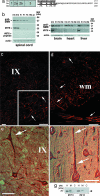
Expression of the a-SMN protein in vivo. (a) Primary structure of human, rat, and mouse a-SMN. Intron 3 epitopes recognized by a-SMN antibodies are underlined. (b) WB analysis of rat tissues. In the spinal cord (Left), the anti-rat a-SMN antibody #976 recognizes a developmentally down-regulated 23-kDa band (arrowhead), which is abolished in preabsorption experiments. Note the similar developmental profile in the brain, heart, and liver (Right). All blots were reprobed with an anti-SMN antibody. (c and d) P1 rat spinal cord. Note the selective a-SMN staining of external cellular membrane and dendrites (arrows in c) and axons (arrows in d) of lamina IX motor neurons. (e and f) Developing human spinal motor neurons (arrows in e) and cortical pyramidal neurons (arrows in f) are intensely a-SMN-immunoreactive. (g) WB analysis of human embryonic spinal cord. Note the a-SMN 20-kDa band in the total homogenate (H) and membrane (M) fraction but not in the cytosol (C). (Scale bars: c and d, 30 μm; e and f, 50 μm.)
Fig. 3.
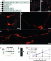
FL-SMN and a-SMN overexpression. (a) WB analysis. Transfected _FL_-SMN (filled arrowheads) and _a_-SMN (open arrowheads) are indicated. Note the two a-SMN bands, suggesting posttranslational processing. (b and c) NSC34 cells transfected with tagged FL-SMN (b) or a-SMN (c). Shown is anti-tag antibody. (b Inset) Few FL-SMN granules in neurites. (c Inset) a-SMN neurites touching neighboring cells. (d–f) Transfection of pIRES-EYFP _a_-SMN. Note the newly formed neurites contacting untransfected NSC34 adjacent cells. (g–j) Transfection in HeLa. (g) WB analysis; protein bands are indicated as above. (h–j) Confocal images of mock-transfected (h), _FL_-_SMN_-transfected (i), and _a_-_SMN_-transfected cells (j) reacted with anti-F-actin antibody. Lane 1, untransfected cells; lane 2, mock transfection; lane 3, _FL_-SMN transfection; lane 4, _a_-SMN transfection. *, endogenous FL-SMN. (Scale bars: b, c, and h–j, 25 μm; d–f, 30 μm; Insets, 10 μm.)
Fig. 4.
Human _a_-SMN (_ha_-SMN) overexpression. (a) WB analysis in NSC34; GFP-a-SMN (Left) and tagged-a-SMN (Right) proteins are indicated. Lane 1, untransfected cells; lane 2 and 4, mock transfection; lane 3, _GFP_-_ha_-SMN transfection; lane 5, tag-ha-SMN transfection. *, endogenous FL-SMN. (b and c) _GFP_-_ha_-SMN expression in cell bodies (b) and neurites (c) of NSC34. (d–f) Confocal images showing human a-SMN (d) (#910 anti-SMN) and F-actin (e) colocalization (f) in newly formed filopodia of HeLa cells transfected with _ha_-SMN. (Scale bars: b, 25 μm; c–f, 10 μm.)
Fig. 5.
Time course of human a-SMN overexpression. (a–d) Confocal images with an anti-tag antibody at 12 (a), 24 (b), 48 (c), and 72 (d) h after transfection. (Scale bar: 30 μm.) (e) Mean axon length at different time points in _a_-SMN and _FL_-SMN transfected motor neurons. Data are mean ± SD of triplicate wells (seven cells per well). Two-way ANOVA and two-tailed t test were used for each _a_-_SMN/FL_-SMN pair at the different time-points (**, P < 0.01). (f) WB analysis. Anti-SMN, anti-tag, and #873 and #910 anti-a-SMN antibodies (arrowheads) show the a-SMN progressive synthesis (lower band). Lane 1, untransfected cells; lane 2, mock transfection; lanes 3–6, a-SMN transfection at 12, 24, 48, and 72 h. *, endogenous FL-SMN.
Fig. 6.
a-SMN functional mapping. Confocal images (a–e) from NSC34 motor neurons transfected with the different N-terminally tagged constructs (upper left). Intervals after transfection are indicated. See text for details. Shown are anti-SMN antibody in a and anti-tag antibody in b–e. (Scale bar: 30 μm.) (f) WB analysis. The anti-SMN antibody reveals the translated a-SMN bands of the expected size (arrowheads). Lane 1, exons 1/2a overexpression (24 h); lanes 2 and 3, exons 1/2a/2b overexpression (24–48 h); lanes 4 and 5, exons 1/2a/2b/3 overexpression (24–48 h). *, endogenous FL-SMN. (g) RT/PCR analysis showing mRNA synthesis (arrowhead) after transfection of construct a. (h) Mean axon length at different times after transfection of FL-SMN, a-SMN, exons 1/2a/2b a-SMN(EX 2b), and exons 1/2a/2b/3 a-SMN(EX 3). Data are mean ± SD of triplicate samples (six cells per well). Two-way ANOVA indicated a significant effect (P < 0.0001) of each _a_-SMN construct compared with _FL_-SMN.
Similar articles
- Survival motor neuron SMN1 and SMN2 gene promoters: identical sequences and differential expression in neurons and non-neuronal cells.
Boda B, Mas C, Giudicelli C, Nepote V, Guimiot F, Levacher B, Zvara A, Santha M, LeGall I, Simonneau M. Boda B, et al. Eur J Hum Genet. 2004 Sep;12(9):729-37. doi: 10.1038/sj.ejhg.5201217. Eur J Hum Genet. 2004. PMID: 15162126 - Smn, the spinal muscular atrophy-determining gene product, modulates axon growth and localization of beta-actin mRNA in growth cones of motoneurons.
Rossoll W, Jablonka S, Andreassi C, Kröning AK, Karle K, Monani UR, Sendtner M. Rossoll W, et al. J Cell Biol. 2003 Nov 24;163(4):801-12. doi: 10.1083/jcb.200304128. Epub 2003 Nov 17. J Cell Biol. 2003. PMID: 14623865 Free PMC article. - Synthesis and biological evaluation of novel 2,4-diaminoquinazoline derivatives as SMN2 promoter activators for the potential treatment of spinal muscular atrophy.
Thurmond J, Butchbach ME, Palomo M, Pease B, Rao M, Bedell L, Keyvan M, Pai G, Mishra R, Haraldsson M, Andresson T, Bragason G, Thosteinsdottir M, Bjornsson JM, Coovert DD, Burghes AH, Gurney ME, Singh J. Thurmond J, et al. J Med Chem. 2008 Feb 14;51(3):449-69. doi: 10.1021/jm061475p. Epub 2008 Jan 19. J Med Chem. 2008. PMID: 18205293 - Pathogenesis of proximal autosomal recessive spinal muscular atrophy.
Simic G. Simic G. Acta Neuropathol. 2008 Sep;116(3):223-34. doi: 10.1007/s00401-008-0411-1. Epub 2008 Jul 16. Acta Neuropathol. 2008. PMID: 18629520 Review. - Spinal muscular atrophy: a deficiency in a ubiquitous protein; a motor neuron-specific disease.
Monani UR. Monani UR. Neuron. 2005 Dec 22;48(6):885-96. doi: 10.1016/j.neuron.2005.12.001. Neuron. 2005. PMID: 16364894 Review.
Cited by
- In Search of Spinal Muscular Atrophy Disease Modifiers.
Chudakova D, Kuzenkova L, Fisenko A, Savostyanov K. Chudakova D, et al. Int J Mol Sci. 2024 Oct 18;25(20):11210. doi: 10.3390/ijms252011210. Int J Mol Sci. 2024. PMID: 39456991 Free PMC article. Review. - A survey of transcripts generated by spinal muscular atrophy genes.
Singh NN, Ottesen EW, Singh RN. Singh NN, et al. Biochim Biophys Acta Gene Regul Mech. 2020 Aug;1863(8):194562. doi: 10.1016/j.bbagrm.2020.194562. Epub 2020 May 6. Biochim Biophys Acta Gene Regul Mech. 2020. PMID: 32387331 Free PMC article. Review. - Selective vulnerability of spinal and cortical motor neuron subpopulations in delta7 SMA mice.
d'Errico P, Boido M, Piras A, Valsecchi V, De Amicis E, Locatelli D, Capra S, Vagni F, Vercelli A, Battaglia G. d'Errico P, et al. PLoS One. 2013 Dec 6;8(12):e82654. doi: 10.1371/journal.pone.0082654. eCollection 2013. PLoS One. 2013. PMID: 24324819 Free PMC article. - Establishment of a molecular diagnostic system for spinal muscular atrophy experience from a clinical laboratory in china.
Zeng J, Lin Y, Yan A, Ke L, Zhu Z, Lan F. Zeng J, et al. J Mol Diagn. 2011 Jan;13(1):41-7. doi: 10.1016/j.jmoldx.2010.11.009. Epub 2010 Dec 23. J Mol Diagn. 2011. PMID: 21227393 Free PMC article. - Cerebellar structural, astrocytic, and neuronal abnormalities in the SMNΔ7 mouse model of spinal muscular atrophy.
Cottam NC, Bamfo T, Harrington MA, Charvet CJ, Hekmatyar K, Tulin N, Sun J. Cottam NC, et al. Brain Pathol. 2023 Sep;33(5):e13162. doi: 10.1111/bpa.13162. Epub 2023 May 22. Brain Pathol. 2023. PMID: 37218083 Free PMC article.
References
- Pearn J. Lancet. 1980;1:919–922. - PubMed
- Lefebvre S, Burglen L, Reboullet S, Clermont O, Burlet P, Viollet L, Benichou B, Cruaud C, Millasseau P, Zeviani M, et al. Cell. 1995;80:155–165. - PubMed
- Gennarelli M, Lucarelli M, Capon F, Pizzuti A, Merlini L, Angelini C, Novelli G, Dallapiccola B. Biochem Biophys Res Commun. 1995;213:342–348. - PubMed
Publication types
MeSH terms
Substances
LinkOut - more resources
Full Text Sources
Other Literature Sources


