Time-specific effects of ethanol exposure on cranial nerve nuclei: gastrulation and neuronogenesis - PubMed (original) (raw)
Time-specific effects of ethanol exposure on cranial nerve nuclei: gastrulation and neuronogenesis
Sandra M Mooney et al. Exp Neurol. 2007 May.
Abstract
During the development of the central nervous system, neurons pass through critical periods or periods of vulnerability. We explored periods of vulnerability for cranial nerve nuclei by determining the effects of acute exposure to ethanol during development on the number of neurons in mature brainstem. Long-Evans rats were injected with 2.9 g ethanol/kg body weight on one day between gestational day (G) 7 and G13, inclusive. Two hours later, animals received a second injection of 1.45 g/kg. Controls were injected with equivalent volumes of saline. Brainstems of 31-day-old offspring were cryosectioned and stained with cresyl violet. Stereological methods were used to determine the volume and numerical density of neurons in three trigeminal sensory nuclei (the principal sensory nucleus of the trigeminal nerve, and the oral and interpolar subnuclei of the spinal trigeminal nuclear complex) and three motor nuclei (the trigeminal, facial, and hypoglossal nuclei). The numbers of neurons in most nuclei were lower following early (on G7 and/or G8) or later (on G12 and/or G13) exposure. Only the trigeminal interpolar nucleus was affected by neither early nor late ethanol exposure. Thus, prenatal exposure to ethanol affects the number of neurons in brainstem nuclei in a time-dependent manner. Windows of vulnerability coincide with gastrulation (G7/G8) and the period of neuronal generation (G12/G13).
Figures
Figure 1
Blood ethanol concentration (BEC). The BEC rose precipitously to reach 233 mg/dl at 1.0 hour after the first injection. Peak BEC was reached two hours later. By eight hours post-injection, the BEC was 0. Arrows show the two injection times. Symbols represent the means of four animals (± standard errors of the means).
Figure 2
Appearance of the sensory cranial nerve nuclei Three sensory trigeminal nuclei were identifiable in horizontal sections stained with cresyl violet. Samples representing the animals treated with saline (controls) or ethanol were taken from cohorts dosed on gestational day 8. No gross differences were apparent between the groups. Rostral is oriented to the left and lateral to the top. Scale bars are 500 μm.
Figure 3
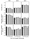
Quantitative measures of the effects of ethanol on sensory nuclei Stereological methods were used to estimate the volume (top) and the neuronal density of each nucleus (middle). Total neuronal number was estimated as the product of volume and density. Bars represent the means and T-bars signify the standard errors of the means. Each mean is based on five or six animals per group. Asterisks identify differences relative to the controls that were statistically significant (p<0.05).
Figure 4
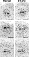
Appearance of the motor cranial nerve nuclei Three motor nuclei (the motor nuclei of the trigeminal (MoV), facial (MoVII), and hypoglossal (MoXII) nerves were evident in horizontal sections. Images are oriented so that rostral is to the left and lateral to the top. Scale bars are 500 μm.
Figure 5
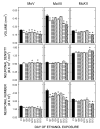
Quantitative measures of the effects of ethanol on motor nuclei Notations as in Figure 3.
Similar articles
- Acute prenatal exposure to ethanol and social behavior: effects of age, sex, and timing of exposure.
Mooney SM, Varlinskaya EI. Mooney SM, et al. Behav Brain Res. 2011 Jan 1;216(1):358-64. doi: 10.1016/j.bbr.2010.08.014. Epub 2010 Aug 20. Behav Brain Res. 2011. PMID: 20728475 Free PMC article. - Vulnerability of macaque cranial nerve neurons to ethanol is time- and site-dependent.
Mooney SM, Miller MW. Mooney SM, et al. Alcohol. 2009 Jun;43(4):323-31. doi: 10.1016/j.alcohol.2009.03.001. Epub 2009 Apr 17. Alcohol. 2009. PMID: 19375881 Free PMC article. - Structure and histogenesis of the principal sensory nucleus of the trigeminal nerve: effects of prenatal exposure to ethanol.
Miller MW, Muller SJ. Miller MW, et al. J Comp Neurol. 1989 Apr 22;282(4):570-80. doi: 10.1002/cne.902820408. J Comp Neurol. 1989. PMID: 2723152 - Episodic exposure to ethanol during development differentially affects brainstem nuclei in the macaque.
Mooney SM, Miller MW. Mooney SM, et al. J Neurocytol. 2001 Dec;30(12):973-82. doi: 10.1023/a:1021832522701. J Neurocytol. 2001. PMID: 12626879 - Anatomy of the brainstem: a gaze into the stem of life.
Angeles Fernández-Gil M, Palacios-Bote R, Leo-Barahona M, Mora-Encinas JP. Angeles Fernández-Gil M, et al. Semin Ultrasound CT MR. 2010 Jun;31(3):196-219. doi: 10.1053/j.sult.2010.03.006. Semin Ultrasound CT MR. 2010. PMID: 20483389 Review.
Cited by
- Low and moderate prenatal ethanol exposures of mice during gastrulation or neurulation delays neurobehavioral development.
Schambra UB, Goldsmith J, Nunley K, Liu Y, Harirforoosh S, Schambra HM. Schambra UB, et al. Neurotoxicol Teratol. 2015 Sep-Oct;51:1-11. doi: 10.1016/j.ntt.2015.07.003. Epub 2015 Jul 11. Neurotoxicol Teratol. 2015. PMID: 26171567 Free PMC article. - Loss of motoneurons in the ventral compartment of the rat hypoglossal nucleus following early postnatal exposure to alcohol.
Stettner GM, Kubin L, Volgin DV. Stettner GM, et al. J Chem Neuroanat. 2013 Sep;52:87-94. doi: 10.1016/j.jchemneu.2013.07.003. Epub 2013 Aug 8. J Chem Neuroanat. 2013. PMID: 23932955 Free PMC article. - Acute prenatal exposure to ethanol and social behavior: effects of age, sex, and timing of exposure.
Mooney SM, Varlinskaya EI. Mooney SM, et al. Behav Brain Res. 2011 Jan 1;216(1):358-64. doi: 10.1016/j.bbr.2010.08.014. Epub 2010 Aug 20. Behav Brain Res. 2011. PMID: 20728475 Free PMC article. - Acute exposure to ethanol on gestational day 15 affects social motivation of female offspring.
Varlinskaya EI, Mooney SM. Varlinskaya EI, et al. Behav Brain Res. 2014 Mar 15;261:106-9. doi: 10.1016/j.bbr.2013.12.016. Epub 2013 Dec 16. Behav Brain Res. 2014. PMID: 24355753 Free PMC article. - Vulnerability of macaque cranial nerve neurons to ethanol is time- and site-dependent.
Mooney SM, Miller MW. Mooney SM, et al. Alcohol. 2009 Jun;43(4):323-31. doi: 10.1016/j.alcohol.2009.03.001. Epub 2009 Apr 17. Alcohol. 2009. PMID: 19375881 Free PMC article.
References
- Abercrombie M. Estimation of nuclear population from microtome sections. Anat. Rec. 1946;94:239–247. - PubMed
- Altman J, Bayer SA. Development of the brain stem in the rat. I. Thymidine-radiographic study of the time of origin of neurons of the lower medulla. J. Comp. Neurol. 1980a;194:1–35. - PubMed
- Altman J, Bayer SA. Development of the brain stem in the rat. II. Thymidine-radiographic study of the time of origin of neurons of the upper medulla, excluding the vestibular and auditory nuclei. J. Comp. Neurol. 1980b;194:37–56. - PubMed
- Altman J, Bayer SA. Development of the brain stem in the rat. IV. Thymidine-radiographic study of the time of origin of neurons in the pontine region. J. Comp. Neurol. 1980c;194:905–929. - PubMed
- Astley SJ, Magnuson SI, Omnell LM, Clarren SK. Fetal alcohol syndrome: changes in craniofacial form with age, cognition, and timing of ethanol exposure in the macaque. Teratology. 1999;59:163–172. - PubMed
Publication types
MeSH terms
Substances
Grants and funding
- AA07568/AA/NIAAA NIH HHS/United States
- AA015413/AA/NIAAA NIH HHS/United States
- R01 AA007568/AA/NIAAA NIH HHS/United States
- R01 AA006916/AA/NIAAA NIH HHS/United States
- R37 AA007568/AA/NIAAA NIH HHS/United States
- AA06916/AA/NIAAA NIH HHS/United States
- R21 AA015413/AA/NIAAA NIH HHS/United States
LinkOut - more resources
Full Text Sources


