Anti-Hebbian long-term potentiation in the hippocampal feedback inhibitory circuit - PubMed (original) (raw)
Anti-Hebbian long-term potentiation in the hippocampal feedback inhibitory circuit
Karri P Lamsa et al. Science. 2007.
Abstract
Long-term potentiation (LTP), which approximates Hebb's postulate of associative learning, typically requires depolarization-dependent glutamate receptors of the NMDA (N-methyl-D-aspartate) subtype. However, in some neurons, LTP depends instead on calcium-permeable AMPA-type receptors. This is paradoxical because intracellular polyamines block such receptors during depolarization. We report that LTP at synapses on hippocampal interneurons mediating feedback inhibition is "anti-Hebbian":Itis induced by presynaptic activity but prevented by postsynaptic depolarization. Anti-Hebbian LTP may occur in interneurons that are silent during periods of intense pyramidal cell firing, such as sharp waves, and lead to their altered activation during theta activity.
Figures
Fig. 1
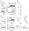
Associative pairing precludes LTP in interneurons in the stratum oriens/alveus. (A) Left: Schematic illustrating in-phase high-frequency burst (HFB) stimulation of weak and strong alveus pathways (filled and open symbols, respectively). Sample traces (1 to 5) show action potentials evoked by pairing in one cell. Right: Baseline-normalized EPSP initial slopes (mean ± SEM). (B) Top: Averaged EPSPs recorded before (blue) and after (red) pairing in one cell, showing the interval used to measure the initial slope. Bottom: Baseline-normalized EPSP initial slopes (25 min after pairing) in the two pathways, plotted against one another. (C) Antiphase pairing of two weak pathways induced LTP in seven out of seven cells. Sample traces (left) are from one cell. (D) EPSPs before and after pairing and summary of results, plotted as in (B). (E) Burst stimulation of one pathway also induced LTP. AMPA/kainate receptors were blocked at the end of the experiment (NBQX), to verify that EPSP initial slopes were not contaminated by monosynaptic inhibition. (F) Effect of HFB stimulation of one pathway (weak 1), plotted as for (B) and (D). Traces (top) also show the effect of NBQX. Data in (C) (right) and (E) (right) are shown as the mean ± SEM. _V_m, membrane potential.
Fig. 2
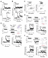
Postsynaptic membrane potential gates anti-Hebbian LTP induction. (A) LTP was evoked by pairing presynaptic stimulation with the hyperpolarizing but not the depolarizing phase of an imposed sinusoidal membrane potential oscillation. Left: Schematic and sample membrane potential traces during pairing in one cell (five sweeps superimposed for each pairing protocol). Right: Baseline-normalized EPSP initial slopes in eight cells showing LTP after anti-Hebbian pairing of HFB stimulation of one pathway with hyperpolarization. Subsequent Hebbian pairing of the other pathway with depolarization was ineffective. AMPA/kainate receptors were blocked at the end of the experiment (NBQX). Data are shown as the mean ± SEM. (B) Averaged EPSPs in one cell taken at the times indicated and after NBQX addition. Top: Anti-Hebbian pairing. Bottom: Hebbian pairing. (C) LTP was induced by pairing single stimuli at 5 Hz with hyperpolarization. Left: Sample traces during pairing. Right: Averages of all cells tested. Data are shown as the mean ± SEM. (D) Pairing with depolarization failed to induce LTP. Left: Sample traces during pairing. Right: Averages of all cells tested. Data are shown as the mean ± SEM. (E) Repatched interneurons recorded in whole-cell voltage-clamp mode show rectifying AMPARs and a negligible NMDAR-mediated component (GABA receptors blocked). Traces: Averaged EPSCs at +60 and −60 mV, showing the times at which the two components were measured. Bottom: current-voltage (I-V) relation of AMPAR-mediated EPSCs in six repatched interneurons (left). I-V relation for the NMDAR-mediated component, normalized by the AMPA EPSC at −60 mV (right).
Fig. 3
Anti-Hebbian LTP occurs in interneurons with rectifying AMPARs in the feedback circuit. (A) High-frequency stimulation (HFS) paired with hyperpolarization evoked LTP in 25 out of 31 interneurons in the oriens/alveus [NMDARs blocked with 100 μM
d
,
l
-2-amino-5-phosphonovalerate (APV)]. Insets: Averaged EPSPs before and after LTP induction, and membrane potential during pairing, in one interneuron. Data are shown as the mean ± SEM. (B) Repatched interneurons recorded in whole-cell voltage-clamp mode revealed strongly rectifying synaptic AMPARs (rectification index < 0.3). Gray and open symbols show cells that did and did not exhibit LTP, respectively. Insets: Averaged EPSCs at −60 and +60 mV in one cell that showed anti-Hebbian LTP. (C) O-LM cells were the commonest identified interneuron type exhibiting anti-Hebbian LTP (left: schematic, with dendritic and axonal arborizations for one cell shown in red and blue, respectively). Three fast spiking perisomatic-projecting neurons were also identified, including a basket cell (right). Scale bar: 200 μm. Firing patterns in response to current injection (_I_c) are shown below. (D) Typical layer- and pathway-specific properties of EPSCs in experiments where NMDARs were not blocked (n, number of repatched interneurons). Interneurons were recorded in the stratum radiatum (1), stratum pyramidale (2), and stratum oriens/alveus (3). (E) Success rates for eliciting Hebbian or anti-Hebbian LTP at synapses made by axons illustrated in (D). (F) Anti-Hebbian LTP requires activation of AMPA/kainate receptors. HFS stimulation of one pathway (filled symbols) was paired with hyperpolarization in NBQX (5 μM) (inset). After wash-out, EPSPs in both pathways recovered at the same rate. Inset: Averaged EPSPs before pairing (blue) and after recovery (red) in the two pathways in one experiment. Data are shown as the mean ± SEM.
Fig. 4
Intracellular polyamines determine the voltage dependence of anti-Hebbian LTP. (A) Postsynaptic depolarization prevents LTP induction. Data from cells recorded in perforated patch mode, showing LTP induced by pairing high-frequency stimulation (HFS) of one pathway with hyperpolarization (top, “anti-Hebbian”), and failure to induce LTP by pairing the other pathway with depolarization (bottom, “Hebbian”). NMDARs were blocked throughout. Data are the mean ± SEM. (B) In six other cells, the second pathway was subsequently paired with hyperpolarization, yielding anti-Hebbian LTP in all cases. Data are the mean ± SEM. (C) Baseline-normalized EPSP slopes plotted against one another 20 min after anti-Hebbian (left) and Hebbian (right) pairing. Insets: Sample membrane potential traces during pairing. (D) Anti-Hebbian LTP was first induced in one pathway (left, filled symbols). The interneuron was then repatched in whole-cell mode with a polyamine-free pipette solution. HFS delivered to the second pathway (right, open symbols) paired with postsynaptic depolarization (+20 mV) induced LTP. Top: Voltage (left) and current (middle, with seal resistance test artefacts) traces during pairing, and one O-LM cell identified among five interneurons in the sample (right; scale bar: 200 μm). (E) Intracellular spermine blocked LTP induction in the second pathway when paired with depolarization. Top: As in (D). (F) LTP was induced with intracellular spermine when paired with hyperpolarization (−90 mV). Top: As in (D) and (E). Data in (D), (E), and (F) (bottom panels) are shown as the mean ± SEM.
Similar articles
- Role of ionotropic glutamate receptors in long-term potentiation in rat hippocampal CA1 oriens-lacunosum moleculare interneurons.
Oren I, Nissen W, Kullmann DM, Somogyi P, Lamsa KP. Oren I, et al. J Neurosci. 2009 Jan 28;29(4):939-50. doi: 10.1523/JNEUROSCI.3251-08.2009. J Neurosci. 2009. PMID: 19176803 Free PMC article. - Induction of Anti-Hebbian LTP in CA1 Stratum Oriens Interneurons: Interactions between Group I Metabotropic Glutamate Receptors and M1 Muscarinic Receptors.
Le Duigou C, Savary E, Kullmann DM, Miles R. Le Duigou C, et al. J Neurosci. 2015 Oct 7;35(40):13542-54. doi: 10.1523/JNEUROSCI.0956-15.2015. J Neurosci. 2015. PMID: 26446209 Free PMC article. - Synaptic plasticity in morphologically identified CA1 stratum radiatum interneurons and giant projection cells.
Christie BR, Franks KM, Seamans JK, Saga K, Sejnowski TJ. Christie BR, et al. Hippocampus. 2000;10(6):673-83. doi: 10.1002/1098-1063(2000)10:6<673::AID-HIPO1005>3.0.CO;2-O. Hippocampus. 2000. PMID: 11153713 - Roles of distinct glutamate receptors in induction of anti-Hebbian long-term potentiation.
Kullmann DM, Lamsa K. Kullmann DM, et al. J Physiol. 2008 Mar 15;586(6):1481-6. doi: 10.1113/jphysiol.2007.148064. Epub 2008 Jan 10. J Physiol. 2008. PMID: 18187472 Free PMC article. Review. - Theta-burst LTP.
Larson J, Munkácsy E. Larson J, et al. Brain Res. 2015 Sep 24;1621:38-50. doi: 10.1016/j.brainres.2014.10.034. Epub 2014 Oct 27. Brain Res. 2015. PMID: 25452022 Free PMC article. Review.
Cited by
- Calcium-permeable AMPA receptors provide a common mechanism for LTP in glutamatergic synapses of distinct hippocampal interneuron types.
Szabo A, Somogyi J, Cauli B, Lambolez B, Somogyi P, Lamsa KP. Szabo A, et al. J Neurosci. 2012 May 9;32(19):6511-6. doi: 10.1523/JNEUROSCI.0206-12.2012. J Neurosci. 2012. PMID: 22573673 Free PMC article. - A role for calcium-permeable AMPA receptors in synaptic plasticity and learning.
Wiltgen BJ, Royle GA, Gray EE, Abdipranoto A, Thangthaeng N, Jacobs N, Saab F, Tonegawa S, Heinemann SF, O'Dell TJ, Fanselow MS, Vissel B. Wiltgen BJ, et al. PLoS One. 2010 Sep 29;5(9):e12818. doi: 10.1371/journal.pone.0012818. PLoS One. 2010. PMID: 20927382 Free PMC article. - Enhanced dendritic action potential backpropagation in parvalbumin-positive basket cells during sharp wave activity.
Chiovini B, Turi GF, Katona G, Kaszás A, Erdélyi F, Szabó G, Monyer H, Csákányi A, Vizi ES, Rózsa B. Chiovini B, et al. Neurochem Res. 2010 Dec;35(12):2086-95. doi: 10.1007/s11064-010-0290-4. Epub 2010 Nov 3. Neurochem Res. 2010. PMID: 21046239 - Group I mGluR agonist-evoked long-term potentiation in hippocampal oriens interneurons.
Le Duigou C, Kullmann DM. Le Duigou C, et al. J Neurosci. 2011 Apr 13;31(15):5777-81. doi: 10.1523/JNEUROSCI.6265-10.2011. J Neurosci. 2011. PMID: 21490219 Free PMC article. - Complex events initiated by individual spikes in the human cerebral cortex.
Molnár G, Oláh S, Komlósi G, Füle M, Szabadics J, Varga C, Barzó P, Tamás G. Molnár G, et al. PLoS Biol. 2008 Sep 2;6(9):e222. doi: 10.1371/journal.pbio.0060222. PLoS Biol. 2008. PMID: 18767905 Free PMC article.
References
- Bliss TV, Collingridge GL. Nature. 1993;361:31. - PubMed
- Jonas P, Racca C, Sakmann B, Seeburg PH, Monyer H. Neuron. 1994;12:1281. - PubMed
- Bowie D, Mayer ML. Neuron. 1995;15:453. - PubMed
Publication types
MeSH terms
Substances
Grants and funding
- G0501424/MRC_/Medical Research Council/United Kingdom
- 071179/WT_/Wellcome Trust/United Kingdom
- G0400627(71256)/MRC_/Medical Research Council/United Kingdom
- G0600368/MRC_/Medical Research Council/United Kingdom
- G0600368(77987)/MRC_/Medical Research Council/United Kingdom
- G0400627/MRC_/Medical Research Council/United Kingdom
- WT_/Wellcome Trust/United Kingdom
- G0400627(76527)/MRC_/Medical Research Council/United Kingdom
- MC_U138135973/MRC_/Medical Research Council/United Kingdom
- G116/147/MRC_/Medical Research Council/United Kingdom
LinkOut - more resources
Full Text Sources
Other Literature Sources


