A hexahistidine-Zn2+-dye label reveals STIM1 surface exposure - PubMed (original) (raw)
A hexahistidine-Zn2+-dye label reveals STIM1 surface exposure
Christina T Hauser et al. Proc Natl Acad Sci U S A. 2007.
Abstract
Site-specific fluorescent labeling of proteins in vivo remains one of the most powerful techniques for imaging complex processes in live cells. Although fluorescent proteins in many colors are useful tools for tracking expression and localization of fusion proteins in cells, these relatively large tags (>220 aa) can perturb protein folding, trafficking and function. Much smaller genetically encodable domains (<15 aa) offer complementary advantages. We introduce a small fluorescent chelator whose membrane-impermeant complex with nontoxic Zn(2+) ions binds tightly but reversibly to hexahistidine (His(6)) motifs on surface-exposed proteins. This live-cell label helps to resolve a current controversy concerning externalization of the stromal interaction molecule STIM1 upon depletion of Ca(2+) from the endoplasmic reticulum. Whereas N-terminal fluorescent protein fusions interfere with surface exposure of STIM1, short His(6) tags are accessible to the dye or antibodies, demonstrating externalization.
Conflict of interest statement
The authors declare no conflict of interest.
Figures
Fig. 1.
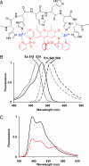
Structure and spectra of HisZiFiT. (A) Structure of HisZiFiT (red) and proposed mode of Zn2+-mediated binding to a His6 sequence (black), in flattened projection. (B) Normalized fluorescence excitation (solid curves) and emission (dashed) spectra of 1 μM HisZiFiT without Zn2+ (gray curves) and with saturating (10 μM) free Zn2+ (black). (C) FRET response of His6-CFP to HisZiFiT binding (ex420). Emission spectrum of 1 μM His6-CFP with 10 μM buffered free Zn2+ before (black curve) and after (red curve) addition of 1 μM HisZiFiT.
Fig. 2.
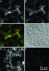
HisZiFiT labeling is specific for cell-surface His6-tagged proteins. (A–D) HEK293T cells expressing His6-CFP-GPI, labeled with 100 nM HisZiFiT in 1 μM buffered free Zn2+. (A) CFP channel, ex420/em475, shows both surface and internal His6-CFP-GPI. (B) HisZiFiT channel, ex540/em595, of the same cells shows only plasma membrane outlines. (C) Merge of A (displayed in green) and B (red). (D) Differential interference contrast image of the same cells. (E and F) Control HEK293T cells expressing nontagged CFP (CFP-GPI), identically exposed to HisZiFiT. (E) CFP channel. (F) HisZiFit channel shows negligible staining of CFP–GPI protein.
Fig. 3.
Orthogonal staining of HEK293T cells expressing two proteins on the PM. (A) Connexin43–4C with an intracellular tetracysteine motif, stained with ReAsH (ex568/55, em653/95). (B) His6 on extracellular PM surface stained and visualized with HisZiFiT (ex495/10, em535/45). (C) Merge of images A and B representing HisZiFiT staining of His6-CFP-GPI (green) and ReAsH staining of Connexin43–4C (red).
Fig. 4.
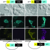
N-terminal tagging with His6 but not CFP allows cell surface exposure of STIM1. (A–L) Images of cells transiently transfected with His6-CFP-STIM1 (A–F) or His6-STIM1-CFP protein (G–L). Cartoons above A–L schematize the expressed polypeptide domains, where SP denotes signal peptide and H6 stands for His6. (A, D, G, and J) Transmitted light images; images in A, D, and J show differential interference contrast. (B, E, H, K) CFP fluorescence (cyan) of the same fields shows that all of the STIM1 fusions are recruited to the plasma membrane after depletion of Ca2+ stores with 2 μM thapsigargin. (C) Lack of specific staining by HisZiFiT (yellow) indicates absence of extracellularly exposed His6-tags on the same cells as in B. (F) Lack of antibody staining for His5 (magenta) confirms absence of extracellularly exposed His6 tags on the same live cell as in E. (I) Surface exposed His6-tags of the same cell as in H are labeled by HisZiFiT (yellow). (L) Live cell His5-Ab staining (detected with Alexa Fluor 568-labeled secondary Ab, magenta) confirms the exposure of N-terminal His6-tags on the same cells as in K. Exposure times with a 5% neutral density filter on excitation were 200, 1,000, and 50 ms for CFP, HisZiFiT, and Alexa Fluor 568, respectively. Cartoons show schematically how His6 remains luminal in His6-CFP-STIM1 (B, C, E, and F) but becomes extracellularly accessible in His6-STIM1-CFP (H, I, K, and L).
Similar articles
- Mutations of the Ca2+-sensing stromal interaction molecule STIM1 regulate Ca2+ influx by altered oligomerization of STIM1 and by destabilization of the Ca2+ channel Orai1.
Kilch T, Alansary D, Peglow M, Dörr K, Rychkov G, Rieger H, Peinelt C, Niemeyer BA. Kilch T, et al. J Biol Chem. 2013 Jan 18;288(3):1653-64. doi: 10.1074/jbc.M112.417246. Epub 2012 Dec 4. J Biol Chem. 2013. PMID: 23212906 Free PMC article. - Stored Ca2+ depletion-induced oligomerization of stromal interaction molecule 1 (STIM1) via the EF-SAM region: An initiation mechanism for capacitive Ca2+ entry.
Stathopulos PB, Li GY, Plevin MJ, Ames JB, Ikura M. Stathopulos PB, et al. J Biol Chem. 2006 Nov 24;281(47):35855-62. doi: 10.1074/jbc.M608247200. Epub 2006 Oct 3. J Biol Chem. 2006. PMID: 17020874 - Cross-talk between N-terminal and C-terminal domains in stromal interaction molecule 2 (STIM2) determines enhanced STIM2 sensitivity.
Emrich SM, Yoast RE, Xin P, Zhang X, Pathak T, Nwokonko R, Gueguinou MF, Subedi KP, Zhou Y, Ambudkar IS, Hempel N, Machaca K, Gill DL, Trebak M. Emrich SM, et al. J Biol Chem. 2019 Apr 19;294(16):6318-6332. doi: 10.1074/jbc.RA118.006801. Epub 2019 Mar 1. J Biol Chem. 2019. PMID: 30824535 Free PMC article. - Recent progress on STIM1 domains controlling Orai activation.
Schindl R, Muik M, Fahrner M, Derler I, Fritsch R, Bergsmann J, Romanin C. Schindl R, et al. Cell Calcium. 2009 Oct;46(4):227-32. doi: 10.1016/j.ceca.2009.08.003. Epub 2009 Sep 4. Cell Calcium. 2009. PMID: 19733393 Review. - Biochemical properties and cellular localisation of STIM proteins.
Dziadek MA, Johnstone LS. Dziadek MA, et al. Cell Calcium. 2007 Aug;42(2):123-32. doi: 10.1016/j.ceca.2007.02.006. Epub 2007 Mar 26. Cell Calcium. 2007. PMID: 17382385 Review.
Cited by
- STIM proteins: dynamic calcium signal transducers.
Soboloff J, Rothberg BS, Madesh M, Gill DL. Soboloff J, et al. Nat Rev Mol Cell Biol. 2012 Sep;13(9):549-65. doi: 10.1038/nrm3414. Nat Rev Mol Cell Biol. 2012. PMID: 22914293 Free PMC article. Review. - Molecular stretching modulates mechanosensing pathways.
Hu X, Margadant FM, Yao M, Sheetz MP. Hu X, et al. Protein Sci. 2017 Jul;26(7):1337-1351. doi: 10.1002/pro.3188. Epub 2017 Jun 6. Protein Sci. 2017. PMID: 28474792 Free PMC article. Review. - Ca-dependent Ras/Erk signaling mediates negative selection of autoreactive B cells.
Limnander A, Weiss A. Limnander A, et al. Small GTPases. 2011 Sep;2(5):282-288. doi: 10.4161/sgtp.2.5.17794. Epub 2011 Sep 1. Small GTPases. 2011. PMID: 22292132 Free PMC article. - Lighting Up and Identifying Metal-Binding Proteins in Cells.
Tiemuer A, Zhao H, Chen J, Li H, Sun H. Tiemuer A, et al. JACS Au. 2024 Nov 29;4(12):4628-4638. doi: 10.1021/jacsau.4c00879. eCollection 2024 Dec 23. JACS Au. 2024. PMID: 39735929 Free PMC article. Review. - ATP depletion induces translocation of STIM1 to puncta and formation of STIM1-ORAI1 clusters: translocation and re-translocation of STIM1 does not require ATP.
Chvanov M, Walsh CM, Haynes LP, Voronina SG, Lur G, Gerasimenko OV, Barraclough R, Rudland PS, Petersen OH, Burgoyne RD, Tepikin AV. Chvanov M, et al. Pflugers Arch. 2008 Nov;457(2):505-17. doi: 10.1007/s00424-008-0529-y. Epub 2008 Jun 10. Pflugers Arch. 2008. PMID: 18542992 Free PMC article.
References
- Giepmans BN, Adams SR, Ellisman MH, Tsien RY. Science. 2006;312:217–224. - PubMed
- Chen I, Ting AY. Curr Opin Biotechnol. 2005;16:35–40. - PubMed
- Griffin BA, Adams SR, Tsien RY. Science. 1998;281:269–272. - PubMed
- Kapanidis AN, Ebright YW, Ebright RH. J Am Chem Soc. 2001;123:12123–12125. - PubMed
- Guignet EG, Hovius R, Vogel H. Nat Biotechnol. 2004;22:440–444. - PubMed
Publication types
MeSH terms
Substances
Grants and funding
- P20 GM072033/GM/NIGMS NIH HHS/United States
- NS 27177/NS/NINDS NIH HHS/United States
- GM 72033/GM/NIGMS NIH HHS/United States
- R01 NS027177/NS/NINDS NIH HHS/United States
- R37 NS027177/NS/NINDS NIH HHS/United States
LinkOut - more resources
Full Text Sources
Other Literature Sources
Miscellaneous


