Amphiregulin is an essential mediator of estrogen receptor alpha function in mammary gland development - PubMed (original) (raw)
Amphiregulin is an essential mediator of estrogen receptor alpha function in mammary gland development
Laura Ciarloni et al. Proc Natl Acad Sci U S A. 2007.
Abstract
Most mammary gland development occurs after birth under the control of systemic hormones. Estrogens induce mammary epithelial cell proliferation during puberty via epithelial estrogen receptor alpha (ERalpha) by a paracrine mechanism. Epidermal growth factor receptor (EGFR) signaling has long been implicated downstream of ERalpha signaling, and several EGFR ligands have been described as estrogen-target genes in tumor cell lines. Here, we show that amphiregulin is the unique EGF family member to be transcriptionally induced by estrogen in the mammary glands of puberal mice at a time of exponential expansion of the ductal system. In fact, we find that estrogens induce amphiregulin through the ERalpha and require amphiregulin to induce proliferation of the mammary epithelium. Like ERalpha, amphiregulin is required in the epithelium of puberal mice for epithelial proliferation, terminal end buds formation, and ductal elongation. Subsequent stages, such as side-branching and alveologenesis, are not affected. When amphiregulin(-/-) mammary epithelial cells are in close vicinity to wild-type cells, they proliferate and contribute to all cell compartments of the ductal outgrowth. Thus, amphiregulin is an important paracrine mediator of estrogen function specifically required for puberty-induced ductal elongation, but not for any earlier or later developmental stages.
Conflict of interest statement
The authors declare no conflict of interest.
Figures
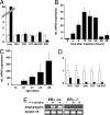
Fig. 1.
Regulation of EGFR ligands' expression by estrogens. Quantitative RT-PCR analysis of mammary gland mRNA for amphiregulin and other EGFR ligands normalized to keratin 18. (A and B) Mice ovariectomized before puberty were injected with either vehicle (open bars) or 17-β-estradiol (filled bars) and analyzed either for EGFR ligand expression 8 h later (A) or for amphiregulin over 48 h (B). (C and D) Mammary glands of 14- to 28-day-old mice were analyzed for amphiregulin expression (C) or for EGFR ligand expression (14- and 21-day-old females, open and filled bars, respectively) (D). Bars report the mean values obtained from three different mice. Error bars indicate standard deviation. Relative increase refers to control treated (A and B) or to the 14-day-old mice (C and D). (E) RT-PCR analysis of amphiregulin and keratin 18 expression in glands from ovariectomized WT and ERα−/− mice 6 h after administration of 17-β-estradiol (+) or vehicle (−).
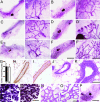
Fig. 2.
Developmental analysis of amphiregulin−/− mammary glands. (A–F) Whole-mount micrographs of inguinal glands from amphiregulin−/− (A, C, and E) and WT (B, D, and F) females were analyzed at the following developmental stages: day 24 (A and B), 6 weeks (C and D), 3 months (E and F). (Scale bars: A–F, 5 mm; A′–F′, 1 mm.) Micrographs are representative of glands from 12 mice analyzed per time point. (G) Number of branching points in inguinal mammary glands of 24-day-old amphiregulin−/− (filled bar, n = 6) and WT (open bar, n = 7) females. (H and I) Histological sections of mammary glands from 24-day-old amphiregulin−/− and WT mice stained by immunohistochemistry with an anti-SMAα antibody to highlight the myoepithelial cells (brown) counterstained with hematoxylin. H&E (J and K) staining of paraffin sections from 6-week-old amphiregulin−/− (J) and WT (K) glands. (Scale bar: 50 μm.) (L and M) Whole-mount micrographs of amphiregulin−/− (L) and WT (M) glands at 16.5 days of pregnancy. (N and O) Histological sections of amphiregulin−/− (N) and WT (O) mammary glands at 18.5 days of pregnancy. (P) Whole-mount micrograph of gland from an amphiregulin−/− female after 12 pregnancies.
Fig. 3.
Estrogen-induced proliferation in amphiregulin−/− and WT mammary glands. Immunohistochemistry was performed on sections of amphiregulin−/− (A and C) or WT (B and D) glands either from 3-week-old ovariectomized females 48 h after 17-β-estradiol stimulation (A and B) or 26-day-old mice (C and D) with an anti-BrdU antibody. Positively stained lymph nodes from the same glands are shown as positive control for BrdU incorporation (A and C Insets). (Scale bars: 50 μm.)
Fig. 4.
Amphiregulin−/− epithelial transplants. Representative outgrowth of amphiregulin−/− (A) or WT (B) GFP+ mammary epithelium transplanted into WT cleared fat pads. The glands were derived from virgin recipients and observed directly under the fluorescence stereomicroscope (B). (Scale bar: 5 mm.)
Fig. 5.
Amphiregulin−/− and WT chimeric mammary epithelia. Whole-mount preparations of a cleared fat pad showing representative mammary reconstitution on injection with a mixture of amphiregulin−/− GFP+ (green), and WT ROSA26+ (blue) epithelial cells in a 1:1 ratio. (A and B) GFP was visualized directly under the fluorescence stereomicroscope (A); β-galactosidase activity revealed after X-Gal staining (B). (C and D) Histological sections of glands reconstituted with amphiregulin−/− _LacZ_+ and WT cells. Amphiregulin−/− _LacZ_+ cells (blue) were found both among the body (C, arrow) and the cap cells (D, arrow). (E and F) Glands reconstituted with amphiregulin−/− GFP+ and WT cells were sectioned and stained with anti-GFP antibody. Both luminal (F, arrow) and myoepithelial cells (E, arrow) show GFP staining (brown). (G) Glands reconstituted with amphiregulin−/− GFP+ and WT cells were sectioned and costained with anti-BrdU (Left), and anti-GFP antibodies (Center) merge (Right) reveals double-positive cells. (Scale bars: A and B, 500 μm; C–G, 50 μm.)
Similar articles
- Amphiregulin: role in mammary gland development and breast cancer.
McBryan J, Howlin J, Napoletano S, Martin F. McBryan J, et al. J Mammary Gland Biol Neoplasia. 2008 Jun;13(2):159-69. doi: 10.1007/s10911-008-9075-7. Epub 2008 Apr 9. J Mammary Gland Biol Neoplasia. 2008. PMID: 18398673 Review. - 2-methoxyestradiol induces mammary gland differentiation through amphiregulin-epithelial growth factor receptor-mediated signaling: molecular distinctions from the mammary gland of pregnant mice.
Huh JI, Qiu TH, Chandramouli GV, Charles R, Wiench M, Hager GL, Catena R, Calvo A, LaVallee TM, Desprez PY, Green JE. Huh JI, et al. Endocrinology. 2007 Mar;148(3):1266-77. doi: 10.1210/en.2006-0964. Epub 2006 Dec 7. Endocrinology. 2007. PMID: 17158205 - Estrogen receptor-alpha expression in the mammary epithelium is required for ductal and alveolar morphogenesis in mice.
Feng Y, Manka D, Wagner KU, Khan SA. Feng Y, et al. Proc Natl Acad Sci U S A. 2007 Sep 11;104(37):14718-23. doi: 10.1073/pnas.0706933104. Epub 2007 Sep 4. Proc Natl Acad Sci U S A. 2007. PMID: 17785410 Free PMC article. - Cyclin D1 determines estrogen signaling in the mammary gland in vivo.
Casimiro MC, Wang C, Li Z, Di Sante G, Willmart NE, Addya S, Chen L, Liu Y, Lisanti MP, Pestell RG. Casimiro MC, et al. Mol Endocrinol. 2013 Sep;27(9):1415-28. doi: 10.1210/me.2013-1065. Epub 2013 Jul 17. Mol Endocrinol. 2013. PMID: 23864650 Free PMC article. - The ADAM17-amphiregulin-EGFR axis in mammary development and cancer.
Sternlicht MD, Sunnarborg SW. Sternlicht MD, et al. J Mammary Gland Biol Neoplasia. 2008 Jun;13(2):181-94. doi: 10.1007/s10911-008-9084-6. Epub 2008 May 10. J Mammary Gland Biol Neoplasia. 2008. PMID: 18470483 Free PMC article. Review.
Cited by
- Cellular Plasticity and Heterotypic Interactions during Breast Morphogenesis and Cancer Initiation.
Ingthorsson S, Traustadottir GA, Gudjonsson T. Ingthorsson S, et al. Cancers (Basel). 2022 Oct 24;14(21):5209. doi: 10.3390/cancers14215209. Cancers (Basel). 2022. PMID: 36358627 Free PMC article. Review. - Progesterone signalling in breast cancer: a neglected hormone coming into the limelight.
Brisken C. Brisken C. Nat Rev Cancer. 2013 Jun;13(6):385-96. doi: 10.1038/nrc3518. Nat Rev Cancer. 2013. PMID: 23702927 Review. - Co-administering Melatonin With an Estradiol-Progesterone Menopausal Hormone Therapy Represses Mammary Cancer Development in a Mouse Model of HER2-Positive Breast Cancer.
Dodda BR, Bondi CD, Hasan M, Clafshenkel WP, Gallagher KM, Kotlarczyk MP, Sethi S, Buszko E, Latimer JJ, Cline JM, Witt-Enderby PA, Davis VL. Dodda BR, et al. Front Oncol. 2019 Jul 9;9:525. doi: 10.3389/fonc.2019.00525. eCollection 2019. Front Oncol. 2019. PMID: 31355130 Free PMC article. - Mammary tumorigenesis induced by fibroblast growth factor receptor 1 requires activation of the epidermal growth factor receptor.
Bade LK, Goldberg JE, Dehut HA, Hall MK, Schwertfeger KL. Bade LK, et al. J Cell Sci. 2011 Sep 15;124(Pt 18):3106-17. doi: 10.1242/jcs.082651. Epub 2011 Aug 24. J Cell Sci. 2011. PMID: 21868365 Free PMC article. - Single-cell and spatial analyses reveal a tradeoff between murine mammary proliferation and lineage programs associated with endocrine cues.
Gray GK, Girnius N, Kuiken HJ, Henstridge AZ, Brugge JS. Gray GK, et al. Cell Rep. 2023 Oct 31;42(10):113293. doi: 10.1016/j.celrep.2023.113293. Epub 2023 Oct 17. Cell Rep. 2023. PMID: 37858468 Free PMC article.
References
- Lyons WR, Li CH, Johnson RE. Recent Prog Horm Res. 1958;14:219–248. - PubMed
- Nandi S. J Natl Cancer Inst. 1958;21:1039–1063. - PubMed
- Shyamala G. J Mammary Gland Biol Neoplasia. 1999;4:89–104. - PubMed
- Brisken C. J Mammary Gland Biol Neoplasia. 2002;7:39–48. - PubMed
Publication types
MeSH terms
Substances
LinkOut - more resources
Full Text Sources
Other Literature Sources
Molecular Biology Databases
Research Materials
Miscellaneous


