Elevated levels of 3-nitrotyrosine in brain from subjects with amnestic mild cognitive impairment: implications for the role of nitration in the progression of Alzheimer's disease - PubMed (original) (raw)
Elevated levels of 3-nitrotyrosine in brain from subjects with amnestic mild cognitive impairment: implications for the role of nitration in the progression of Alzheimer's disease
D Allan Butterfield et al. Brain Res. 2007.
Abstract
A number of studies reported that oxidative and nitrosative damage may be important in the pathogenesis of Alzheimer's disease (AD). However, whether oxidative damage precedes, contributes directly, or is secondary to AD pathogenesis is not known. Amnestic mild cognitive impairment (MCI) is a clinical condition that is a transition between normal aging and dementia and AD, characterized by a memory deficit without loss of general cognitive and functional abilities. Analysis of nitrosative stress in MCI could be important to determine whether nitrosative damage directly contributes to AD. In the present study, we measured the level of total protein nitration to determine if excess protein nitration occurs in brain samples from subjects with MCI compared to that in healthy controls. We demonstrated using slot blot that protein nitration is higher in the inferior parietal lobule (IPL) and hippocampus in MCI compared to those regions from control subjects. Immunohistochemistry analysis of hippocampus confirmed this result. These findings suggest that nitrosative damage occurs early in the course of MCI, and that protein nitration may be important for conversion of MCI to AD.
Figures
Figure 1
Tyrosine nitration as indexed by 3-NT immunoreactivity. ‘A’ represents the histogram obtained from Control and MCI hippocampus and IPL. ‘B’ is the slot blot results for IPL, while ‘C’ is the slot blot for hippocampus. (D) Slot blots showing standards that consist of BSA treated with defined concentrations of peroxynitrite along with control and MCI IPL *p<0.01. (E) Standard graph prepared with nitration of BSA with known concentration of peroxynitrite. The figure also shows the mean level of 3-NT in MCI hippocampus and IPL and associated concentration of peroxynitrite needed to produce these levels. Data are presented as the mean ± SEM. N=6.
Figure 2
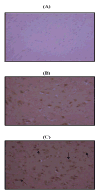
Immunohistochemical staining. ‘A’ represents the negative control using rabbit IgG. ‘B’ and ‘C’ are representative micrographs of immunohistochemistry obtained with a polyclonal antibody for 3-nitrotyrosine in control hippocampus and MCI hippocampus, respectively (×20 magnification). Intense nitrotyrosine staining is present in MCI hippocampus, whereas staining is far less prominent in control hippocampus. Nitrotyrosine is localized predominantly in neurons (arrows).
Similar articles
- Elevated levels of pro-apoptotic p53 and its oxidative modification by the lipid peroxidation product, HNE, in brain from subjects with amnestic mild cognitive impairment and Alzheimer's disease.
Cenini G, Sultana R, Memo M, Butterfield DA. Cenini G, et al. J Cell Mol Med. 2008 Jun;12(3):987-94. doi: 10.1111/j.1582-4934.2008.00163.x. J Cell Mol Med. 2008. PMID: 18494939 Free PMC article. - Effects of oxidative and nitrosative stress in brain on p53 proapoptotic protein in amnestic mild cognitive impairment and Alzheimer disease.
Cenini G, Sultana R, Memo M, Butterfield DA. Cenini G, et al. Free Radic Biol Med. 2008 Jul 1;45(1):81-5. doi: 10.1016/j.freeradbiomed.2008.03.015. Epub 2008 Apr 8. Free Radic Biol Med. 2008. PMID: 18439434 Free PMC article. - Proteomic identification of nitrated brain proteins in amnestic mild cognitive impairment: a regional study.
Sultana R, Reed T, Perluigi M, Coccia R, Pierce WM, Butterfield DA. Sultana R, et al. J Cell Mol Med. 2007 Jul-Aug;11(4):839-51. doi: 10.1111/j.1582-4934.2007.00065.x. J Cell Mol Med. 2007. PMID: 17760844 Free PMC article. - Oxidative damage in mild cognitive impairment and early Alzheimer's disease.
Lovell MA, Markesbery WR. Lovell MA, et al. J Neurosci Res. 2007 Nov 1;85(14):3036-40. doi: 10.1002/jnr.21346. J Neurosci Res. 2007. PMID: 17510979 Review. - Biomarkers of oxidative and nitrosative damage in Alzheimer's disease and mild cognitive impairment.
Mangialasche F, Polidori MC, Monastero R, Ercolani S, Camarda C, Cecchetti R, Mecocci P. Mangialasche F, et al. Ageing Res Rev. 2009 Oct;8(4):285-305. doi: 10.1016/j.arr.2009.04.002. Epub 2009 Apr 17. Ageing Res Rev. 2009. PMID: 19376275 Review.
Cited by
- Kinetic and mechanistic considerations to assess the biological fate of peroxynitrite.
Carballal S, Bartesaghi S, Radi R. Carballal S, et al. Biochim Biophys Acta. 2014 Feb;1840(2):768-80. doi: 10.1016/j.bbagen.2013.07.005. Epub 2013 Jul 18. Biochim Biophys Acta. 2014. PMID: 23872352 Free PMC article. Review. - Elevated oxidative stress and decreased antioxidant function in the human hippocampus and frontal cortex with increasing age: implications for neurodegeneration in Alzheimer's disease.
Venkateshappa C, Harish G, Mahadevan A, Srinivas Bharath MM, Shankar SK. Venkateshappa C, et al. Neurochem Res. 2012 Aug;37(8):1601-14. doi: 10.1007/s11064-012-0755-8. Epub 2012 Mar 30. Neurochem Res. 2012. PMID: 22461064 - Honey and Alzheimer's Disease-Current Understanding and Future Prospects.
Shaikh A, Ahmad F, Teoh SL, Kumar J, Yahaya MF. Shaikh A, et al. Antioxidants (Basel). 2023 Feb 9;12(2):427. doi: 10.3390/antiox12020427. Antioxidants (Basel). 2023. PMID: 36829985 Free PMC article. Review. - Phosphoproteomics of Alzheimer disease brain: Insights into altered brain protein regulation of critical neuronal functions and their contributions to subsequent cognitive loss.
Butterfield DA. Butterfield DA. Biochim Biophys Acta Mol Basis Dis. 2019 Aug 1;1865(8):2031-2039. doi: 10.1016/j.bbadis.2018.08.035. Epub 2018 Aug 29. Biochim Biophys Acta Mol Basis Dis. 2019. PMID: 31167728 Free PMC article. Review. - Apolipoprotein E and oxidative stress in brain with relevance to Alzheimer's disease.
Butterfield DA, Mattson MP. Butterfield DA, et al. Neurobiol Dis. 2020 May;138:104795. doi: 10.1016/j.nbd.2020.104795. Epub 2020 Feb 6. Neurobiol Dis. 2020. PMID: 32036033 Free PMC article. Review.
References
- Aksenova MV, Aksenov MY, Payne RM, Trojanowski JQ, Schmidt ML, Carney JM, Butterfield DA, Markesbery WR. Oxidation of cytosolic proteins and expression of creatine kinase BB in frontal lobe in different neurodegenerative disorders. Dement Geriatr Cogn Disord. 1999;10:158–65. - PubMed
- Beckman JS. Oxidative damage and tyrosine nitration from peroxynitrite. Chem Res Toxicol. 1996;9:836–44. - PubMed
- Butterfield DA, Stadtman ER. Protein oxidation processes in aging brain. Adv Cell Aging Gerontol. 1997;2:161–191.
- Butterfield DA, Kanski J. Brain protein oxidation in age-related neurodegenerative disorders that are associated with aggregated proteins. Mech Ageing Dev. 2001;122:945–62. - PubMed
- Butterfield DA. Proteomics: a new approach to investigate oxidative stress in Alzheimer's disease brain. Brain Research. 2004;1000:1–7. - PubMed
Publication types
MeSH terms
Substances
Grants and funding
- P01 AG010836-130011/AG/NIA NIH HHS/United States
- AG-05144/AG/NIA NIH HHS/United States
- P01 AG005119/AG/NIA NIH HHS/United States
- AG-05119/AG/NIA NIH HHS/United States
- P01 AG010836/AG/NIA NIH HHS/United States
- P01 AG005119-150006/AG/NIA NIH HHS/United States
- P50 AG005144/AG/NIA NIH HHS/United States
- AG-10836/AG/NIA NIH HHS/United States
LinkOut - more resources
Full Text Sources
Other Literature Sources
Medical

