How the brain translates money into force: a neuroimaging study of subliminal motivation - PubMed (original) (raw)
How the brain translates money into force: a neuroimaging study of subliminal motivation
Mathias Pessiglione et al. Science. 2007.
Abstract
Unconscious motivation in humans is often inferred but rarely demonstrated empirically. We imaged motivational processes, implemented in a paradigm that varied the amount and reportability of monetary rewards for which subjects exerted physical effort. We show that, even when subjects cannot report how much money is at stake, they nevertheless deploy more force for higher amounts. Such a motivational effect is underpinned by engagement of a specific basal forebrain region. Our findings thus reveal this region as a key node in brain circuitry that enables expected rewards to energize behavior, without the need for the subjects;awareness.
Figures
Fig. 1
The incentive force task. Successive screens displayed in one trial are shown from left to right, with durations in ms. Coin images, either one pound (£1) or one penny (1p), indicate the monetary value attributed to the top of the thermometer image. The fluid level in the thermometer represents the online force exerted on the hand grip. The last screen indicates cumulative total of the money won so far.
Fig. 2
SPMs of brain activity. Voxels displayed in gray on glass brains showed a significant effect at P < 0.05 after correction for multiple comparisons over the entire brain. The [x, y, z] coordinates of the different maxima refer to the Montreal Neurological Institute (MNI) space. Axial and coronal slices were taken at global maxima of interest indicated by red symbols on the glass brains. SPMs are shown at a lower threshold (P < 0.001, uncorrected) and were superimposed on the average structural scan to localize significant activations. The images in the left column show regression with the amount of force produced, whatever the condition. The images in the middle column show contrast between conscious pounds and pennies trials (£1 to 1p, 100 ms). For this contrast, SPMs were coregistered with an atlas of the basal ganglia (right column). Caudate, putamen, and accumbens are shown in green; external and internal pallidum are shown in blue, with limbic sectors in violet.
Fig. 3
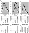
Main effects of stimulus duration. (A) Incentive force task. Time courses were averaged across trials for the different stimuli (black lines indicate £1 and white lines indicate 1p) and durations (thin, intermediate, and thick lines indicate 17, 50, and 100 ms, respectively). Time 0 corresponds to the moment of stimulus display. The histograms indicate the effect of motivation (£1 to 1p), and the error bars indicate SEM. Pallidal activation is expressed as percentage of blood oxygen level-dependent signal change. Force and skin conductance are expressed in proportion of the highest measure. (B) Perception task. Stimuli were the same as in (A). Possible responses were “seen £1,” “seen 1p,” “guess £1,” and “guess 1p.” A “correct” response means that the subject chose the stimulus that had been displayed. A “seen” response means that the subject perceived all or part of the stimulus. Error bars indicate SEM.
Similar articles
- Disconnecting force from money: effects of basal ganglia damage on incentive motivation.
Schmidt L, d'Arc BF, Lafargue G, Galanaud D, Czernecki V, Grabli D, Schüpbach M, Hartmann A, Lévy R, Dubois B, Pessiglione M. Schmidt L, et al. Brain. 2008 May;131(Pt 5):1303-10. doi: 10.1093/brain/awn045. Epub 2008 Mar 15. Brain. 2008. PMID: 18344560 - Preserved Unconscious Processing in Schizophrenia: The Case of Motivation.
Berkovitch L, Gaillard R, Abdel-Ahad P, Smadja S, Gauthier C, Attali D, Beaucamps H, Plaze M, Pessiglione M, Vinckier F. Berkovitch L, et al. Schizophr Bull. 2022 Sep 1;48(5):1094-1103. doi: 10.1093/schbul/sbac076. Schizophr Bull. 2022. PMID: 35751516 Free PMC article. - The effect of subliminal incentives on goal-directed eye movements.
Hinze VK, Uslu O, Antono JE, Wilke M, Pooresmaeili A. Hinze VK, et al. J Neurophysiol. 2021 Dec 1;126(6):2014-2026. doi: 10.1152/jn.00414.2021. Epub 2021 Nov 10. J Neurophysiol. 2021. PMID: 34758270 Free PMC article. - Distinct neurofunctional alterations during motivational and hedonic processing of natural and monetary rewards in depression - a neuroimaging meta-analysis.
Bore MC, Liu X, Gan X, Wang L, Xu T, Ferraro S, Li L, Zhou B, Zhang J, Vatansever D, Biswal B, Klugah-Brown B, Becker B. Bore MC, et al. Psychol Med. 2024 Mar;54(4):639-651. doi: 10.1017/S0033291723003410. Epub 2023 Nov 24. Psychol Med. 2024. PMID: 37997708 Review. - Mapping social reward and punishment processing in the human brain: A voxel-based meta-analysis of neuroimaging findings using the social incentive delay task.
Martins D, Rademacher L, Gabay AS, Taylor R, Richey JA, Smith DV, Goerlich KS, Nawijn L, Cremers HR, Wilson R, Bhattacharyya S, Paloyelis Y. Martins D, et al. Neurosci Biobehav Rev. 2021 Mar;122:1-17. doi: 10.1016/j.neubiorev.2020.12.034. Epub 2021 Jan 6. Neurosci Biobehav Rev. 2021. PMID: 33421544 Review.
Cited by
- Psychomotor Symptoms in Chronic Cocaine Users: An Interpretative Model.
Cenci D, Carbone MG, Callegari C, Maremmani I. Cenci D, et al. Int J Environ Res Public Health. 2022 Feb 8;19(3):1897. doi: 10.3390/ijerph19031897. Int J Environ Res Public Health. 2022. PMID: 35162918 Free PMC article. - The tempted brain eats: pleasure and desire circuits in obesity and eating disorders.
Berridge KC, Ho CY, Richard JM, DiFeliceantonio AG. Berridge KC, et al. Brain Res. 2010 Sep 2;1350:43-64. doi: 10.1016/j.brainres.2010.04.003. Epub 2010 Apr 11. Brain Res. 2010. PMID: 20388498 Free PMC article. Review. - The limit to exercise tolerance in humans: mind over muscle?
Marcora SM, Staiano W. Marcora SM, et al. Eur J Appl Physiol. 2010 Jul;109(4):763-70. doi: 10.1007/s00421-010-1418-6. Epub 2010 Mar 11. Eur J Appl Physiol. 2010. PMID: 20221773 Clinical Trial. - Expectation of reward differentially modulates executive inhibition.
Herrera PM, Van Meerbeke AV, Speranza M, Cabra CL, Bonilla M, Canu M, Bekinschtein TA. Herrera PM, et al. BMC Psychol. 2019 Aug 23;7(1):55. doi: 10.1186/s40359-019-0332-x. BMC Psychol. 2019. PMID: 31443739 Free PMC article. - How does reward expectation influence cognition in the human brain?
Rowe JB, Eckstein D, Braver T, Owen AM. Rowe JB, et al. J Cogn Neurosci. 2008 Nov;20(11):1980-92. doi: 10.1162/jocn.2008.20140. J Cogn Neurosci. 2008. PMID: 18416677 Free PMC article.
References
- Robbins TW, Everitt BJ. Curr. Opin. Neurobiol. 1996;6:228. - PubMed
- Berridge KC. Physiol. Behav. 2004;81:179. - PubMed
- Schultz W. Annu. Rev. Psychol. 2006;57:87. - PubMed
- Dehaene S, et al. Trends Cognit. Sci. 2006;10:204. - PubMed
- Dehaene S, et al. Nature. 1998;395:597. - PubMed
Publication types
MeSH terms
LinkOut - more resources
Full Text Sources
Other Literature Sources


