Selective chemical probe inhibitor of Stat3, identified through structure-based virtual screening, induces antitumor activity - PubMed (original) (raw)
. 2007 May 1;104(18):7391-6.
doi: 10.1073/pnas.0609757104. Epub 2007 Apr 26.
Shumin Zhang, Wayne C Guida, Michelle A Blaskovich, Benjamin Greedy, Harshani R Lawrence, M L Richard Yip, Richard Jove, Mark M McLaughlin, Nicholas J Lawrence, Said M Sebti, James Turkson
Affiliations
- PMID: 17463090
- PMCID: PMC1863497
- DOI: 10.1073/pnas.0609757104
Selective chemical probe inhibitor of Stat3, identified through structure-based virtual screening, induces antitumor activity
Khandaker Siddiquee et al. Proc Natl Acad Sci U S A. 2007.
Abstract
S3I-201 (NSC 74859) is a chemical probe inhibitor of Stat3 activity, which was identified from the National Cancer Institute chemical libraries by using structure-based virtual screening with a computer model of the Stat3 SH2 domain bound to its Stat3 phosphotyrosine peptide derived from the x-ray crystal structure of the Stat3beta homodimer. S3I-201 inhibits Stat3.Stat3 complex formation and Stat3 DNA-binding and transcriptional activities. Furthermore, S3I-201 inhibits growth and induces apoptosis preferentially in tumor cells that contain persistently activated Stat3. Constitutively dimerized and active Stat3C and Stat3 SH2 domain rescue tumor cells from S3I-201-induced apoptosis. Finally, S3I-201 inhibits the expression of the Stat3-regulated genes encoding cyclin D1, Bcl-xL, and survivin and inhibits the growth of human breast tumors in vivo. These findings strongly suggest that the antitumor activity of S3I-201 is mediated in part through inhibition of aberrant Stat3 activation and provide the proof-of-concept for the potential clinical use of Stat3 inhibitors such as S3I-201 in tumors harboring aberrant Stat3.
Conflict of interest statement
The authors declare no conflict of interest.
Figures
Fig. 1.
Application of computational modeling in in silico screening (virtual screening) to identify the compound S3I-201 from a chemical database. (A) S3I-201 (NSC 74859) docked to the SH2 domain of Stat3; a solvent-accessible surface of the protein (rendered on a 6-Å shell of residues surrounding the ligand) is shown. It is color coded according to the electrostatic potential, with red most negative and dark blue most positive. Hydrogen bonding to Lys-591, Ser-611, Ser-613, and Arg-609 is shown by white dashed lines. Carbon atoms of S3I-201 are shown in green. (B) S3I-201 docked to the SH2 domain of Stat3 along with pTyr peptide. Coloring for pTyr peptide is as follows: Ala, yellow; Pro, red; pTyr, magenta; Leu, orange; and Lys, green.
Fig. 2.
Evaluation for effects of S3I-201 on STATs, Shc, Src, and Erks activation, and on Lck-SH2 domain phosphopeptide binding. (A) Nuclear extracts containing activated STAT proteins were preincubated with or without S3I-201 for 30 min at room temperature before incubation with radiolabeled hSIE probe, which binds Stat1 and Stat3, or MGFe probe, which binds Stat1 and Stat5, or nuclear extracts were incubated with anti-Stat1, anti-Stat3, or anti-Stat5A antibody for 30 min at room temperature before incubation with radiolabeled probes, and were subjected to EMSA analysis. (B) (i) Cell lysates containing activated Stat3 were preincubated with S3I-201 or without (control, 0.05% DMSO) in the presence or absence of increasing amount of cell lysates containing inactive Stat3 protein (monomer), inactive Stat1 protein (monomer), inactive Stat5 protein (monomer), or Src protein before incubation with radiolabeled hSIE probe followed by EMSA analysis. (ii) SDS/PAGE and Western blot analysis of cell lysate preparations of equal amounts of total protein containing inactive Stat3 (monomer), Stat1 (monomer), or Stat5 (monomer), or Src protein and probed with anti-Stat3, anti-Stat1, anti-Stat5, or anti-Src antibody. (C) (i) SDS/PAGE and Western blot analysis of Stat3-yellow fluorescent protein (YFP) immunoprecipitates (i.p. YFP, Left) probing for FLAG-Stat3 (anti-FLAG, Upper) or Stat3-YFP (anti-YFP, Lower), or of FLAG-Stat3 immunoprecipitates (i.p. FLAG, Right) probing for Stat3-YFP (anti-YFP, Lower) or FLAG-Stat3 (anti-FLAG, Upper); (ii) SDS/PAGE and Western blot analysis of whole-cell lysates from FLAG-ST3-transiently transfected cells (input) treated with S3I-201 or untreated (control) probing for FLAG. (D) In vitro ELISA for the binding of Lck-SH2-GST to the conjugate biotinylated pTyr-peptide (EPQpYEEIEL), and effects of increasing concentration of S3I-201. Values are the mean ± SD of three independent determinations. (E) EMSA analysis of Stat3 DNA-binding activity in nuclear extract preparations from v-Src-transformed mouse fibroblasts (NIH 3T3/v-Src) treated for the indicated times or from the human breast cancer MDA-MB-231, MDA-MB-435, and MDA-MB-468 cells treated for 48 h with 100 μM S3I-201 and incubated with radiolabeled hSIE probe. (F) SDS/PAGE and Western blot analysis of whole-cell lysates from NIH 3T3/v-Src fibroblasts untreated (control) or treated with S3I-201 (100 μM, 24 h) probing with anti-pTyr-705-Stat3 or anti-Stat3 antibody. Positions of STAT·DNA complexes or proteins in gel are labeled; control lanes represent nuclear extracts treated with 0.05% DMSO, and nuclear extracts or cell lysates prepared from 0.05% DMSO-treated cells; asterisks (∗) indicate supershifted complexes of STAT·probe·antibody in the presence of anti-Stat1, anti-Stat3, or anti-Stat5 antibody.
Fig. 3.
S3I-201 suppresses Stat3-dependent but not Stat3-independent transcriptional activity. Shown are luciferase reporter activities in cytosolic extracts prepared from normal mouse fibroblasts (NIH 3T3) transiently cotransfected with the Stat3-dependent (pLucTKS3), or the Stat3-independent (pLucSRE or β-casein promoter-driven Luc) luciferase reporters together with a plasmid expressing the v-Src oncoprotein that activates all three reporters, and untreated (0.05% DMSO, control) or treated with 100 μM S3I-201 for 24 h. Values are the mean + SD of three independent determinations.
Fig. 4.
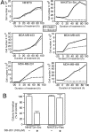
S3I-201 inhibits anchorage-dependent and -independent growth only of cells that contain persistently active Stat3. (A) Normal mouse fibroblasts (NIH 3T3) and v-Src transformed counterparts (NIH 3T3/v-Src), as well as the human breast carcinoma cells (MDA-MB-453, MDA-MB-435, MDA-MB-231, or MDA-MB-468) were untreated (0.05% DMSO, control) or treated with 100 μM S3I-201 and counted by trypan blue exclusion on each of 4 days for viable cell number. Values are the mean ± SD of three independent determinations. (B) v-Src-transformed mouse fibroblasts (NIH 3T3/v-Src) and their v-Ras-transformed counterparts (NIH 3T3/v-Ras) were grown in soft-agar suspension and untreated (0.05% DMSO, control) or treated with 100 μM S3I-201 every 3 days until large colonies were visible, which were enumerated. Values are the mean + SD of three or four independent determinations.
Fig. 5.
S3I-201 inhibits cyclin D1, Bcl-xL, and survivin expression and induces apoptosis in a Stat3-dependent manner. (A) Normal NIH 3T3 mouse fibroblasts and their v-Src-transformed counterparts (NIH 3T3/v-Src) and the human breast carcinoma MDA-MB-453 and MDA-MB-435 cell lines were untreated (0.05% DMSO) or treated with 30–300 μM S3I-201 for 48 h and subjected to annexin V staining and flow cytometry. 7AAD, 7-aminoactinomycin D. (B) Human breast carcinoma MDA-MB-231 cells were transfected with pcDNA3 (Mock) or Stat3C or were untransfected (Non) (Left), or the v-Src-transformed mouse fibroblasts (NIH 3T3/v-Src) were transfected with pcDNA3 (Mock), the N terminus of Stat3 (ST3-NT), or the Stat3 SH2 domain (ST3-SH2) for 4 h (Right). Twenty-four hours after transfection, cells were untreated [0.05% DMSO (−)] or treated (+) with 100 μM S3I-201 for an additional 24 h and subjected to annexin V staining and flow cytometry. For A and B, values are the mean + SD of six independent determinations. (C) SDS/PAGE and Western blot analysis of whole-cell lysates prepared from the v-Src-transformed mouse fibroblasts (NIH 3T3/v-Src) or the human breast cancer MDA-MB-231 cells untreated (DMSO, control) or treated with 100 μM S3I-201 for 48 h. Probing was with anti-cyclin D1, anti-Bcl-xL, and anti-survivin antibodies. Western blot data are representative of three independent analyses.
Fig. 6.
Tumor growth inhibition by S3I-201. (A) Human breast (MDA-MB-231) tumor-bearing mice were given S3I-201 (5 mg/kg) i.v. every 2 or every 3 days. Tumor sizes were monitored every 2–3 days, converted to tumor volumes, and plotted. Values are the mean ± SD of eight tumor-bearing mice each. (B) Three days after the last S3I-201 injection, animals were killed, tumors from one control animal (DMSO-treated) or residual tumor tissue from two S3I-201-treated (T1 and T2) mice were extracted, and lysate preparations with equal total proteins were analyzed for Stat3 activation by incubating with radiolabeled hSIE probe and subjecting the products to EMSA analysis (lanes 1–3), or lysates from control tumor tissue of equal total proteins were preincubated with or without increasing concentrations of S3I-201 before incubation with radiolabeled hSIE probe and subjecting the products to EMSA analysis (lanes 1, 4, 5, and 6). Positions of Stat3·DNA complexes are shown.
Similar articles
- A novel small-molecule disrupts Stat3 SH2 domain-phosphotyrosine interactions and Stat3-dependent tumor processes.
Zhang X, Yue P, Fletcher S, Zhao W, Gunning PT, Turkson J. Zhang X, et al. Biochem Pharmacol. 2010 May 15;79(10):1398-409. doi: 10.1016/j.bcp.2010.01.001. Epub 2010 Jan 11. Biochem Pharmacol. 2010. PMID: 20067773 Free PMC article. - An oxazole-based small-molecule Stat3 inhibitor modulates Stat3 stability and processing and induces antitumor cell effects.
Siddiquee KA, Gunning PT, Glenn M, Katt WP, Zhang S, Schrock C, Sebti SM, Jove R, Hamilton AD, Turkson J. Siddiquee KA, et al. ACS Chem Biol. 2007 Dec 21;2(12):787-98. doi: 10.1021/cb7001973. ACS Chem Biol. 2007. PMID: 18154266 - A cell-permeable Stat3 SH2 domain mimetic inhibits Stat3 activation and induces antitumor cell effects in vitro.
Zhao W, Jaganathan S, Turkson J. Zhao W, et al. J Biol Chem. 2010 Nov 12;285(46):35855-65. doi: 10.1074/jbc.M110.154088. Epub 2010 Aug 31. J Biol Chem. 2010. PMID: 20807764 Free PMC article. - Strategies and Approaches of Targeting STAT3 for Cancer Treatment.
Furtek SL, Backos DS, Matheson CJ, Reigan P. Furtek SL, et al. ACS Chem Biol. 2016 Feb 19;11(2):308-18. doi: 10.1021/acschembio.5b00945. Epub 2016 Jan 15. ACS Chem Biol. 2016. PMID: 26730496 Review. - STAT 3 as a target for cancer drug discovery.
Costantino L, Barlocco D. Costantino L, et al. Curr Med Chem. 2008;15(9):834-43. doi: 10.2174/092986708783955464. Curr Med Chem. 2008. PMID: 18473793 Review.
Cited by
- Hydrazinocurcumin Encapsuled nanoparticles "re-educate" tumor-associated macrophages and exhibit anti-tumor effects on breast cancer following STAT3 suppression.
Zhang X, Tian W, Cai X, Wang X, Dang W, Tang H, Cao H, Wang L, Chen T. Zhang X, et al. PLoS One. 2013 Jun 25;8(6):e65896. doi: 10.1371/journal.pone.0065896. Print 2013. PLoS One. 2013. PMID: 23825527 Free PMC article. - Meteorin-like facilitates skeletal muscle repair through a Stat3/IGF-1 mechanism.
Baht GS, Bareja A, Lee DE, Rao RR, Huang R, Huebner JL, Bartlett DB, Hart CR, Gibson JR, Lanza IR, Kraus VB, Gregory SG, Spiegelman BM, White JP. Baht GS, et al. Nat Metab. 2020 Mar;2(3):278-289. doi: 10.1038/s42255-020-0184-y. Epub 2020 Mar 16. Nat Metab. 2020. PMID: 32694780 Free PMC article. - Targeting the DNA-binding activity of the human ERG transcription factor using new heterocyclic dithiophene diamidines.
Nhili R, Peixoto P, Depauw S, Flajollet S, Dezitter X, Munde MM, Ismail MA, Kumar A, Farahat AA, Stephens CE, Duterque-Coquillaud M, David Wilson W, Boykin DW, David-Cordonnier MH. Nhili R, et al. Nucleic Acids Res. 2013 Jan 7;41(1):125-38. doi: 10.1093/nar/gks971. Epub 2012 Oct 23. Nucleic Acids Res. 2013. PMID: 23093599 Free PMC article. - CTRP4/interleukin-6 receptor signaling ameliorates autoimmune encephalomyelitis by suppressing Th17 cell differentiation.
Cao L, Deng J, Chen W, He M, Zhao N, Huang H, Ling L, Li Q, Zhu X, Wang L. Cao L, et al. J Clin Invest. 2023 Nov 28;134(4):e168384. doi: 10.1172/JCI168384. J Clin Invest. 2023. PMID: 38015631 Free PMC article. - Modulation of reactivation of latent herpes simplex virus 1 in ganglionic organ cultures by p300/CBP and STAT3.
Du T, Zhou G, Roizman B. Du T, et al. Proc Natl Acad Sci U S A. 2013 Jul 9;110(28):E2621-8. doi: 10.1073/pnas.1309906110. Epub 2013 Jun 20. Proc Natl Acad Sci U S A. 2013. PMID: 23788661 Free PMC article.
References
- Darnell JE., Jr Nat Rev Cancer. 2002;2:740–749. - PubMed
- Yu H, Jove R. Nat Rev Cancer. 2004;4:97–105. - PubMed
- Bromberg J, Darnell JE., Jr Oncogene. 2000;19:2468–2473. - PubMed
- Bowman T, Garcia R, Turkson J, Jove R. Oncogene. 2000;19:2474–2488. - PubMed
Publication types
MeSH terms
Substances
LinkOut - more resources
Full Text Sources
Other Literature Sources
Chemical Information
Molecular Biology Databases
Research Materials
Miscellaneous


