Oesophageal atresia - PubMed (original) (raw)
Review
Oesophageal atresia
Lewis Spitz. Orphanet J Rare Dis. 2007.
Abstract
Oesophageal atresia (OA) encompasses a group of congenital anomalies comprising of an interruption of the continuity of the oesophagus with or without a persistent communication with the trachea. In 86% of cases there is a distal tracheooesophageal fistula, in 7% there is no fistulous connection, while in 4% there is a tracheooesophageal fistula without atresia. OA occurs in 1 in 2500 live births. Infants with OA are unable to swallow saliva and are noted to have excessive salivation requiring repeated suctioning. Associated anomalies occur in 50% of cases, the majority involving one or more of the VACTERL association (vertebral, anorectal, cardiac, tracheooesophageal, renal and limb defects). The aetiology is largely unknown and is likely to be multifactorial, however, various clues have been uncovered in animal experiments particularly defects in the expression of the gene Sonic hedgehog (Shh). The vast majority of cases are sporadic and the recurrence risk for siblings is 1%. The diagnosis may be suspected prenatally by a small or absent stomach bubble on antenatal ultrasound scan at around 18 weeks gestation. The likelihood of an atresia is increased by the presence of polyhydramnios. A nasogastric tube should be passed at birth in all infants born to a mother with polyhydramnios as well as to infants who are excessively mucusy soon after delivery to establish or refute the diagnosis. In OA the tube will not progress beyond 10 cm from the mouth (confirmation is by plain X-ray of the chest and abdomen). Definitive management comprises disconnection of the tracheooesophageal fistula, closure of the tracheal defect and primary anastomosis of the oesophagus. Where there is a "long gap" between the ends of the oesophagus, delayed primary repair should be attempted. Only very rarely will an oesophageal replacement be required. Survival is directly related to birth weight and to the presence of a major cardiac defect. Infants weighing over 1500 g and having no major cardiac problem should have a near 100% survival, while the presence of one of the risk factors reduces survival to 80% and further to 30-50% in the presence of both risk factors.
Figures
Figure 1
Common anatomical types of oesophageal atresia. a) Oesophageal atresia with distal tracheooesophageal fistula (86%). b) Isolated esophageal atresia without tracheooesophageal fistula (7%). c) H-type tracheooesophageal fistula (4%)
Figure 2
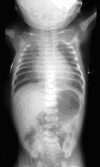
Plain X-ray of the chest and abdomen showing the radio-opaque tube in the blind upper oesophageal pouch. Air in the stomach indicates the presence of a distal tracheooesophageal fistula.
Figure 3
The operative repair of an oesophageal atresia and distal tracheooesophageal fistula.
Similar articles
- Genetic syndrome suspicion: examples of clinical approach in the neonatal unit.
Giuffrè M, De Sanctis L. Giuffrè M, et al. Minerva Pediatr. 2010 Jun;62(3 Suppl 1):199-201. Minerva Pediatr. 2010. PMID: 21089741 Review. - Phenotypic presentation and outcome of esophageal atresia in the era of the Spitz classification.
Driver CP, Shankar KR, Jones MO, Lamont GA, Turnock RR, Lloyd DA, Losty PD. Driver CP, et al. J Pediatr Surg. 2001 Sep;36(9):1419-21. doi: 10.1053/jpsu.2001.26389. J Pediatr Surg. 2001. PMID: 11528619 - Tracheal agenesis and esophageal atresia with proximal and distal bronchoesophageal fistulas.
Demircan M, Aksoy T, Ceran C, Kafkasli A. Demircan M, et al. J Pediatr Surg. 2008 Aug;43(8):e1-3. doi: 10.1016/j.jpedsurg.2008.04.015. J Pediatr Surg. 2008. PMID: 18675618 - An overview of isolated and syndromic oesophageal atresia.
Geneviève D, de Pontual L, Amiel J, Sarnacki S, Lyonnet S. Geneviève D, et al. Clin Genet. 2007 May;71(5):392-9. doi: 10.1111/j.1399-0004.2007.00798.x. Clin Genet. 2007. PMID: 17489843 Review. - Care of infants with esophageal atresia, tracheoesophageal fistula, and associated anomalies.
Holder TM, Ashcraft KW, Sharp RJ, Amoury RA. Holder TM, et al. J Thorac Cardiovasc Surg. 1987 Dec;94(6):828-35. J Thorac Cardiovasc Surg. 1987. PMID: 3682853
Cited by
- Quality improvement program reduces venous thromboembolism in infants and children with long-gap esophageal atresia (LGEA).
Kelly DP, Bairdain S, Zurakowski D, Dodson B, Harney KM, Jennings RW, Trenor CC. Kelly DP, et al. Pediatr Surg Int. 2016 Jul;32(7):691-6. doi: 10.1007/s00383-016-3902-5. Epub 2016 Jun 4. Pediatr Surg Int. 2016. PMID: 27262479 - Endoscopic interventional therapies for tracheoesophageal fistulas in children: A systematic review.
Ling Y, Sun B, Li J, Ma L, Li D, Yin G, Meng F, Gao M. Ling Y, et al. Front Pediatr. 2023 Feb 22;11:1121803. doi: 10.3389/fped.2023.1121803. eCollection 2023. Front Pediatr. 2023. PMID: 36911034 Free PMC article. Review. - In-utero gastric perforation from combined duodenal and esophageal atresia without consistent polyhydramnios.
Lyttle BD, Liechty K, Corkum K, Galan H, Behrendt N, Zaretsky M, Bruny J, Derderian SC. Lyttle BD, et al. J Surg Case Rep. 2021 Dec 28;2021(12):rjab551. doi: 10.1093/jscr/rjab551. eCollection 2021 Dec. J Surg Case Rep. 2021. PMID: 34987752 Free PMC article. - An approach to the identification of anomalies and etiologies in neonates with identified or suspected VACTERL (vertebral defects, anal atresia, tracheo-esophageal fistula with esophageal atresia, cardiac anomalies, renal anomalies, and limb anomalies) association.
Solomon BD, Baker LA, Bear KA, Cunningham BK, Giampietro PF, Hadigan C, Hadley DW, Harrison S, Levitt MA, Niforatos N, Paul SM, Raggio C, Reutter H, Warren-Mora N. Solomon BD, et al. J Pediatr. 2014 Mar;164(3):451-7.e1. doi: 10.1016/j.jpeds.2013.10.086. Epub 2013 Dec 12. J Pediatr. 2014. PMID: 24332453 Free PMC article. Review. No abstract available. - Maternal hyperthyroidism increases the prevalence of foregut atresias in fetal rats exposed to adriamycin.
Fragoso AC, Martinez L, Estevão-Costa J, Tovar JA. Fragoso AC, et al. Pediatr Surg Int. 2014 Feb;30(2):151-7. doi: 10.1007/s00383-013-3445-y. Pediatr Surg Int. 2014. PMID: 24363086
References
- Gibson T. In: The anatomy of humane bodies epitomized. 5th. Awnsham , Churchill J, editor. London ; 1697.
- Hill TP. Congenital malformation. Boston Med Surg J. 1840;21:320–321.
- Holmes T. Cattive conformazioni nel collo, in chiusura congenita dell'esofagao. 1869.
- Richter HM. Congenital atresia of the oesophagus: an operation designed for its cure. Surg Gynecol Obstet. 1913;17:397–402.
Publication types
MeSH terms
LinkOut - more resources
Full Text Sources
Other Literature Sources
Medical
Miscellaneous


