Functional characterization of human PFTK1 as a cyclin-dependent kinase - PubMed (original) (raw)
Functional characterization of human PFTK1 as a cyclin-dependent kinase
Fang Shu et al. Proc Natl Acad Sci U S A. 2007.
Abstract
Cyclin-dependent kinases (CDKs) are crucial regulators of the eukaryotic cell cycle whose activities are controlled by associated cyclins. PFTK1 shares limited homology to CDKs, but its ability to associate with any cyclins and its biological functions remain largely unknown. Here, we report the functional characterization of human PFTK1 as a CDK. PFTK1 specifically interacted with cyclin D3 (CCND3) and formed a ternary complex with the cell cycle inhibitor p21(Cip1) in mammalian cells. We demonstrated that the kinase activity of PFTK1 depended on CCND3 and was negatively regulated by p21(Cip1). Moreover, we identified the tumor suppressor Rb as a potential downstream substrate for the PFTK1/CCND3 complex. Importantly, knocking down PFTK1 expression by using siRNA caused cell cycle arrest at G(1), whereas ectopic expression of PFTK1 promoted cell proliferation. Taken together, our data strongly suggest that PFTK1 acts as a CDK that regulates cell cycle progression and cell proliferation.
Conflict of interest statement
The authors declare no conflict of interest.
Figures
Fig. 1.
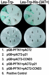
PFTK1 interacts with p21Cip1 and CCND3 in the yeast two-hybrid assay. pGB or pGB-PFTK1 was cotransformed with pACT2, pACT2-p21Cip1, or pACT2-CCND3 into the yeast strain Y190 along with a positive control pair, and tranformants were streaked on an SD/Leu-Trp plate (Left) and an SD/Leu-Trp-His plate with 3-AT (Right). β-Galactosidase assay was also performed (Upper).
Fig. 2.
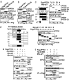
PFTK1 forms a ternary complex with p21Cip1 and CCND3. (a) The 293T cells were transfected with indicated constructs of PFTK1. Lysates were immunoprecipitated with anti-p21Cip1 and blotted with anti-p21Cip1 and anti-Flag. (b) Anti-Flag was used to immunoprecipitate the same lysates used in a. The immunoprecipitants were blotted with anti-p21Cip1 and anti-Flag. (c Left) Schematic representation of Flag-tagged PFTK1 truncations. (c Right) The 293T cells were transfected with indicated constructs. Lysates were immunoprecipitated with anti-Myc and blotted with anti-Flag. Lysates were also blotted with anti-Myc and anti-Flag. (d Left) Schematic representation of Myc-tagged p21Cip1 truncations. (d Right) PFTK1 interacts with full-length p21Cip1, D1, D2, and N fragments of p21Cip1. (e) The 293T cells were transfected with indicated constructs. Lysates were immunoprecipitated with anti-Myc and blotted with anti-Myc, anti-Flag, and anti-HA. (f) The 293T cells were transfected with indicated constructs. Lysates were immunoprecipitated with anti-Flag. Both lysates and immunoprecipitants were blotted with anti-Flag-HRP, anti-HA-HRP, and anti-Myc-HRP separately.
Fig. 3.
Endogenous interaction between PFTK1 and CCND3. (a) Expression of endogenous PFTK1 in different cell lines. Lysates of different cell lines were blotted with monoclonal anti-PFTK1 and anti-β-actin. (b) U2OS cells were arrested in G1/G0 phase by treatment with 0.5 mM
l
-mimosine for 24 h. Cells were then released and harvested at indicated time points. Immunoblotting for endogenous PFTK1 was performed along with an anti-β-actin blot. (c) Lysates from 1×10⁸ SH-SY5Y cells were immunoprecipitated with anti-PFTK1 or anti-CDK6, and the immunoprecipitants were blotted with anti-PFTK1, anti-CDK6, anti-CCND1, or anti-CCND3. (d) Lysates from SH-SY5Y cells were immunoprecipitated by using anti-CCND3 or mIgG as the control. The resulting supernatant was blotted with anti-PFTK1, anti-CCND3, or anti-β-actin.
Fig. 4.
PFTK1 is an active kinase that phosphorylates Rb. (a) The 293T cells transfected with indicated constructs were lysed, and lysates were immunoprecipitated with anti-Flag. Half of the precipitates were blotted with anti-Flag, and the other half were used for kinase assay. (b) The kinase activity of PFTK1 is enhanced by CCND3 and inhibited by p21Cip1. The 293T cells transfected with indicated constructs were harvested and analyzed as described above. The p21Cip1 derivatives used were the same as described in Fig. 2_d Left_. (c) Phosphorylation of His-RbC137 by PFTK1. The 293T cells transfected with indicated constructs were harvested and analyzed as described above. Two micrograms of His-RbC137 protein was added to each reaction as the substrate. (d) Activity of endogenous PFTK1 in U2OS cells. U2OS cells (1×10⁸) were harvested and immunoprecipitated with mouse anti-PFTK1 (mouse IgG was used as the control). Half of the precipitates were used for blotting with rabbit anti-PFTK1 (Lower), and the other half were used for kinase assay containing 2 μg of RbC137 protein as the substrate (Upper).
Fig. 5.
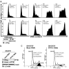
PFTK1 regulates G1/S transition of cell cycle. (a) Stably expressed PFTK1 promotes cell cycle progression. A stable PFTK1-expressing cell line (U2OS) and a control cell line were synchronized by 0.5 mM
l
-mimosine for 24 h. The drug-containing medium was removed, and fresh medium was added (marked as 0 h) to allow cells to exit from G0/G1 arrest. Cells were collected for FACS analysis every 4 h. Lysates were blotted with anti-Flag to confirm the expression of PFTK1. (b) siRNA-mediated knockdown of PFTK1 in SH-SY5Y cells. Cells were harvested 48 h after transfection. The knockdown effect of siRNA against PFTK1 was examined by using an anti-PFTK1 monoclonal antibody. The blot was also probed with anti-β-actin antibody. (c) siRNA against PFTK1 leads to cell cycle arrest at G1. Representative histograms for cell cycle distribution in SH-SY5Y cells transfected with control siRNA (Left) and PFTK1-specific siRNA (Right) are shown.
Similar articles
- Characterization of murine gammaherpesvirus 68 v-cyclin interactions with cellular cdks.
Upton JW, van Dyk LF, Speck SH. Upton JW, et al. Virology. 2005 Oct 25;341(2):271-83. doi: 10.1016/j.virol.2005.07.014. Epub 2005 Aug 15. Virology. 2005. PMID: 16102793 - Inhibition of the melanoma cell cycle and regulation at the G1/S transition by 12-O-tetradecanoylphorbol-13-acetate (TPA) by modulation of CDK2 activity.
Coppock DL, Buffolino P, Kopman C, Nathanson L. Coppock DL, et al. Exp Cell Res. 1995 Nov;221(1):92-102. doi: 10.1006/excr.1995.1356. Exp Cell Res. 1995. PMID: 7589260 - Tumor suppressor stars in yeast G1/S transition.
Li P, Hao Z, Zeng F. Li P, et al. Curr Genet. 2021 Apr;67(2):207-212. doi: 10.1007/s00294-020-01126-3. Epub 2020 Nov 11. Curr Genet. 2021. PMID: 33175222 Review. - The role of cyclin Y in normal and pathological cells.
Opacka A, Żuryń A, Krajewski A, Mikołajczyk K. Opacka A, et al. Cell Cycle. 2023 Apr;22(8):859-869. doi: 10.1080/15384101.2022.2162668. Epub 2022 Dec 28. Cell Cycle. 2023. PMID: 36576166 Free PMC article. Review.
Cited by
- Aberrant regulation of Wnt signaling in hepatocellular carcinoma.
Liu LJ, Xie SX, Chen YT, Xue JL, Zhang CJ, Zhu F. Liu LJ, et al. World J Gastroenterol. 2016 Sep 7;22(33):7486-99. doi: 10.3748/wjg.v22.i33.7486. World J Gastroenterol. 2016. PMID: 27672271 Free PMC article. Review. - Cyclin-dependent kinases: a family portrait.
Malumbres M, Harlow E, Hunt T, Hunter T, Lahti JM, Manning G, Morgan DO, Tsai LH, Wolgemuth DJ. Malumbres M, et al. Nat Cell Biol. 2009 Nov;11(11):1275-6. doi: 10.1038/ncb1109-1275. Nat Cell Biol. 2009. PMID: 19884882 Free PMC article. No abstract available. - A Potential Role for the Gsdf-eEF1α Complex in Inhibiting Germ Cell Proliferation: A Protein-Interaction Analysis in Medaka (Oryzias latipes) From a Proteomics Perspective.
Zhang X, Chang Y, Zhai W, Qian F, Zhang Y, Xu S, Guo H, Wang S, Hu R, Zhong X, Zhao X, Chen L, Guan G. Zhang X, et al. Mol Cell Proteomics. 2021;20:100023. doi: 10.1074/mcp.RA120.002306. Epub 2021 Jan 7. Mol Cell Proteomics. 2021. PMID: 33293461 Free PMC article. - Wnt Signaling and Its Impact on Mitochondrial and Cell Cycle Dynamics in Pluripotent Stem Cells.
Rasmussen ML, Ortolano NA, Romero-Morales AI, Gama V. Rasmussen ML, et al. Genes (Basel). 2018 Feb 19;9(2):109. doi: 10.3390/genes9020109. Genes (Basel). 2018. PMID: 29463061 Free PMC article. Review. - Cyclin dependent kinase 14 as a paclitaxel-resistant marker regulated by the TGF-β signaling pathway in human ovarian cancer.
Guan W, Yuan J, Li X, Gao X, Wang F, Liu H, Shi J, Xu G. Guan W, et al. J Cancer. 2023 Aug 15;14(13):2538-2551. doi: 10.7150/jca.86842. eCollection 2023. J Cancer. 2023. PMID: 37670966 Free PMC article.
References
- Sherr CJ, Roberts JM. Genes Dev. 2004;18:2699–2711. - PubMed
- Malumbres M, Barbacid M. Trends Biochem Sci. 2005;30:630–641. - PubMed
- Besset V, Rhee K, Wolgemuth DJ. Mol Reprod Dev. 1998;50:18–29. - PubMed
- Brambilla R, Draetta G. Oncogene. 1994;9:3037–3041. - PubMed
- Charrasse S, Carena I, Hagmann J, Woods-Cook K, Ferrari S. Cell Growth Differ. 1999;10:611–620. - PubMed
Publication types
MeSH terms
Substances
LinkOut - more resources
Full Text Sources
Molecular Biology Databases


