Polymer-based elemental tags for sensitive bioassays - PubMed (original) (raw)
Polymer-based elemental tags for sensitive bioassays
Xudong Lou et al. Angew Chem Int Ed Engl. 2007.
No abstract available
Figures
Figure 1
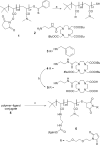
Experimental design for tagging antibodies with metal-chelating polymers. The antibody of interest is subjected to selective reduction of -S—S-groups to produce reactive -SH groups, which are reacted with the terminal maleimide groups of a polymer bearing metal-chelating ligands along its backbone. The polymer-bearing antibodies are purified, treated with a given lanthanide ion, and then purified again. Each type of antibody is labeled with a different element.
Scheme 1
a) Et3N, DMF, benzylamine or 2, 14 h; b) TFA 95 %, 14 h; c) 1) DTT (20 m
m
), Na2HPO4 (50 m
m
, pH 8.5), 50 °C, 1 h; 2) 2,2′-(ethylenedioxy)bis(ethylmaleimide), DMF/H2O, 1 h, 22 °C.
Figure 2
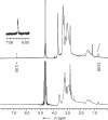
1H NMR spectrum of maleimide conjugate 6 in D2O prior to (top) and after (bottom) reaction with 2-aminoethanethiol.
Figure 3
Titration of element-tagged antibody against cell surface antigen. KG-1a cells (1 × 10⁶ cells per sample, run in triplicate) were incubated with increasing concentrations of CD45-Eu antibody. Separately, the same number of KG-1a cells were treated with mouse IgG-Eu.
Figure 4
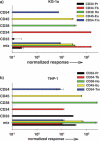
Multiplex analysis of antigen expression by two acute leukemia cell lines. a) KG-1a cells were probed with five element-tagged antibodies to cell surface antigens: CD33-Pr, CD34-Tb, CD38-Ho, CD45-Eu, and CD54-Tm. Background controls included element-tagged mouse IgG-Pr, IgG-Tb, IgG-Ho, IgG-Eu, and IgG-Tm. Triplicate samples with 1 × 10⁶ cells per tube were set up for reaction with each antibody separately and with a mix of all five antibodies together as well as controls. The cells were stained, fixed, and then treated with a RhIII-containing metallointercalator for cell enumeration and signal normalization. Washed cell pellets were dissolved in concentrated HCl, combined with an equal volume of Ir standard solution (1 ppb), and analyzed by ICP-MS. Results are presented as normalized response with respect to Ir, Rh, and background signals from nonspecific IgG binding. b) THP-1 cell line treated as described for KG-1a.
Similar articles
- Inductively coupled plasma mass spectrometry-based immunoassay: a review.
Liu R, Wu P, Yang L, Hou X, Lv Y. Liu R, et al. Mass Spectrom Rev. 2014 Sep-Oct;33(5):373-93. doi: 10.1002/mas.21391. Epub 2013 Nov 22. Mass Spectrom Rev. 2014. PMID: 24272753 Review. - Element-tagged immunoassay with inductively coupled plasma mass spectrometry for multianalyte detection.
Careri M, Elviri L, Mangia A. Careri M, et al. Anal Bioanal Chem. 2009 Jan;393(1):57-61. doi: 10.1007/s00216-008-2419-8. Epub 2008 Oct 23. Anal Bioanal Chem. 2009. PMID: 18946666 Review. - [Application of monoclonal antibody to laboratory tests--immunoassay].
Miyai K, Endo Y, Ichihara K, Amino N. Miyai K, et al. Rinsho Byori. 1986 Feb;34(2):125-32. Rinsho Byori. 1986. PMID: 3009924 Review. Japanese. No abstract available. - Use of alpha-N,N-bis[carboxymethyl]lysine-modified peroxidase in immunoassays.
Jin L, Wei X, Gomez J, Datta M, Birkett A, Peterson DL. Jin L, et al. Anal Biochem. 1995 Jul 20;229(1):54-60. doi: 10.1006/abio.1995.1378. Anal Biochem. 1995. PMID: 8533895 - Immunoreagents based on polymer dispersions for immunochemical assays.
Lukin YuV, Pavlova IS, Generalova AN, Zubov VP, Zhorov OV, Martsev SP. Lukin YuV, et al. J Mol Recognit. 1998 Winter;11(1-6):185-7. doi: 10.1002/(SICI)1099-1352(199812)11:1/6<185::AID-JMR419>3.0.CO;2-7. J Mol Recognit. 1998. PMID: 10076836
Cited by
- IMMUNOLOGY. An interactive reference framework for modeling a dynamic immune system.
Spitzer MH, Gherardini PF, Fragiadakis GK, Bhattacharya N, Yuan RT, Hotson AN, Finck R, Carmi Y, Zunder ER, Fantl WJ, Bendall SC, Engleman EG, Nolan GP. Spitzer MH, et al. Science. 2015 Jul 10;349(6244):1259425. doi: 10.1126/science.1259425. Science. 2015. PMID: 26160952 Free PMC article. - Multiplexed protease assays using element-tagged substrates.
Lathia US, Ornatsky O, Baranov V, Nitz M. Lathia US, et al. Anal Biochem. 2011 Jan 1;408(1):157-9. doi: 10.1016/j.ab.2010.09.008. Epub 2010 Sep 16. Anal Biochem. 2011. PMID: 20849809 Free PMC article. - Staggered starts in the race to T cell activation.
Richard AC, Frazer GL, Ma CY, Griffiths GM. Richard AC, et al. Trends Immunol. 2021 Nov;42(11):994-1008. doi: 10.1016/j.it.2021.09.004. Epub 2021 Oct 11. Trends Immunol. 2021. PMID: 34649777 Free PMC article. Review. - Element-tagged immunoassay with ICP-MS detection: evaluation and comparison to conventional immunoassays.
Razumienko E, Ornatsky O, Kinach R, Milyavsky M, Lechman E, Baranov V, Winnik MA, Tanner SD. Razumienko E, et al. J Immunol Methods. 2008 Jul 20;336(1):56-63. doi: 10.1016/j.jim.2008.03.011. Epub 2008 Apr 22. J Immunol Methods. 2008. PMID: 18456275 Free PMC article. - Bioimaging of metallothioneins in ocular tissue sections by laser ablation-ICP-MS using bioconjugated gold nanoclusters as specific tags.
Cruz-Alonso M, Fernandez B, Álvarez L, González-Iglesias H, Traub H, Jakubowski N, Pereiro R. Cruz-Alonso M, et al. Mikrochim Acta. 2017 Dec 18;185(1):64. doi: 10.1007/s00604-017-2597-1. Mikrochim Acta. 2017. PMID: 29594525
References
- Etzioni R, Urban N, Ramsey S, McIntosh M, Schwartz S, Reid B, Radich J, Anderson G, Hartwell L. Nat. Rev. Cancer. 2003;3:243. - PubMed
- Melton L. Nature. 2004;429:101. - PubMed
- Baranov VI, Quinn Z, Bandura DR, Tanner SD. Anal. Chem. 2002;74:1629. - PubMed
- Baranov VI, Quinn ZA, Bandura DR, Tanner SD. J. Anal. At. Spectrom. 2002;17:1148.
- Ornatsky O, Baranov VI, Bandura DR, Tanner SD, Dick J. J. Immunol. Methods. 2006;308:68. - PubMed
Publication types
MeSH terms
Substances
LinkOut - more resources
Full Text Sources
Other Literature Sources


