Mapping genes that contribute to daunorubicin-induced cytotoxicity - PubMed (original) (raw)
Mapping genes that contribute to daunorubicin-induced cytotoxicity
Shiwei Duan et al. Cancer Res. 2007.
Abstract
Daunorubicin is an anthracycline antibiotic agent used in the treatment of hematopoietic malignancies. Toxicities associated with this agent include myelosuppression and cardiotoxicity; however, the genes or genetic determinants that contribute to these toxicities are unknown. We present an unbiased genome-wide approach that incorporates heritability, whole-genome linkage analysis, and linkage-directed association to uncover genetic variants contributing to the sensitivity to daunorubicin-induced cytotoxicity. Cell growth inhibition in 324 Centre d' Etude du Polymorphisme Humain lymphoblastoid cell lines (24 pedigrees) was evaluated following treatment with daunorubicin for 72 h. Heritability analysis showed a significant genetic component contributing to the cytotoxic phenotypes (h2 = 0.18-0.63 at 0.0125, 0.025, 0.05, 0.1, 0.2, and 1.0 mumol/L daunorubicin and at the IC50, the dose required to inhibit 50% cell growth). Whole-genome linkage scans at all drug concentrations and IC50 uncovered 11 regions with moderate peak LOD scores (> 1.5), including 4q28.2 to 4q32.3 with a maximum LOD score of 3.18. The quantitative transmission disequilibrium tests were done using 31,312 high-frequency single-nucleotide polymorphisms (SNP) located in the 1 LOD confidence interval of these 11 regions. Thirty genes were identified as significantly associated with daunorubicin-induced cytotoxicity (P < or = 2.0 x 10(-4), false discovery rate < or = 0.1). Pathway and functional gene ontology analysis showed that these genes were overrepresented in the phosphatidylinositol signaling system, axon guidance pathway, and GPI-anchored proteins family. Our findings suggest that a proportion of susceptibility to daunorubicin-induced cytotoxicity may be controlled by genetic determinants and that analysis using linkage-directed association studies with dense SNP markers can be used to identify the genetic variants contributing to cytotoxicity.
Figures
Figure 1
Boxplots for 324 cell lines within 24 pedigrees are shown for various concentrations of daunorubicin, illustrating interfamily and intrafamily variance. The mean for each family’s percentage survival after daunorubicin treatment for 72 h with (A) 0.0125 µmol/L; (B) 0.025 µmol/L; (C) 0.2 µmol/L, and (D) 1.0 µmol/L. Line, mean phenotypic response within each family; box, mean ± SE; whiskers, mean ± 1.97 × SE.
Figure 2
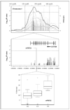
Linkage-directed association studies on chromosome 4 to identify SNPs conferring sensitivity to daunorubicin-induced cytotoxicity. Top, results from linkage analysis based on 24 CEPH families. The 1 LOD confidence interval of the peak on chromosome 4 from multipoint linkage of daunorubicin-induced cytotoxicity (0.05 µmol/L: black solid curve) and from the linkage analysis of 0.1 µmol/L, IC50, 0.2 µmol/L, 0.025 µmol/L, and 0.0125 µmol/L(dotted curves with peaks in descending order). Vertical dashed lines, 1 LOD confidence interval of the linkage peaks for the follow-up association studies; horizontal dashed lines, P value of 2 × 10⁻⁴. Results of QTDT analysis using genotypes for 30 CEU trios from the HapMap Project (gray bars) and associated genotypes within INPP4B (black bars). Middle, results of QTDT analysis illustrating associated genotypes within INPP4B. Bottom, SNP (rs978752) located in the 16th intron of INPP4B gene shows suggestive association evidence with daunorubicin (1 µmol/L)–induced cytotoxicity (FDR = 0.66, P = 3 × 10⁻⁵).This SNP is also modestly associated with other daunorubicin phenotypes (0.1 µmol/L: FDR = 0.1, P = 2 × 10⁻⁴;0.2 µmol/L: FDR = 0.22, P = 4 × 10⁻⁵).
Figure 3
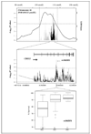
Linkage-directed association studies on chromosome 16 to identify SNPs conferring sensitivity to daunorubicin-induced cytotoxicity. Top, results from linkage analysis based on 24 CEPH families. The 1 LOD confidence interval of the peak on chromosome 16 from multipoint (solid curve) linkage of daunorubicin (0.0125 µmol/L)–induced cytotoxicity. Vertical dashed lines, 1 L OD confidence interval of the linkage peaks for the follow-up association studies; horizontal dashed lines, P value of 2 × 10⁻⁴. Results of QTDT analysis using genotypes for 30 CEU trios from the HapMap Project (gray bars) and associated genotypes within CDH13 (black bars). Middle, results of QTDT analysis illustrating associated genotypes within CDH13. Bottom, SNP rs1862831 in the fifth intron of CDH13 gene are associated with daunorubicin (0.0125 µmol/L)–induced cytotoxicity (FDR = 0.03, P = 1 × 10⁻⁶).
Similar articles
- Genetic variants contributing to daunorubicin-induced cytotoxicity.
Huang RS, Duan S, Kistner EO, Bleibel WK, Delaney SM, Fackenthal DL, Das S, Dolan ME. Huang RS, et al. Cancer Res. 2008 May 1;68(9):3161-8. doi: 10.1158/0008-5472.CAN-07-6381. Cancer Res. 2008. PMID: 18451141 Free PMC article. - Susceptibility loci involved in cisplatin-induced cytotoxicity and apoptosis.
Shukla SJ, Duan S, Badner JA, Wu X, Dolan ME. Shukla SJ, et al. Pharmacogenet Genomics. 2008 Mar;18(3):253-62. doi: 10.1097/FPC.0b013e3282f5e605. Pharmacogenet Genomics. 2008. PMID: 18300947 Free PMC article. - Heritability and linkage analysis of sensitivity to cisplatin-induced cytotoxicity.
Dolan ME, Newbold KG, Nagasubramanian R, Wu X, Ratain MJ, Cook EH Jr, Badner JA. Dolan ME, et al. Cancer Res. 2004 Jun 15;64(12):4353-6. doi: 10.1158/0008-5472.CAN-04-0340. Cancer Res. 2004. PMID: 15205351 - Identification of genomic regions contributing to etoposide-induced cytotoxicity.
Bleibel WK, Duan S, Huang RS, Kistner EO, Shukla SJ, Wu X, Badner JA, Dolan ME. Bleibel WK, et al. Hum Genet. 2009 Mar;125(2):173-80. doi: 10.1007/s00439-008-0607-4. Epub 2008 Dec 17. Hum Genet. 2009. PMID: 19089452 Free PMC article. - Use of CEPH and non-CEPH lymphoblast cell lines in pharmacogenetic studies.
Shukla SJ, Dolan ME. Shukla SJ, et al. Pharmacogenomics. 2005 Apr;6(3):303-10. doi: 10.1517/14622416.6.3.303. Pharmacogenomics. 2005. PMID: 16013961 Review.
Cited by
- Novel approaches to the prediction, diagnosis and treatment of cardiac late effects in survivors of childhood cancer: a multi-centre observational study.
Skitch A, Mital S, Mertens L, Liu P, Kantor P, Grosse-Wortmann L, Manlhiot C, Greenberg M, Nathan PC. Skitch A, et al. BMC Cancer. 2017 Aug 3;17(1):519. doi: 10.1186/s12885-017-3505-0. BMC Cancer. 2017. PMID: 28774277 Free PMC article. - An update of the molecular mechanisms underlying anthracycline induced cardiotoxicity.
Xie S, Sun Y, Zhao X, Xiao Y, Zhou F, Lin L, Wang W, Lin B, Wang Z, Fang Z, Wang L, Zhang Y. Xie S, et al. Front Pharmacol. 2024 Jun 26;15:1406247. doi: 10.3389/fphar.2024.1406247. eCollection 2024. Front Pharmacol. 2024. PMID: 38989148 Free PMC article. Review. - Targeted Delivery of Epirubicin to Cancer Cells by Polyvalent Aptamer System in vitro and in vivo.
Yazdian-Robati R, Ramezani M, Jalalian SH, Abnous K, Taghdisi SM. Yazdian-Robati R, et al. Pharm Res. 2016 Sep;33(9):2289-97. doi: 10.1007/s11095-016-1967-4. Epub 2016 Jun 9. Pharm Res. 2016. PMID: 27283831 - Identifying genetic variants that contribute to chemotherapy-induced cytotoxicity.
Hartford CM, Dolan ME. Hartford CM, et al. Pharmacogenomics. 2007 Sep;8(9):1159-68. doi: 10.2217/14622416.8.9.1159. Pharmacogenomics. 2007. PMID: 17924831 Free PMC article. Review. - Chemotherapeutic-induced apoptosis: a phenotype for pharmacogenomics studies.
Wen Y, Gorsic LK, Wheeler HE, Ziliak DM, Huang RS, Dolan ME. Wen Y, et al. Pharmacogenet Genomics. 2011 Aug;21(8):476-88. doi: 10.1097/FPC.0b013e3283481967. Pharmacogenet Genomics. 2011. PMID: 21642893 Free PMC article.
References
- Davis HL, Davis TE. Daunorubicin and Adriamycin in cancer treatment: an analysis of their roles and limitations. Cancer treatment reports. 1979;63:809–815. - PubMed
- Chaires JB, Fox KR, Herrera JE, Britt M, Waring MJ. Site and sequence specificity of the daunomycin-DNA interaction. Biochemistry. 1987;26:8227–8236. - PubMed
- Bachur NR, Yu F, Johnson R, Hickey R, Wu Y, Malkas L. Helicase inhibition by anthracycline anticancer agents. Mol Pharmacol. 1992;41:993–998. - PubMed
- Palayoor ST, Stein JM, Hait WN. Inhibition of protein kinase C by antineoplastic agents: implications for drug resistance. Biochem Biophys Res Commun. 1987;148:718–725. - PubMed
- Ohmori T, Podack ER, Nishio K, et al. Apoptosis of lung cancer cells caused by some anti-cancer agents (MMC, CPT-11, ADM) is inhibited by bcl-2. Biochem Biophys Res Commun. 1993;192:30–36. - PubMed
Publication types
MeSH terms
Substances
Grants and funding
- U01 GM061393-090005/GM/NIGMS NIH HHS/United States
- U01 GM061393/GM/NIGMS NIH HHS/United States
- U01 GM061393-090010/GM/NIGMS NIH HHS/United States
- U01 GM061393-070005/GM/NIGMS NIH HHS/United States
- U01GM61374/GM/NIGMS NIH HHS/United States
- GM61393/GM/NIGMS NIH HHS/United States
- U01 GM061393-080005/GM/NIGMS NIH HHS/United States
- U01 GM061374/GM/NIGMS NIH HHS/United States
- U01 GM061393-060010/GM/NIGMS NIH HHS/United States
- U01 GM061393-070010/GM/NIGMS NIH HHS/United States
- U01 GM061393-060005/GM/NIGMS NIH HHS/United States
- U01 GM061393-080010/GM/NIGMS NIH HHS/United States
LinkOut - more resources
Full Text Sources
Other Literature Sources
Research Materials


