Lentiviral Vpr usurps Cul4-DDB1[VprBP] E3 ubiquitin ligase to modulate cell cycle - PubMed (original) (raw)
Lentiviral Vpr usurps Cul4-DDB1[VprBP] E3 ubiquitin ligase to modulate cell cycle
Kasia Hrecka et al. Proc Natl Acad Sci U S A. 2007.
Abstract
The replication of viruses depends on the cell cycle status of the infected cells. Viruses have evolved functions that alleviate restrictions imposed on their replication by the host. Vpr, an accessory factor of primate lentiviruses, arrests cells at the DNA damage checkpoint in G2 phase of the cell cycle, but the mechanism underlying this effect has remained elusive. Here we report that Vpr proteins of both the human (HIV-1) and the distantly related simian (SIVmac) immunodeficiency viruses specifically associate with a protein complex comprising subunits of E3 ubiquitin ligase assembled on Cullin-4 scaffold (Cul4-DDB1[VprBP]). We show that Vpr binding to Cul4-DDB1[VprBP] leads to increased neddylation and elevated intrinsic ubiquitin ligase activity of this E3. This effect is mediated through the VprBP subunit of the complex, which recently has been suggested to function as a substrate receptor for Cul4. We also demonstrate that VprBP regulates G1 phase and is essential for the completion of DNA replication in S phase. Furthermore, the ability of Vpr to arrest cells in G2 phase correlates with its ability to interact with Cul4-DDB1[VprBP] E3 complex. Our studies identify the Cul4-DDB1[VprBP] E3 ubiquitin ligase complex as the downstream effector of lentiviral Vpr for the induction of cell cycle arrest in G2 phase and suggest that Vpr may use this complex to perturb other aspects of the cell cycle and DNA metabolism in infected cells.
Conflict of interest statement
The authors declare no conflict of interest.
Figures
Fig. 1.
Vpr binds DDA1–DDB1–VprBP complex. (A) Biochemical interactions among Vpr, VprBP, DDB1, and DDA1. Detergent extracts from HEK293T cells transiently expressing FLAG-tagged HIV-1 Vpr (lane 2), VprBP (lane 3), DDB1 (lane 4), or DDA1 (lane 5) subunits were immunoprecipitated (IP) with α-FLAG beads, and immunoprecipitates were analyzed by immunoblotting for VprBP, DDB1, DDA1, Vpr, and COP9 signalosome subunits CSN3 and CSN8. (B) Vpr binds the DDA1–DDB1–VprBP complex. FLAG-VprBP (Upper) and FLAG-Vpr (Lower) containing complexes were sedimented through 10–40% glycerol gradients. Aliquots of fractions collected from the tops of the gradients were immunoblotted for the indicated proteins.
Fig. 2.
Vpr elevates neddylation and intrinsic ubiquitin-ligase activity of the Cul4–DDB1[VprBP] E3 complex. (A) Vpr binds to and specifically elevates neddylation of Cul4A associated with VprBP. Myc-tagged Cullin 4A (_m_-Cul4) was coexpressed with Vpr, VprBP, and DDB2, in various combinations, in HEK293T cells. FLAG-tagged (f) versions of these proteins were used in some experiments to facilitate their immunoprecipitation. _m_-Cul4A (arrow) and its neddylated form (∗) were detected in detergent extracts (Extr) and α-FLAG immune complexes (IP) by immunoblotting with α-myc and α-Nedd8 antibodies, respectively. (B) Vpr stimulates intrinsic ubiquitin ligase activity of the Cul4–DDB1[VprBP] E3 complex. _m_-Cul4–DDB1[VprBP] complexes were assembled with or without Vpr in HEK293T cells, purified via their FLAG-VprBP subunits, and incubated with E1, ubiquitin, and/or E2 as indicated. Cul4A and its ubiquitinated forms were detected by immunoblotting for Cul4.
Fig. 3.
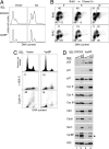
VprBP depletion leads to G1 and G2 phase arrests. (A) The effect of RNAi to VprBP on the ability of Vpr to arrest cells in G2. U2OS cells expressing shRNAs to VprBP (Lower) or DOCK2 (Upper) were transduced with a lentiviral TEIG vector expressing HIV-1 Vpr (Vpr) or a control empty vector (Vector). Three days later, the cells were stained with PI, and the DNA content was analyzed by flow cytometry. (B) VprBP-depleted cells arrest in G1 and G2. VprBP-depleted (Lower) and control cell populations (Upper) were labeled with BrdU for 1 h. BrdU incorporation and DNA content were analyzed by flow cytometry either immediately after BrdU labeling or after 6 and 12 h chase. Bivariant distributions of BrdU incorporation and DNA content are shown. The percent fractions of cells in the G1 (Lower Left), S (Upper), and G2 (Lower Right) phases are indicated. (C) Histone γ-H2A.X and cyclin A expression in VprBP-depleted (VprBP) and control cells (vector) were revealed by indirect fluorescence. Cells were counterstained with DAPI, and fluorescent signals were imaged with an iCys laser scanning cytometer. Histograms of DNA content and bivariate distributions of γ-H2A.X or cyclin A fluorescence versus DNA content are shown. (D) Levels of cyclins, checkpoint, and replication licensing proteins in lysates of VprBP-depleted and control cells were analyzed by immunoblotting with antibodies to the indicated proteins. Splicing factor 2 (SF2) was used as a loading control. The asterisk indicates a band reacting nonspecifically with the α-VprBP IgG. Lanes 1 and 3 contain 3-fold dilutions of the amounts loaded in lanes 2 and 4, respectively.
Fig. 4.
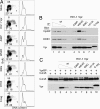
Effects of mutations on Vpr abilities to arrest cells in the G2 phase, bind VprBP and DDB1, and elevate Cul4 neddylation. (A) Mutations disrupt the ability of Vpr to arrest cells in G2. U2OS cells were transduced with retroviral vectors expressing wild type (HIV-1 Vpr), the indicated mutant forms of HIV-1 Vpr, or a control empty vector (mock). Two days later, cells were labeled with BrdU, and their cell cycle profiles were analyzed by flow cytometry. (Left) Quantification of cells in the G1, S, and G2/M phases is shown. Cells with >4N DNA content are boxed. (Right) DNA content histograms also are shown. (B) Vpr mutations disrupt binding to DDB1 and VprBP. FLAG-tagged wild-type and mutant HIV-1 Vpr proteins were immunoprecipitated from transiently transfected HEK293T cells, and immune complexes were analyzed by immunoblotting for VprBP, DDB1, and Vpr. (C) Vpr mutants unable to arrest cells in G2 do not increase Cul4 neddylation. Wild-type and mutant HIV-1 Vpr proteins were transiently coexpressed with myc-Cul4A and VprBP in HEK293T cells, as described in the legend for Fig. 2_A_. Detergent extracts were immunoblotted for Cul4A and its neddylated form with α-myc antibody.
Similar articles
- Lentiviral Vpx accessory factor targets VprBP/DCAF1 substrate adaptor for cullin 4 E3 ubiquitin ligase to enable macrophage infection.
Srivastava S, Swanson SK, Manel N, Florens L, Washburn MP, Skowronski J. Srivastava S, et al. PLoS Pathog. 2008 May 9;4(5):e1000059. doi: 10.1371/journal.ppat.1000059. PLoS Pathog. 2008. PMID: 18464893 Free PMC article. - HIV-1 Vpr Protein Enhances Proteasomal Degradation of MCM10 DNA Replication Factor through the Cul4-DDB1[VprBP] E3 Ubiquitin Ligase to Induce G2/M Cell Cycle Arrest.
Romani B, Shaykh Baygloo N, Aghasadeghi MR, Allahbakhshi E. Romani B, et al. J Biol Chem. 2015 Jul 10;290(28):17380-9. doi: 10.1074/jbc.M115.641522. Epub 2015 Jun 1. J Biol Chem. 2015. PMID: 26032416 Free PMC article. - HIV-1 Vpr-mediated G2 arrest involves the DDB1-CUL4AVPRBP E3 ubiquitin ligase.
Belzile JP, Duisit G, Rougeau N, Mercier J, Finzi A, Cohen EA. Belzile JP, et al. PLoS Pathog. 2007 Jul;3(7):e85. doi: 10.1371/journal.ppat.0030085. PLoS Pathog. 2007. PMID: 17630831 Free PMC article. - The functions of the HIV1 protein Vpr and its action through the DCAF1.DDB1.Cullin4 ubiquitin ligase.
Casey L, Wen X, de Noronha CM. Casey L, et al. Cytokine. 2010 Jul;51(1):1-9. doi: 10.1016/j.cyto.2010.02.018. Epub 2010 Mar 27. Cytokine. 2010. PMID: 20347598 Free PMC article. Review. - Lentivirus Vpr and Vpx accessory proteins usurp the cullin4-DDB1 (DCAF1) E3 ubiquitin ligase.
Romani B, Cohen EA. Romani B, et al. Curr Opin Virol. 2012 Dec;2(6):755-63. doi: 10.1016/j.coviro.2012.09.010. Epub 2012 Oct 10. Curr Opin Virol. 2012. PMID: 23062609 Free PMC article. Review.
Cited by
- HIV/simian immunodeficiency virus (SIV) accessory virulence factor Vpx loads the host cell restriction factor SAMHD1 onto the E3 ubiquitin ligase complex CRL4DCAF1.
Ahn J, Hao C, Yan J, DeLucia M, Mehrens J, Wang C, Gronenborn AM, Skowronski J. Ahn J, et al. J Biol Chem. 2012 Apr 6;287(15):12550-8. doi: 10.1074/jbc.M112.340711. Epub 2012 Feb 23. J Biol Chem. 2012. PMID: 22362772 Free PMC article. - Human immunodeficiency virus type 1 Vpr inhibits axonal outgrowth through induction of mitochondrial dysfunction.
Kitayama H, Miura Y, Ando Y, Hoshino S, Ishizaka Y, Koyanagi Y. Kitayama H, et al. J Virol. 2008 Mar;82(5):2528-42. doi: 10.1128/JVI.02094-07. Epub 2007 Dec 19. J Virol. 2008. PMID: 18094160 Free PMC article. - Exposed hydrophobic residues in human immunodeficiency virus type 1 Vpr helix-1 are important for cell cycle arrest and cell death.
Barnitz RA, Chaigne-Delalande B, Bolton DL, Lenardo MJ. Barnitz RA, et al. PLoS One. 2011;6(9):e24924. doi: 10.1371/journal.pone.0024924. Epub 2011 Sep 16. PLoS One. 2011. PMID: 21949789 Free PMC article. - HIV-1 Vpr Protein Induces Proteasomal Degradation of Chromatin-associated Class I HDACs to Overcome Latent Infection of Macrophages.
Romani B, Baygloo NS, Hamidi-Fard M, Aghasadeghi MR, Allahbakhshi E. Romani B, et al. J Biol Chem. 2016 Feb 5;291(6):2696-711. doi: 10.1074/jbc.M115.689018. Epub 2015 Dec 17. J Biol Chem. 2016. PMID: 26679995 Free PMC article. - Inhibition of Vpx-Mediated SAMHD1 and Vpr-Mediated Host Helicase Transcription Factor Degradation by Selective Disruption of Viral CRL4 (DCAF1) E3 Ubiquitin Ligase Assembly.
Wang H, Guo H, Su J, Rui Y, Zheng W, Gao W, Zhang W, Li Z, Liu G, Markham RB, Wei W, Yu XF. Wang H, et al. J Virol. 2017 Apr 13;91(9):e00225-17. doi: 10.1128/JVI.00225-17. Print 2017 May 1. J Virol. 2017. PMID: 28202763 Free PMC article.
References
- Yamashita M, Emerman M. Virology. 2006;344:88–93. - PubMed
- Goh WC, Rogel ME, Kinsey CM, Michael SF, Fultz PN, Nowak MA, Hahn BH, Emerman M. Nat Med. 1998;4:65–71. - PubMed
- Turlure F, Devroe E, Silver PA, Engelman A. Front Biosci. 2004;9:3187–3208. - PubMed
- Frankel AD, Young JA. Annu Rev Biochem. 1998;67:1–25. - PubMed
Publication types
MeSH terms
Substances
LinkOut - more resources
Full Text Sources
Other Literature Sources
Molecular Biology Databases


