Eps15 and Dap160 control synaptic vesicle membrane retrieval and synapse development - PubMed (original) (raw)
. 2007 Jul 16;178(2):309-22.
doi: 10.1083/jcb.200701030. Epub 2007 Jul 9.
Viktor I Korolchuk, Yogesh P Wairkar, Wei Jiao, Emma Evergren, Hongling Pan, Yi Zhou, Koen J T Venken, Oleg Shupliakov, Iain M Robinson, Cahir J O'Kane, Hugo J Bellen
Affiliations
- PMID: 17620409
- PMCID: PMC2064449
- DOI: 10.1083/jcb.200701030
Eps15 and Dap160 control synaptic vesicle membrane retrieval and synapse development
Tong-Wey Koh et al. J Cell Biol. 2007.
Abstract
Epidermal growth factor receptor pathway substrate clone 15 (Eps15) is a protein implicated in endocytosis, endosomal protein sorting, and cytoskeletal organization. Its role is, however, still unclear, because of reasons including limitations of dominant-negative experiments and apparent redundancy with other endocytic proteins. We generated Drosophila eps15-null mutants and show that Eps15 is required for proper synaptic bouton development and normal levels of synaptic vesicle (SV) endocytosis. Consistent with a role in SV endocytosis, Eps15 moves from the center of synaptic boutons to the periphery in response to synaptic activity. The endocytic protein, Dap160/intersectin, is a major binding partner of Eps15, and eps15 mutants phenotypically resemble dap160 mutants. Analyses of eps15 dap160 double mutants suggest that Eps15 functions in concert with Dap160 during SV endocytosis. Based on these data, we hypothesize that Eps15 and Dap160 promote the efficiency of endocytosis from the plasma membrane by maintaining high concentrations of multiple endocytic proteins, including dynamin, at synapses.
Figures
Figure 1.
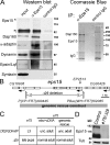
Binding partners of Drosophila Eps15 and generation of two null alleles of fly eps15. (A, left) Anti-Eps15 antiserum coimmunoprecipitates Eps15 with Dap160, α-adaptin, dynamin, and epsin/Lqf, but not Syntaxin (input is 5%). Asterisks mark the positions of Eps15-specific bands. (right) Coomassie staining shows that Dap160 is the major binding partner of Eps15 in the coimmunoprecipitate; bands were identified by mass spectroscopy (see Table S1, available at
http://www.jcb.org/cgi/content/full/jcb.200701030/DC1
, for peptide sequences). (B) The eps15 genomic locus is tagged with three different transposable elements, EP2513, _P{XP_=FRT}d00445, and _PBac{WH_=FRT}f02085. EP2513 was excised to generate an imprecise deletion allele eps15e75 (e75). The FRT sites in _P{XP_=FRT}d00445 and _PBac{WH_=FRT}f02085 were used to generate an FLP-mediated site-specific deletion of the entire eps15 coding region, resulting in the allele eps15Δ29 (Δ29). The gray bar indicates the genomic region that was used to rescue the eps15 mutants. This region contains the entire eps15 locus and partial sequences of two flanking genes. (C) Lethal phase analysis of mutants in heteroallelic combinations and in the presence or absence of rescue constructs driven by elavC155-Gal4 (see Materials and methods). (D) The eps15e75 and eps15Δ29 mutations cause loss of Eps15 protein expression as revealed by Western blot analysis using rabbit anti-Eps15. Each lane contains proteins extracted from 10 brains of eps15e75/eps15Δ29 (e75/Δ29) and w control larvae. See also Fig. S1 for Western blot and immunohistochemistry of homozygous e75 larvae.
Figure 2.
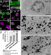
Eps15 is enriched at synapses. (A) Guinea pig anti-Eps15 detects Eps15 protein at the muscle 4 NMJ of a w control larva but not that of an eps15e75/eps15Δ29 mutant. (B) Eps15 (green) is closely associated with Dlg (magenta) in the neuropil of the larval ventral nerve cord. (bottom) Merge of anti-Eps15 and anti-Dlg staining patterns. (C) Dlg (green), which labels the postsynaptic compartment of the NMJ at this resolution, shows a pattern that envelopes that of Eps15 (magenta). This indicates that Eps15 is enriched in the presynaptic compartment at the NMJ. (D) Eps15 (magenta) is excluded from puncta decorated by Bruchpilot (nc82 antigen; green), which marks T-bars. (E) Eps15 (magenta; left) and Dap160 (green; right in merged panel) largely colocalize in the boutons, resulting in large areas of white in the merged image (right). A and C–E show single confocal slices, and B shows a projection view. (F) Eps15 immunogold labeling in two zones located within 500 nm lateral to T-bars (thick arrows in H and I). The first zone (gray bars) refers to areas 0–200 nm from the plasma membrane, and the second zone (white bars) refers to areas 200–400 nm from the plasma membrane. Relative distribution shows the percentage of total immunogold labeling distributed in either zones. Error bars indicate standard deviation. **, P < 0.01; ***, P < 0.001 (t test). Bar graphs represent mean values from 15 resting wild-type boutons, 18 wild-type boutons stimulated with 60 mM K+, and 20 shibirets1 boutons stimulated with 60 mM K+. (G) Silver-enhanced immunogold labeling (black precipitates; see Materials and methods) of Eps15 using guinea pig anti-Eps15 reveals that Eps15 is associated with vesicle-rich regions of a wild-type resting bouton. (H) A high-magnification view of anti-Eps15 immunogold labeling in a wild-type resting bouton shows that Eps15 is associated with vesicles but largely excluded from the area around the synaptic dense body or T-bar (short wide arrow). (I) Upon stimulation with 60 mM K+, anti-Eps15 immunogold labeling becomes concentrated at plasma membrane regions adjacent to the active zone (long arrows). Bars: (G) 1 μm; (H and I) 200 nm. Guinea pig anti-Eps15 was used in A, B, and E–I, and rabbit anti-Eps15 was used in C and D.
Figure 3.
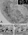
High K+ stimulation of shibirets1 NMJs at the restrictive temperature leads to a dramatic redistribution of Eps15 in boutons independent of SVs. (A) Silver-enhanced immunogold labeling of Eps15 using guinea pig anti-Eps15 reveals that Eps15 is predominantly localized to the plasma membrane (thin arrows) in areas surrounding the T-bars (thick arrow; the 60 mM K+ stimulus did not completely deplete the preexisting pool of vesicles in the shibirets1 bouton). As reported previously in shibirets1 mutants, this stimulus leads to mild depletions of the vesicle pool in a subset of boutons (A) and more severe depletions in other boutons (not depicted), even though endocytosis is blocked at the restrictive temperature (Estes et al., 1996). (Inset) Confocal imaging of guinea pig anti-Eps15 staining reveals that Eps15 is localized in the lumen of a resting bouton (rest) and is redistributed to the periphery in a bouton stimulated with 60 mM K+ (stim). m, mitochondria; a, axoplasm. (B) Boxed area in A is shown at higher magnification. (C) Electron micrograph of the same area from an adjacent ultrathin section. Gold particles were often located close to the rims of invaginating pits at the plasma membrane. Small arrows indicate fuzzy coats on vesicles. Although the areas that are immunolabeled for Eps15 seem to be reduced after stimulation, levels of Eps15 proteins are unchanged in resting and stimulated control and shibirets1 terminals (quantification by immunofluorescent microscopy; not depicted). Bars: (A) 300 nm; (B and C) 100 nm.
Figure 4.
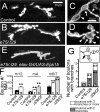
eps15 mutants show bouton overgrowth at the NMJ. Muscle 12 NMJ of eps15e75/eps15Δ29 L3 larva (B) possess more boutons and branches than a control larva (A) as revealed by mouse anti-Dlg staining. A close-up view of anti-HRP–stained muscle 4 type IB boutons reveals that there are more branchpoints (arrows) in eps15e75/eps15Δ29 (D) than in controls (C). A bouton that was connected to more than one distal neurite or bouton (arrowheads) was counted as a branchpoint (arrow). (E) Neuronal expression of the full-length eps15 cDNA rescues the supernumerary boutons at the muscle 12 in the eps15e75/eps15Δ29 background (see Fig. 9 B for quantification). (F) Comparison of mean bouton numbers per NMJ at muscle 12, 4, and 6/7 in eps15e75/eps15Δ29 (e75/ Δ 29), eps15Δ29/Df(2L)Dll-MP (Δ29/Df), and control larvae reveals that eps15 mutant NMJs possess significantly more boutons per synapse than control NMJs. In addition, bouton numbers at Δ_29/Df_ NMJs are significantly more than those at e75/Δ29 NMJs (m4, P < 0.01; m6/7, P < 0.05). (G) Comparison of branchpoint numbers per NMJ (arrow in inset shows an example of a bouton with three branches) reveals that _eps15_ mutants muscle 4 NMJs possess more boutons with two branches (gray bar) and three or more branches (white bar) than controls. In addition, Δ_29/Df_ NMJs exhibit somewhat more branchpoints than _e75/Δ29_, but the difference is not significant (P > 0.05). w larvae were used as controls. Bars: (A, B, and E) 20 μm; (C and D) 10 μm. *, P < 0.05; **, P < 0.01 (controls vs. mutants; Mann-Whitney U-test). Error bars indicate SEM, and the numbers in histograms indicate the number of larvae.
Figure 5.
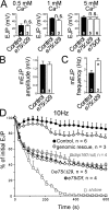
eps15 mutants show normal neurotransmitter release when stimulated at low frequency but are unable to sustain release under high-frequency stimulation. (A) Mean EJP measured at the NMJ reveals that neurotransmitter release is normal at eps15e75/eps15Δ29 (e75/Δ29) and eps15e75/Df(2L)Dll-MP (e75/Df) during 0.1 Hz of stimulation (0.5 and 1 mM Ca2+) and single nerve stimulations (5 mM Ca2+). (B) Mean mEJP amplitudes recorded in 0.5 mM Ca2+ and 3 μM tetrodotoxin were not different between e75/Δ29 and w controls. (C) However, mean mEJP frequency of e75/Δ29 is higher than controls. (D) When stimulated at 10 Hz, eps15 mutant NMJs show defects in maintaining release. Specifically, the mean EJPs of both e75/Δ29 and e75/Df were significantly different from controls (P < 0.01) after 60 s. Introducing one copy of _eps15_ genomic fragment in the _e75/Δ29_ background rescued this defect (genomic vs. control, P > 0.1; genomic vs. e75/Δ29, P < 0.01). Note that the two different _eps15_-null transheterozygotes show defects that are similar to that of dap160 Δ1/Df(2L)bur-K1 (dap160 null) but less severe than that of shibirets1 (shibirets1 data is adapted from Koh et al. [2004] for comparison). w larvae were used as controls. *, P < 0.05 (controls vs. mutants; Mann-Whitney U-test). Error bars indicate SEM, and the numbers in histograms indicate the number of larvae.
Figure 6.
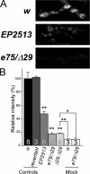
eps15 loss-of-function mutants show impaired FM1-43FX uptake during nerve stimulation. (A) FM1-43FX, a lipophilic fluorescent dye was applied to the NMJ during stimulation with 90 mM K+ and 5 mM Ca2+. The “mock” control was performed by incubating the NMJ in FM1-43FX dissolved in a Ca2+-free, low-K+ medium. (B) FM1-43FX uptake expressed as the mean fluorescence intensity relative to w controls. The NMJ of L3 larvae homozygous for the hypomorphic allele EP2513 show a mildly reduced dye uptake and that of the eps15e75/_eps15Δ29_-null mutants shows severely reduced dye uptake. In our hands, EP2513 retains considerable levels of endocytic activities, which contrasts with the observation of little or no dye uptake in a recent characterization of the same allele (Majumdar et al., 2006). *, P < 0.05; **: P < 0.01 (controls vs. mutants; Mann-Whitney U-test). Error bars indicate SEM, and the numbers in histograms indicate the number of larvae.
Figure 7.
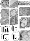
TEM reveals a membrane retrieval defect at eps15 mutant NMJs. Compared with w controls (A), eps15e75/eps15Δ29 (e75/Δ29) mutants (B) have normal numbers of vesicles at rest but are inefficient in vesicle budding during endocytosis when stimulated with 90 mM K+/5 mM Ca2+ (C, control; D, e75/Δ29). Upon incubation in normal HL3 without Ca2+ for 1 min, vesicle density recovers to near normal levels in controls (E) but not mutants (F). (G) Mean vesicle density in boutons at rest, after stimulation and recovery. Compared with controls, e75/Δ29 mutant NMJs are much less efficient in the recovery of vesicles during stimulation. Vesicle densities of resting e75/Δ29 and control boutons are not significantly different (P > 0.05). However, vesicle densities of e75/Δ29 boutons during stimulation and after recovery are less than controls. Mean size distribution of vesicles at rest (H) and after stimulation (I) and recovery (J). e75/Δ29 NMJ boutons accumulate a greater proportion of large membraneous bodies or cisternae during stimulation. (K) A high-magnification view of the boxed area in D shows a cisterna that appears to emanate from the plasma membrane (arrowhead). (L) Another mutant bouton showing large membraneous bodies (arrowheads) are apparently contiguous with the plasma membrane. Similarly, cisternae (M) and membrane invaginations (N) are seen in dap160Δ1/Df(2L)bur-K1 mutants when stimulated with 60 mM K+ at 34°C. Vesicle quantification was performed on at least 15 boutons using two or three larvae per genotype for each condition. Vesicle size distribution was quantified using 300–800 vesicles for each genotype and condition. *, P < 0.05; **, P < 0.01 (controls vs. mutants; t test). Error bars indicate SEM, and the numbers in histograms indicate the number of boutons examined.
Figure 8.
Synaptic markers are reduced at eps15 mutant NMJs. Anti-Dlg outlines the NMJ boutons (magenta). The synaptic markers stained in green are quantified. (A) Dynamin and Dap160 are strongly reduced at eps15 mutant NMJs. (B) Stoned B, synaptotagmin I, α-adaptin, and endophilin are mildly reduced. (C) Csp and FasII are not significantly reduced at the eps15 mutant NMJs (P > 0.05). (D) Eps15 level is slightly but not significantly reduced at dap160 Δ1/Df(2L)bur-K1 NMJs compared with controls (P > 0.05). w larvae were used as controls. At least 10 muscle 12 type I boutons from abdominal segment 3 of each larva were used to obtain a pixel intensity for each larva, and at least three larvae were used to arrive at mean values for statistical comparisons. *, P < 0.05; **, P < 0.01 (controls vs. mutants; t test). Error bars indicate SEM.
Figure 9.
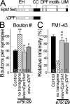
An N-terminal fragment of Eps15 plays a major role in NMJ development and SV endocytosis. (A) The ΔDPF truncation mutant of Eps15 contains the three EH domains and the coiled coil (CC) domain but lacks the C-terminal regions containing the DPF motifs (vertical black bars) and the UIMs. (B) Mean bouton numbers per synapse at muscle 6/7 synapses in control (revertant), eps15e75/e_ps15_Δ29 (e75/Δ29), and e75/Δ29 larvae expressing Eps15wt and ΔDPF. (C) Mean values of FM1-43FX uptake levels in e75/Δ29 and e75/Δ29 larvae expressing Eps15wt and ΔDPF relative to levels in control larvae (revertant). Expression of Eps15wt and ΔDPF are driven by elavC155-Gal4 in B and by elav-Gal43A4 in C. *, P < 0.05; **, P < 0.01 (controls vs. mutants; t test). Error bars indicate SEM, and the numbers in histograms indicate the number of larvae.
Figure 10.
Eps15 is a functional binding partner of Dap160. (A) With single stimuli at 5 mM Ca2+, dap160 Δ1 eps15e75/Df(2L)bur-K1 eps15Δ29 double mutants (dap160 and eps15 null) show normal EJP. (B) When stimulated at 10 Hz in 5 mM Ca2+, _dap160_- and _eps15_-null double mutants show synaptic depression kinetics that overlap with eps15e75/e_ps15_Δ29 (eps15 null) and dap160 Δ1/_Df(2L)bur-_K1(dap160 null) single mutants. (shibirets1 34°C recordings are taken from Koh et al. [2004] for comparison.) (C) _dap160_- and _eps15_-null double mutants show reductions in FM1-43FX uptake similar to _eps15_- and _dap160_-null single mutants. w adults or larvae were used as controls. Error bars indicate SEM, and the numbers in histograms indicate the number of larvae.
Similar articles
- Dap160/intersectin acts as a stabilizing scaffold required for synaptic development and vesicle endocytosis.
Koh TW, Verstreken P, Bellen HJ. Koh TW, et al. Neuron. 2004 Jul 22;43(2):193-205. doi: 10.1016/j.neuron.2004.06.029. Neuron. 2004. PMID: 15260956 - Dap160/intersectin scaffolds the periactive zone to achieve high-fidelity endocytosis and normal synaptic growth.
Marie B, Sweeney ST, Poskanzer KE, Roos J, Kelly RB, Davis GW. Marie B, et al. Neuron. 2004 Jul 22;43(2):207-19. doi: 10.1016/j.neuron.2004.07.001. Neuron. 2004. PMID: 15260957 - An Endocytic Scaffolding Protein together with Synapsin Regulates Synaptic Vesicle Clustering in the Drosophila Neuromuscular Junction.
Winther ÅM, Vorontsova O, Rees KA, Näreoja T, Sopova E, Jiao W, Shupliakov O. Winther ÅM, et al. J Neurosci. 2015 Nov 4;35(44):14756-70. doi: 10.1523/JNEUROSCI.1675-15.2015. J Neurosci. 2015. PMID: 26538647 Free PMC article. - Synapse scaffolding: intersection of endocytosis and growth.
Broadie K. Broadie K. Curr Biol. 2004 Oct 5;14(19):R853-5. doi: 10.1016/j.cub.2004.09.042. Curr Biol. 2004. PMID: 15458667 Review. - Stonins--specialized adaptors for synaptic vesicle recycling and beyond?
Maritzen T, Podufall J, Haucke V. Maritzen T, et al. Traffic. 2010 Jan;11(1):8-15. doi: 10.1111/j.1600-0854.2009.00971.x. Traffic. 2010. PMID: 19732400 Review.
Cited by
- Spartin regulates synaptic growth and neuronal survival by inhibiting BMP-mediated microtubule stabilization.
Nahm M, Lee MJ, Parkinson W, Lee M, Kim H, Kim YJ, Kim S, Cho YS, Min BM, Bae YC, Broadie K, Lee S. Nahm M, et al. Neuron. 2013 Feb 20;77(4):680-95. doi: 10.1016/j.neuron.2012.12.015. Neuron. 2013. PMID: 23439121 Free PMC article. - Abnormal synaptic vesicle biogenesis in Drosophila synaptogyrin mutants.
Stevens RJ, Akbergenova Y, Jorquera RA, Littleton JT. Stevens RJ, et al. J Neurosci. 2012 Dec 12;32(50):18054-67, 18067a. doi: 10.1523/JNEUROSCI.2668-12.2012. J Neurosci. 2012. PMID: 23238721 Free PMC article. - Brain tumor regulates neuromuscular synapse growth and endocytosis in Drosophila by suppressing mad expression.
Shi W, Chen Y, Gan G, Wang D, Ren J, Wang Q, Xu Z, Xie W, Zhang YQ. Shi W, et al. J Neurosci. 2013 Jul 24;33(30):12352-63. doi: 10.1523/JNEUROSCI.0386-13.2013. J Neurosci. 2013. PMID: 23884941 Free PMC article. - Dynamin I phosphorylation by GSK3 controls activity-dependent bulk endocytosis of synaptic vesicles.
Clayton EL, Sue N, Smillie KJ, O'Leary T, Bache N, Cheung G, Cole AR, Wyllie DJ, Sutherland C, Robinson PJ, Cousin MA. Clayton EL, et al. Nat Neurosci. 2010 Jul;13(7):845-51. doi: 10.1038/nn.2571. Epub 2010 Jun 6. Nat Neurosci. 2010. PMID: 20526333 Free PMC article.
References
- Bache, K.G., C. Raiborg, A. Mehlum, and H. Stenmark. 2003. STAM and Hrs are subunits of a multivalent ubiquitin-binding complex on early endosomes. J. Biol. Chem. 278:12513–12521. - PubMed
- Bean, A.J., S. Davanger, M.F. Chou, B. Gerhardt, S. Tsujimoto, and Y. Chang. 2000. Hrs-2 regulates receptor-mediated endocytosis via interactions with Eps15. J. Biol. Chem. 275:15271–15278. - PubMed
- Bellen, H.J., and V. Budnik. 2000. The neuromuscular junction. In Drosophila Protocols. W. Sullivan, M. Ashburner, and R.S. Hawley, editors. Cold Spring Harbor Laboratory Press, Cold Spring Harbor, NY. 175–199.
Publication types
MeSH terms
Substances
LinkOut - more resources
Full Text Sources
Other Literature Sources
Molecular Biology Databases
Research Materials
Miscellaneous
