Sulcal variability, stereological measurement and asymmetry of Broca's area on MR images - PubMed (original) (raw)
Sulcal variability, stereological measurement and asymmetry of Broca's area on MR images
Simon Sean Keller et al. J Anat. 2007 Oct.
Abstract
Leftward volume asymmetry of the pars opercularis and pars triangularis may exist in the human brain, frequently referred to as Broca's area, given the functional asymmetries observed in this region with regard to language expression. However, post-mortem and magnetic resonance imaging (MRI) studies have failed to consistently identify such a volumetric asymmetry. In the present study, an analysis of the asymmetry of sulco-gyral anatomy and volume of this anterior speech region was performed in combination with an analysis of the morphology and volume asymmetry of the planum temporale, located within the posterior speech region, in 50 healthy subjects using MRI. Variations in sulcal anatomy were documented according to strict classification schemes and volume estimation of the grey matter within the brain structures was performed using the Cavalieri method of stereology. Results indicated great variation in the morphology of and connectivity between the inferior frontal, inferior precentral and diagonal sulci. There were significant inter-hemispheric differences in the presence of (1) the diagonal sulcus within the pars opercularis, and (2) horizontal termination of the posterior Sylvian fissure (relative to upward oblique termination), both with an increased leftward incidence. Double parallel inferior precentral sulci and absent anterior rami of the Sylvian fissure prevented stereological measurements in five subjects. Therefore volumes were obtained from 45 subjects. There was a significant leftward volume asymmetry of the pars opercularis (P = 0.02), which was significantly related to the asymmetrical presence of the diagonal sulcus (P < 0.01). Group-wise pars opercularis volume asymmetry did not exist when a diagonal sulcus was present in both or neither hemispheres. There was no significant volume asymmetry of the pars triangularis. There was a significant leftward volume asymmetry of the planum temporale (P < 0.001), which was significantly associated with the shape of the posterior Sylvian fissure as a unilateral right or left upward oblique termination was always associated with leftward or rightward volume asymmetry respectively (P < 0.01). There was no relationship between volume asymmetries of the anterior and posterior speech regions. Our findings illustrate the extent of morphological variability of the anterior speech region and demonstrate the difficulties encountered when determining volumetric asymmetries of the inferior frontal gyrus, particularly when sulci are discontinuous, absent or bifid. When the intrasulcal grey matter of this region is exhaustively sampled according to strict anatomical landmarks, the volume of the pars opercularis is leftward asymmetrical. This manuscript illustrates the importance of simultaneous consideration of brain morphology and morphometry in studies of cerebral asymmetry.
Figures
Fig. 1
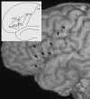
The major sulcal contours defining the pars opercularis (po) and pars triangularis (ptr) of the inferior frontal gyrus. The main image shows the sulcal contours from the lateral convexity of a brain studied in the present investigation. The insert presents the major sulcal contours defining the pars opercularis and pars triangularis schematically (connections and precise sulcal morphology do not necessarily correspond to the main image). The pars opercularis is the region of cortex located between the ventral segment of the inferior precentral sulcus [ipcs(v)], the inferior frontal sulcus (ifs) and the anterior ascending ramus of the Sylvian fissure (ar). The pars triangularis is the region of cortex located between the anterior ascending ramus, inferior frontal sulcus and the anterior horizontal ramus of the Sylvian fissure (hr). In this example, a diagonal sulcus (ds) is present, and connects to the ventral segment of the inferior precentral sulcus. A triangular sulcus (ts) is also present, and connects to the inferior frontal sulcus. The inferior precentral sulcus consists of three segments, the ventral segment, a horizontal segment [ipcs(h)] and a dorsal vertical segment [ipcs(d)]. Cs, central sulcus.
Fig. 2
Location of the lateral frontomarginal sulcus (lfms) with respect to the major sulcal contours that define the pars opercularis and pars triangularis. This sulcus may give the impression of an anterior extension of the inferior frontal sulcus, but navigation through orthogonal sections clearly differentiates the two sulci. ar, anterior ascending ramus; ds, diagonal sulcus; hr, anterior horizontal ramus; ifs, inferior frontal sulcus; ipcs, inferior precentral sulcus.
Fig. 3
The four connections of the diagonal sulcus. The connections are shown in subjects studied in the present investigation (left) and are also schematically illustrated (right). (a) Connection with the ascending horizontal ramus of the Sylvian fissure. (b) Connection with the inferior precentral sulcus. (c) Connection with the inferior frontal sulcus. (d) No connection with surrounding sulci. ar, anterior ascending ramus; ds, diagonal sulcus; hr, anterior horizontal ramus; ifs, inferior frontal sulcus; ipcs, inferior precentral sulcus.
Fig. 4
Termination of the posterior Sylvian fissure: horizontal (left) and upward oblique (right).
Fig. 5
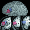
The sulcal contours defining the pars opercularis (blue) and pars triangularis (red) on MRI. Orthogonal sagittal (bottom left) and coronal (bottom middle/right) sections are shown in conjunction with a lateral rendering of the cerebral hemisphere in the same individual. ar, anterior ascending ramus; cis, circular insular sulcus; ds, diagonal sulcus; hr, anterior horizontal ramus; ifs, inferior frontal sulcus; ipcs, inferior precentral sulcus; ts, triangular sulcus.
Fig. 6
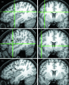
Defining the posterior most region of the pars opercularis. The same coronal section is shown on the right (a–c) indicating the last section that the frontal operculum can be visualised in this particular right pars opercularis (on the left side of image). The crosshairs indicate the last section on which the dorsomedial (a) and ventrolateral (b) operculum can be visualised, immediately anterior to the inferior precentral sulcus (see corresponding sagittal sections). (c) is the same sagittal and coronal sections as (a) without the crosshairs. Markers were placed to separate the deep operculum from the laterally overlapping precentral gyrus (pcg), which are indicated by the arrows on the coronal section. The broken white line indicates the point of separation. ifs, inferior frontal sulcus; ipcs, inferior precentral sulcus.
Fig. 7
Connecting the segments of a discontinuous inferior frontal sulcus. The orthogonal sagittal and coronal sections at the top show the point at which a bridge of cortex interrupts the sulcus (crosshairs). The same sections below indicate the two segments of the inferior frontal sulcus (ifs1, ifs2) and the point at which these segments are connected (broken white line).
Fig. 8
Point counting for stereological analysis of the pars triangularis (1), pars opercularis (2) and planum temporale (3). The approximate locations of the coronal sections are indicated on the lateral renderings of the same brain. The section areas of the three structures are indicated by the yellow points in the left and right hemispheres. ar, anterior ascending ramus; hr, anterior horizontal ramus; ifs, inferior frontal sulcus; ipcs, inferior precentral sulcus.
Fig. 9
Rare features of the inferior precentral sulcus. (a) Double parallel inferior precentral gyrus. (b) Ventral inferior precentral sulcus connecting with the Sylvian fissure on the surface of the brain (white arrow). This connection is lateral/superficial, given that the posterior frontal operculum always connects with the precentral gyrus more medially. ar, anterior ascending ramus; ds, diagonal sulcus; hr, anterior horizontal ramus; ifs, inferior frontal sulcus; ipcs, inferior precentral sulcus.
Fig. 10
Asymmetry coefficients (%) for the pars opercularis, pars triangularis and planum temporale.
Fig. 11
Scatterplot of pars opercularis asymmetry against pars triangularis asymmetry. Negative values of pars opercularis asymmetry are associated with positive values of pars triangularis asymmetry, and vice versa.
Fig. 12
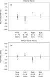
Distribution of pars opercularis volume (pop) asymmetry relative to the presence of a diagonal sulcus (12a) and planum temporale volume (pt) asymmetry relative to presence of upward oblique Sylvian fissure termination (12b). Note that in contrast to the data presented in Tables 2 and 3, the data presented here are for the 45 subjects in whom structure volumes could be estimated. Negative values are leftward volume asymmetry. Asymmetry coefficients are broken down in subgroups: none = morphological feature not present in either hemisphere, left = present only in left hemisphere, right = present only in right hemisphere, both = present in both hemispheres. These figures indicate that leftward or rightward volume asymmetry of the pars opercularis is related to the presence of the diagonal sulcus in the left or right hemisphere respectively (top), and that leftward or rightward planum temporale asymmetry is driven by the presence of an upward oblique Sylvian fissure in the right or left hemisphere respectively (bottom).
Similar articles
- Pars triangularis asymmetry and language dominance.
Foundas AL, Leonard CM, Gilmore RL, Fennell EB, Heilman KM. Foundas AL, et al. Proc Natl Acad Sci U S A. 1996 Jan 23;93(2):719-22. doi: 10.1073/pnas.93.2.719. Proc Natl Acad Sci U S A. 1996. PMID: 8570622 Free PMC article. - MRI asymmetries of Broca's area: the pars triangularis and pars opercularis.
Foundas AL, Eure KF, Luevano LF, Weinberger DR. Foundas AL, et al. Brain Lang. 1998 Oct 1;64(3):282-96. doi: 10.1006/brln.1998.1974. Brain Lang. 1998. PMID: 9743543 - Broca's area: nomenclature, anatomy, typology and asymmetry.
Keller SS, Crow T, Foundas A, Amunts K, Roberts N. Keller SS, et al. Brain Lang. 2009 Apr;109(1):29-48. doi: 10.1016/j.bandl.2008.11.005. Epub 2009 Jan 19. Brain Lang. 2009. PMID: 19155059 Review. - Functional anatomy of dominance for speech comprehension in left handers vs right handers.
Tzourio N, Crivello F, Mellet E, Nkanga-Ngila B, Mazoyer B. Tzourio N, et al. Neuroimage. 1998 Jul;8(1):1-16. doi: 10.1006/nimg.1998.0343. Neuroimage. 1998. PMID: 9698571 Review.
Cited by
- Overt naming fMRI pre- and post-TMS: Two nonfluent aphasia patients, with and without improved naming post-TMS.
Martin PI, Naeser MA, Ho M, Doron KW, Kurland J, Kaplan J, Wang Y, Nicholas M, Baker EH, Alonso M, Fregni F, Pascual-Leone A. Martin PI, et al. Brain Lang. 2009 Oct;111(1):20-35. doi: 10.1016/j.bandl.2009.07.007. Epub 2009 Aug 19. Brain Lang. 2009. PMID: 19695692 Free PMC article. - Protocol for an observational cohort study investigating biomarkers predicting seizure recurrence following a first unprovoked seizure in adults.
Adan GH, de Bézenac C, Bonnett L, Pridgeon M, Biswas S, Das K, Richardson MP, Laiou P, Keller SS, Marson T. Adan GH, et al. BMJ Open. 2022 Dec 5;12(12):e065390. doi: 10.1136/bmjopen-2022-065390. BMJ Open. 2022. PMID: 36576179 Free PMC article. - A surface-based analysis of language lateralization and cortical asymmetry.
Greve DN, Van der Haegen L, Cai Q, Stufflebeam S, Sabuncu MR, Fischl B, Brysbaert M. Greve DN, et al. J Cogn Neurosci. 2013 Sep;25(9):1477-92. doi: 10.1162/jocn_a_00405. Epub 2013 Apr 22. J Cogn Neurosci. 2013. PMID: 23701459 Free PMC article. - Is the human left middle longitudinal fascicle essential for language? A brain electrostimulation study.
De Witt Hamer PC, Moritz-Gasser S, Gatignol P, Duffau H. De Witt Hamer PC, et al. Hum Brain Mapp. 2011 Jun;32(6):962-73. doi: 10.1002/hbm.21082. Epub 2010 Jun 24. Hum Brain Mapp. 2011. PMID: 20578169 Free PMC article. - A comparative magnetic resonance imaging study of the anatomy, variability, and asymmetry of Broca's area in the human and chimpanzee brain.
Keller SS, Roberts N, Hopkins W. Keller SS, et al. J Neurosci. 2009 Nov 18;29(46):14607-16. doi: 10.1523/JNEUROSCI.2892-09.2009. J Neurosci. 2009. PMID: 19923293 Free PMC article.
References
- Albanese E, Merlo A, Albanese A, Gomez E. Anterior speech region. Asymmetry and weight-surface correlation. Arch Neurol. 1989;46:307–310. - PubMed
- Alexander MP, Naeser MA, Palumbo C. Broca's area aphasias: aphasia after lesions including the frontal operculum. Neurology. 1990;40:353–362. - PubMed
- Amunts K, Schleicher A, Burgel U, Mohlberg H, Uylings HB, Zilles K. Broca's region revisited: cytoarchitecture and intersubject variability. J Comp Neurol. 1999;412:319–341. - PubMed
- Amunts K, Schleicher A, Ditterich A, Zilles K. Broca's region: cytoarchitectonic asymmetry and developmental changes. J Comp Neurol. 2003;465:72–89. - PubMed
- Amunts K, Weiss PH, Mohlberg H, et al. Analysis of neural mechanisms underlying verbal fluency in cytoarchitectonically defined stereotaxic space – the roles of Brodmann areas 44 and 45. Neuroimage. 2004;22:42–56. - PubMed
MeSH terms
LinkOut - more resources
Full Text Sources
Medical
Miscellaneous


