Arterial response to shear stress critically depends on endothelial TRPV4 expression - PubMed (original) (raw)
Arterial response to shear stress critically depends on endothelial TRPV4 expression
Veronika Hartmannsgruber et al. PLoS One. 2007.
Abstract
Background: In blood vessels, the endothelium is a crucial signal transduction interface in control of vascular tone and blood pressure to ensure energy and oxygen supply according to the organs' needs. In response to vasoactive factors and to shear stress elicited by blood flow, the endothelium secretes vasodilating or vasocontracting autacoids, which adjust the contractile state of the smooth muscle. In endothelial sensing of shear stress, the osmo- and mechanosensitive Ca(2+)-permeable TRPV4 channel has been proposed to be candidate mechanosensor. Using TRPV4(-/-) mice, we now investigated whether the absence of endothelial TRPV4 alters shear-stress-induced arterial vasodilation.
Methodology/principal findings: In TRPV4(-/-) mice, loss of the TRPV4 protein was confirmed by Western blot, immunohistochemistry and by in situ-patch-clamp techniques in carotid artery endothelial cells (CAEC). Endothelium-dependent vasodilation was determined by pressure myography in carotid arteries (CA) from TRPV4(-/-) mice and wild-type littermates (WT). In WT CAEC, TRPV4 currents could be elicited by TRPV4 activators 4alpha-phorbol-12,13-didecanoate (4alphaPDD), arachidonic acid (AA), and by hypotonic cell swelling (HTS). In striking contrast, in TRPV4(-/-) mice, 4alphaPDD did not produce currents and currents elicited by AA and HTS were significantly reduced. 4alphaPDD caused a robust and endothelium-dependent vasodilation in WT mice, again conspicuously absent in TRPV4(-/-) mice. Shear stress-induced vasodilation could readily be evoked in WT, but was completely eliminated in TRPV4(-/-) mice. In addition, flow/reperfusion-induced vasodilation was significantly reduced in TRPV4(-/-) vs. WT mice. Vasodilation in response to acetylcholine, vasoconstriction in response to phenylephrine, and passive mechanical compliance did not differ between genotypes, greatly underscoring the specificity of the above trpv4-dependent phenotype for physiologically relevant shear stress.
Conclusions/significance: Genetically encoded loss-of-function of trpv4 results in a loss of shear stress-induced vasodilation, a response pattern critically dependent on endothelial TRPV4 expression. Thus, Ca(2+)-influx through endothelial TRPV4 channels is a molecular mechanism contributing significantly to endothelial mechanotransduction.
Conflict of interest statement
Competing Interests: The authors have declared that no competing interests exist.
Figures
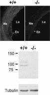
Figure 1. Immunohistochemistry for TRPV4 in CAs (upper panel) and Western blot analysis of TRPV4 protein expression in kidneys (lower panel) of WT and TRPV4−/− mice.
Note the absence of specific staining in CA of TRPV4−/− mice. Me = media; Lu = lumen; En = endothelium. In a kidney extract obtained from WT mice, the anti-TRPV4 antibody detected a protein of ∼95 kDa, which is in good agreement with the calculated molecular weight (98 kDa). Furthermore, an additional band of ∼107 kDa, presumably representing the glycosylated protein, is detected by the antibody. In extracts from TRPV4−/− mice, no signals were present. Equal protein loading of the blots was validated by visualization of tubulin.
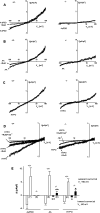
Figure 2. Electrophysiological properties of TRPV4 currents in carotid artery endothelial cells (CAEC) from WT and TRPV4−/− mice.
A, left panel, Representative recording of 4αPDD (1 µmol/L)-inducible TRPV4-currents in CAEC of WT. Voltage-dependent inhibition by RuR (1 µmol/L). Right panel, 4αPDD-inducible currents were undetectable in CAEC of TRPV4−/− mice. B, left panel, Representative recording of AA (10 µmol/L)-inducible TRPV4 currents in CAEC of WT and inhibition by RuR (1 µmol/L). Right panel, small AA-inducible cation-currents in CAEC of TRPV4−/− mice. C, left panel, HTS (206 mosmol/L)-inducible TRPV4-currents in CAEC of WT. Right panel, HTS-inducible cation currents of smaller amplitude in CAEC of TRPV4−/− mice. D, left panel, Partial voltage-dependent inhibition of HTS-inducible TRPV4 currents in CAEC of WT and almost complete inhibition by the combination of RuR and Gd3+ (10 µmol/L). Right panel, RuR insensitivity and Gd3+ sensitivity of HTS-inducible cation currents in CAEC of TRPV4−/− mice. E, Mean 4αPDD-, AA-, and HTS-inducible TRPV4 and other cation currents in CAEC of WT and TRPV4−/− mice. Numbers in brackets indicate the number of cells investigated. Values are given as means±SEM; * P<0.05, ** P<0.01, t test.
Figure 3. Compliance of carotid arteries (CA) of WT and TRPV4−/− mice.
A and B, Diameter of CA pressurized to 80 mmHg (basal) and in the extravascular presence of PE, K+, and SNP in the absence and presence of L-NNA (300 µmol/L) and INDO (10 µmol/L); WT CA, -L-NNA/INDO: n = 11 and+L-NNA/INDO: n = 8; TRPV4−/− CA, -L-NNA/INDO: n = 7 and+L-NNA/INDO: n = 8. C, PE-induced contraction of CA from WT (CA, n = 4) and TRPV4−/− mice (CA, n = 4) in the presence of L-NNA and INDO. D, Change in passive diameter (normalized to body weight) of CA from WT (CA, n = 13) and TRPV4−/− mice (CA, n = 10) in response to increasing intravascular pressure and in the presence of SNP. Values are given as means±SEM.
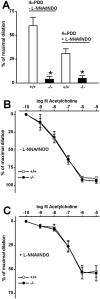
Figure 4. 4αPDD and ACh-induced vasodilation in CA of WT and TRPV4−/− mice.
A, 4αPDD (1 µmol/L)-induced vasodilation in CA of WT and TRPV4−/− mice in the absence and presence of L-NNA and INDO. WT CA, -L-NNA/INDO: n = 12 and+L-NNA/INDO: n = 9; TRPV4−/− CA, -L-NNA/INDO: n = 6 and+L-NNA/INDO: n = 8. B, ACh-induced vasodilation in CA of WT (n = 7) and TRPV4−/− (n = 6) in the absence of L-NNA and INDO and B, in their presence; WT CA (n = 6); TRPV4−/− CA (n = 6). * P<0.001, t test.
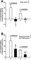
Figure 5. Shear stress- and flow/reperfusion-induced vasodilation in CA of WT and TRPV4−/− mice.
A, Shear stress-induced vasodilation in CA of WT and TRPV4−/− mice in the absence and presence of L-NNA and INDO; WT CA, n = 12 and n = 9; TRPV4−/− CA, n = 6 and n = 8, respectively. Shear stress-induced vasodilation was elicited by switching to a perfusion medium containing 5% dextran (5% Dex). B, Flow/reperfusion-induced vasodilation of CA of WT and TRPV4−/− mice in the absence and presence of L-NNA and INDO. WT CA, n = 9 and n = 8; TRPV4−/− CA, n = 7 and n = 7, respectively. * P<0.05, ** P<0.01, t test.
Similar articles
- Evidence for a functional role of endothelial transient receptor potential V4 in shear stress-induced vasodilatation.
Köhler R, Heyken WT, Heinau P, Schubert R, Si H, Kacik M, Busch C, Grgic I, Maier T, Hoyer J. Köhler R, et al. Arterioscler Thromb Vasc Biol. 2006 Jul;26(7):1495-502. doi: 10.1161/01.ATV.0000225698.36212.6a. Epub 2006 May 4. Arterioscler Thromb Vasc Biol. 2006. PMID: 16675722 - Role of cytochrome P450-dependent transient receptor potential V4 activation in flow-induced vasodilatation.
Loot AE, Popp R, Fisslthaler B, Vriens J, Nilius B, Fleming I. Loot AE, et al. Cardiovasc Res. 2008 Dec 1;80(3):445-52. doi: 10.1093/cvr/cvn207. Epub 2008 Aug 5. Cardiovasc Res. 2008. PMID: 18682435 - TRPV4-mediated endothelial Ca2+ influx and vasodilation in response to shear stress.
Mendoza SA, Fang J, Gutterman DD, Wilcox DA, Bubolz AH, Li R, Suzuki M, Zhang DX. Mendoza SA, et al. Am J Physiol Heart Circ Physiol. 2010 Feb;298(2):H466-76. doi: 10.1152/ajpheart.00854.2009. Epub 2009 Dec 4. Am J Physiol Heart Circ Physiol. 2010. PMID: 19966050 Free PMC article. - Endothelium-dependent cerebral artery dilation mediated by transient receptor potential and Ca2+-activated K+ channels.
Earley S. Earley S. J Cardiovasc Pharmacol. 2011 Feb;57(2):148-53. doi: 10.1097/FJC.0b013e3181f580d9. J Cardiovasc Pharmacol. 2011. PMID: 20729757 Review. - Role of TRPV4 channel in vasodilation and neovascularization.
Chen M, Li X. Chen M, et al. Microcirculation. 2021 Aug;28(6):e12703. doi: 10.1111/micc.12703. Epub 2021 May 24. Microcirculation. 2021. PMID: 33971061 Review.
Cited by
- Impaired TRPV4-eNOS signaling in trabecular meshwork elevates intraocular pressure in glaucoma.
Patel PD, Chen YL, Kasetti RB, Maddineni P, Mayhew W, Millar JC, Ellis DZ, Sonkusare SK, Zode GS. Patel PD, et al. Proc Natl Acad Sci U S A. 2021 Apr 20;118(16):e2022461118. doi: 10.1073/pnas.2022461118. Proc Natl Acad Sci U S A. 2021. PMID: 33853948 Free PMC article. - Trpv4 induces collateral vessel growth during regeneration of the arterial circulation.
Troidl C, Troidl K, Schierling W, Cai WJ, Nef H, Möllmann H, Kostin S, Schimanski S, Hammer L, Elsässer A, Schmitz-Rixen T, Schaper W. Troidl C, et al. J Cell Mol Med. 2009 Aug;13(8B):2613-2621. doi: 10.1111/j.1582-4934.2008.00579.x. Epub 2009 Feb 6. J Cell Mol Med. 2009. PMID: 19017361 Free PMC article. - Reduced Post-ischemic Brain Injury in Transient Receptor Potential Vanilloid 4 Knockout Mice.
Tanaka K, Matsumoto S, Yamada T, Yamasaki R, Suzuki M, Kido MA, Kira JI. Tanaka K, et al. Front Neurosci. 2020 May 12;14:453. doi: 10.3389/fnins.2020.00453. eCollection 2020. Front Neurosci. 2020. PMID: 32477057 Free PMC article. - TRPV channels and vascular function.
Baylie RL, Brayden JE. Baylie RL, et al. Acta Physiol (Oxf). 2011 Sep;203(1):99-116. doi: 10.1111/j.1748-1716.2010.02217.x. Epub 2010 Dec 9. Acta Physiol (Oxf). 2011. PMID: 21062421 Free PMC article. Review. - Endothelial transcriptomics reveals activation of fibrosis-related pathways in hypertension.
Nelson JW, Ferdaus MZ, McCormick JA, Minnier J, Kaul S, Ellison DH, Barnes AP. Nelson JW, et al. Physiol Genomics. 2018 Feb 1;50(2):104-116. doi: 10.1152/physiolgenomics.00111.2017. Epub 2018 Jan 8. Physiol Genomics. 2018. PMID: 29212850 Free PMC article.
References
- Furchgott RF, Zawadzki JV. The obligatory role of endothelial cells in the relaxation of arterial smooth muscle by acetylcholine. Nature. 1980;288:373–376. - PubMed
- Moncada S, Gryglewski R, Bunting S, Vane JR. An enzyme isolated from arteries transforms prostaglandin endoperoxides to an unstable substance that inhibits platelet aggregation. Nature. 1976;263:663–665. - PubMed
- Nilius B, Droogmans G. Ion channels and their functional role in vascular endothelium. Physiol Rev. 2001;81:1415–1459. - PubMed
- Feletou M, Vanhoutte PM. Endothelium-derived hyperpolarizing factor: where are we now? Arterioscler Thromb Vasc Biol. 2006;26:1215–1225. - PubMed
- Köhler R, Hoyer J. The endothelium-derived hyperpolarizing factor: insights from genetic animal models. Kidney Int Apr. 2007;25; [Epub ahead of print]; doi:10.1038/sj.ki.5002303 - PubMed
Publication types
MeSH terms
Substances
LinkOut - more resources
Full Text Sources
Other Literature Sources
Molecular Biology Databases
Miscellaneous


