S-nitrosothiols signal hypoxia-mimetic vascular pathology - PubMed (original) (raw)
S-nitrosothiols signal hypoxia-mimetic vascular pathology
Lisa A Palmer et al. J Clin Invest. 2007 Sep.
Abstract
NO transfer reactions between protein and peptide cysteines have been proposed to represent regulated signaling processes. We used the pharmaceutical antioxidant N-acetylcysteine (NAC) as a bait reactant to measure NO transfer reactions in blood and to study the vascular effects of these reactions in vivo. NAC was converted to S-nitroso-N-acetylcysteine (SNOAC), decreasing erythrocytic S-nitrosothiol content, both during whole-blood deoxygenation ex vivo and during a 3-week protocol in which mice received high-dose NAC in vivo. Strikingly, the NAC-treated mice developed pulmonary arterial hypertension (PAH) that mimicked the effects of chronic hypoxia. Moreover, systemic SNOAC administration recapitulated effects of both NAC and hypoxia. eNOS-deficient mice were protected from the effects of NAC but not SNOAC, suggesting that conversion of NAC to SNOAC was necessary for the development of PAH. These data reveal an unanticipated adverse effect of chronic NAC administration and introduce a new animal model of PAH. Moreover, evidence that conversion of NAC to SNOAC during blood deoxygenation is necessary for the development of PAH in this model challenges conventional views of oxygen sensing and of NO signaling.
Figures
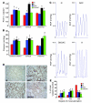
Figure 1. Systemic NAC and SNOAC cause hypoxia-mimetic PAH in mice.
C57BL/6/129SvEv, C57BL/6, and eNOS–/– male mice were maintained in normoxia (N, red; 21% O2) or hypoxia (H, black; 10% O2); or were treated with NAC (blue) or SNOAC (green) in their drinking water for 3 weeks. (A) Relative RV weight was determined as the ratio of the weight of the RV to the LV+S weight. (B) RV systolic pressures were measured in the closed chest using a Millar 1.4 F catheter/transducer. (C) Representative RV pressure (RVP) tracings (each = 1 s). (D) Lung section images from C57BL/6/129SvEv mice immunostained for von Willebrand factor and α-SMA to illustrate changes in muscularization after 3 weeks of exposure to normoxia, hypoxia, NAC, or SNOAC. Scale bar: 100 μm (applies to all panels). (E) Changes in muscularization in C57BL/6/129SvEv mice in the small (<80-μm) vessels from histological sections (as in D) counted by an observer blinded to the protocol. FM, fully muscular; PM, partly muscular, NM, nonmuscular. Significant increases in muscularization in each treatment group were seen in comparison to normoxic controls. Data are mean ± SEM. ζ_P_ < 0.02, *P < 0.001, †P < 0.003, by 1-way ANOVA followed by pairwise comparison, all compared with normoxic control.
Figure 2. Three weeks of NAC treatment or hypoxia increases the whole-lung expression of certain genes associated with the development of PAH in mice.
The expression of fibronectin (A), HIMF (B), eNOS (C and D), VEGF-A (E), and endothelin (F) in whole-lung homogenates from NAC-treated mice was examined by immunoblot. Fold increase in density relative to MAPK (equal loading control) was determined for each condition. The increases in fibronectin, HIMF, and eNOS (n = 3–5 each) were significant. (G) Three weeks of NAC treatment also increased whole-lung mRNA, assayed relative to 18S RNA by RT-PCR, for HIMF and VEGF-A but not fibronectin or Glut-1 (n = 3 each). Time course analysis of NAC-treated mice (D) revealed that the increase in whole-lung eNOS mRNA (filled squares, left axis) preceded the increase in eNOS protein expression (open circles, right axis) but decreased by 3 weeks. *P < 0.05; #P < 0.01.
Figure 3. SNOAC is formed from NAC in blood ex vivo and in vivo.
(A) The SNOrbc in heparinized LV blood (black bars), measured by reductive chemiluminescence (11), was lower than normal following 3 weeks of treatment with 10 mg/ml NAC (n = 3–4 each). In the same mice, plasma SNOAC levels (gray bars; measured by MS) increased from undetectable to approximately 2 μM over the same time (*P < 0.05). (B) Serum SNOAC, measured by MS, formed in NAC-treated mice (3 weeks). Left: liquid chromatogram; right: MS spectrum. NAC-treated mice had a SNOAC peak (m/z 193; red) coeluting with 15N-labeled SNOAC standard (m/z 194; black) that was absent in untreated animals (green) and was not detected in NAC-treated mice after serum pretreatment with HgCl2 to displace NO+ from the thiolate (blue). (C) Oxygenated erythrocytes were deoxygenated ex vivo (argon; ref. 11) in the presence of 100 μM NAC; supernatant SNOAC was measured by MS (above). SNOAC concentration increased with oxyhemoglobin (Oxy Hb) desaturation (co-oximetry: inset), being maximal at 59.3% saturation (blue), less at 77.2% saturation (green), and undetectable at 98% saturation. (D) SNOAC (filled circles) accumulated as the concentration of _S_-nitrosothiol–modified Hb (SNOHb; open circles) and oxyhemoglobin saturation (Hb SO2; blue line) both decreased in heparinized whole blood using argon with 5% CO2 (pH 7.3) in a tonometer. Both the increase in SNOAC and the loss of SNOrbc between 0 and 20 minutes were significant (P < 0.01 by ANOVA followed by pairwise comparison to the maximum value; n = 3). #SNOAC levels were below the limit of detection when the oxyhemoglobin saturation was greater than 80%.
Figure 4. SNOAC recapitulates in primary pulmonary arterial endothelial cells the hypoxia-mimetic whole-lung effect of chronic NAC administration on Sp3 expression in vivo.
(A) One micromolar SNOAC, but not 50 μM NAC, treatment (4 hours each) increased intracellular _S_-nitrosothiol levels (assayed by Cu/cysteine chemiluminescence; ref. 11) in primary murine pulmonary endothelial cells (*P < 0.05 compared with SNOAC treatment). (B) Immunoblot showing increased Sp3 expression relative to MAPK in the whole-lung homogenates of mice treated for 3 weeks with 10 mg/ml NAC but not in those of control mice. By densitometry, this increase was significant (P < 0.01). (C) Paradoxically, however, NAC (50 μM; 4 hours) did not increase Sp3 expression relative to β-actin in primary murine pulmonary endothelial cells in vitro, while both SNOAC (1 μM; 4 hours) and hypoxia (10%; 4 hours) did.
Figure 5. _S_-Nitrosothiols prevent normoxic ubiquitination and degradation of HIF 1α.
(A) NAC treatment (10 mg/ml; 3 weeks) increased whole-lung HIF 1–DNA binding activity. Complexes were supershifted (ss) with anti–HIF 1β and eliminated with excess cold probe (P). (B) SNOAC (1 μM), like GSNO (5, 13, 38), increased normoxic HIF 1α expression in nuclear extracts isolated from primary murine pulmonary endothelial cells. NAC alone (50 μM) did not affect HIF 1α expression. β-Actin was used as a protein load control. (C) In BPAECs transfected with HA-tagged HIF 1α and dominant-negative His-6-Myc–tagged ubiquitin (DN-Ub), ubiquitinated proteins were isolated using a nickel column and immunoblotted for HIF 1α. Both hypoxia and GSNO (G; 10 μM) inhibited HIF 1α ubiquitination relative to normoxia. (D) In COS cells cotransfected with HA-tagged HIF 1α and FLAG-tagged pVHL, GSNO (10 μM) prevented the coimmunoprecipitation of HIF 1α with pVHL. (E) _S_-nitrosylation of pVHL by SNOAC (5 μM) in equal protein aliquots isolated from HeLa cells was identified by biotin substitution (49); in the absence of ascorbate, _S_-nitrosylated pVHL was not detected. (F) Similarly, SNOAC and GSNO (5 μM) increased pVHL _S_-nitrosylation in pVHL-overexpressing 786-O cells. (G) C162, but not C77, was identified by biotin substitution to be _S_-nitrosylated in BPAECs transfected with wild-type cysteine 77 to serine mutant (C77S), C162S, or combined C77S/C162S pVHL exposed to SNOAC (1 μM). Native pVHL and MAPK immunoblots represented the pVHL expression and protein load controls, respectively. All in vitro treatments were for 4 hours.
Comment in
- Low-molecular-weight S-nitrosothiols and blood vessel injury.
Marsden PA. Marsden PA. J Clin Invest. 2007 Sep;117(9):2377-80. doi: 10.1172/JCI33136. J Clin Invest. 2007. PMID: 17786231 Free PMC article.
Similar articles
- Low-molecular-weight S-nitrosothiols and blood vessel injury.
Marsden PA. Marsden PA. J Clin Invest. 2007 Sep;117(9):2377-80. doi: 10.1172/JCI33136. J Clin Invest. 2007. PMID: 17786231 Free PMC article. - The relaxation induced by S-nitroso-glutathione and S-nitroso-N-acetylcysteine in rat aorta is not related to nitric oxide production.
Ceron PI, Cremonez DC, Bendhack LM, Tedesco AC. Ceron PI, et al. J Pharmacol Exp Ther. 2001 Aug;298(2):686-94. J Pharmacol Exp Ther. 2001. PMID: 11454932 - Spontaneous liberation of nitric oxide cannot account for in vitro vascular relaxation by S-nitrosothiols.
Kowaluk EA, Fung HL. Kowaluk EA, et al. J Pharmacol Exp Ther. 1990 Dec;255(3):1256-64. J Pharmacol Exp Ther. 1990. PMID: 2175799 - [Pulmonary hypertension in endothelial NO synthase knockout mice].
Hirata Y. Hirata Y. Nihon Rinsho. 2001 Jun;59(6):1081-5. Nihon Rinsho. 2001. PMID: 11411117 Review. Japanese. - S-nitrosothiols control breathing and oxygen homeostasis.
Ponka P, Kim S. Ponka P, et al. Redox Rep. 2002;7(1):5-7. doi: 10.1179/135100002125000118. Redox Rep. 2002. PMID: 11981448 Review. No abstract available.
Cited by
- Enzymatic mechanisms regulating protein S-nitrosylation: implications in health and disease.
Anand P, Stamler JS. Anand P, et al. J Mol Med (Berl). 2012 Mar;90(3):233-44. doi: 10.1007/s00109-012-0878-z. Epub 2012 Feb 24. J Mol Med (Berl). 2012. PMID: 22361849 Free PMC article. Review. - Erythrocyte-dependent regulation of human skeletal muscle blood flow: role of varied oxyhemoglobin and exercise on nitrite, S-nitrosohemoglobin, and ATP.
Dufour SP, Patel RP, Brandon A, Teng X, Pearson J, Barker H, Ali L, Yuen AH, Smolenski RT, González-Alonso J. Dufour SP, et al. Am J Physiol Heart Circ Physiol. 2010 Dec;299(6):H1936-46. doi: 10.1152/ajpheart.00389.2010. Epub 2010 Sep 17. Am J Physiol Heart Circ Physiol. 2010. PMID: 20852046 Free PMC article. - Stimuli-responsive star poly(ethylene glycol) drug conjugates for improved intracellular delivery of the drug in neuroinflammation.
Navath RS, Wang B, Kannan S, Romero R, Kannan RM. Navath RS, et al. J Control Release. 2010 Mar 19;142(3):447-56. doi: 10.1016/j.jconrel.2009.10.035. Epub 2009 Nov 5. J Control Release. 2010. PMID: 19896998 Free PMC article. - N-acetyl cysteine treatment reduces mercury-induced neurotoxicity in the developing rat hippocampus.
Falluel-Morel A, Lin L, Sokolowski K, McCandlish E, Buckley B, DiCicco-Bloom E. Falluel-Morel A, et al. J Neurosci Res. 2012 Apr;90(4):743-50. doi: 10.1002/jnr.22819. J Neurosci Res. 2012. PMID: 22420031 Free PMC article. - N-acetylcysteine in psychiatry: current therapeutic evidence and potential mechanisms of action.
Dean O, Giorlando F, Berk M. Dean O, et al. J Psychiatry Neurosci. 2011 Mar;36(2):78-86. doi: 10.1503/jpn.100057. J Psychiatry Neurosci. 2011. PMID: 21118657 Free PMC article. Review.
References
- Kim S.F., Huri D.A., Snyder S.H. Inducible nitric oxide synthase binds, S-nitrosylates and activates cyclooxygenase-2. Science. 2005;310:1966. - PubMed
- Lipton A.J., et al. S-Nitrosothiols signal the ventilatory response to hypoxia. Nature. 2001;413:171–174. - PubMed
- Jia L., Bonaventura C., Bonaventura J., Stamler J.S. S-Nitrosohaemoglobin: a dynamic activity of blood involved in vascular control. Nature. 1996;380:221–226. - PubMed
- Palmer L.A., Gaston B., Johns R.A. Normoxic stabilization of hypoxia-inducible factor-1 expression and activity: redox-dependent effect of nitrogen oxides. Mol. Pharmacol. 2000;58:1197–1203. - PubMed
Publication types
MeSH terms
Substances
Grants and funding
- HL059337/HL/NHLBI NIH HHS/United States
- R01 HL068173/HL/NHLBI NIH HHS/United States
- R56 HL059337/HL/NHLBI NIH HHS/United States
- 1K08GM069977/GM/NIGMS NIH HHS/United States
- HL068173/HL/NHLBI NIH HHS/United States
- K08 GM069977/GM/NIGMS NIH HHS/United States
- R01 HL059337/HL/NHLBI NIH HHS/United States
LinkOut - more resources
Full Text Sources
Other Literature Sources
Molecular Biology Databases


