Synaptic integration of adult-generated olfactory bulb granule cells: basal axodendritic centrifugal input precedes apical dendrodendritic local circuits - PubMed (original) (raw)
Comparative Study
Synaptic integration of adult-generated olfactory bulb granule cells: basal axodendritic centrifugal input precedes apical dendrodendritic local circuits
Mary C Whitman et al. J Neurosci. 2007.
Abstract
The adult mammalian olfactory bulb (OB) receives a continuing influx of new interneurons. Neuroblasts from the subventricular zone (SVZ) migrate into the OB and differentiate into granule cells and periglomerular cells that are presumed to integrate into the synaptic circuits of the OB. We have used retroviral infection into the SVZ of mice to label adult-generated granule cells and follow their differentiation and integration into OB circuitry. Using synaptic markers and electron microscopy, we show new granule cells integrating into the reciprocal circuitry of the external plexiform layer (EPL), beginning at 21 d postinfection (dpi). We further show that synapses are formed earlier, beginning at 10 dpi, on the somata and basal dendrites of new cells in the granule cell layer (GCL), before dendritic elaboration in the EPL. In the EPL, elaborate dendritic arbors with spines are first evident at 14 dpi. The density of spines increases from 14 to 28 dpi, and then decreases by 56 dpi. Despite the initial appearance of dendritic spines at 14 dpi in the EPL, no expression of presynaptic or postsynaptic markers is seen until 21 dpi. These data suggest that adult-generated granule cells are first innervated by centrifugal or mitral/tufted cell axon collaterals in the GCL and that these inputs may contribute to their differentiation, maturation, and synaptic integration into the dendrodendritic local circuits found in the EPL.
Figures
Figure 1.
Maturation of adult-generated granule cells. A, Ten days after viral labeling in the SVZ, cells have migrated into the GCL, and extended an apical dendrite toward the EPL. B, By 14 dpi, the apical dendrites have developed considerably, with several branches in the EPL, as well as spines along the dendrites. C–E, The basal dendrites have also developed spines. At 21 dpi (C), 28 dpi (D), and 42 dpi (E), cells look remarkably similar, with one long apical dendrite with three to four branches in the EPL, and several short basal dendrites. All images are maximum projections of confocal stacks through the full thickness of the cell. Green is GFP, and blue is DRAQ5, a nuclear marker. Scale bar: (in A) A–E, 50 μm.
Figure 2.
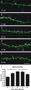
Spine density increases and then decreases as the cells mature. A, Fourteen days after viral labeling in the SVZ, dendrites in the EPL have spines, but at a low density along the shaft. B, Spine density increases by 21 dpi, as does the apparent maturity of the spines (more pedunculated, less filopodial). C–E, At 28 dpi (C) and 42 dpi (D), spines are very dense along the dendrite, but spine density decreases by 56 dpi (E). Quantification is shown in F. One-way ANOVA, p ≤ 0.0007, followed by Bonferroni's multiple comparison test; *p ≤ 0.05; **p ≤ 0.001. Scale bar: (in E) A–E, 10 μm. Error bars indicate SEM.
Figure 3.
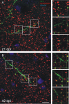
Adult-generated cells express synaptoporin, a presynaptic marker of granule cells. A, Synaptoporin (red) puncta in the EPL first colocalize with spine heads on labeled cells (green) at 21 dpi. B, By 42 dpi, the colocalization of GFP and synaptoporin is evident throughout the EPL and repeatedly along the same dendritic process. Images on the left are maximum projections of confocal images, and images on the right are higher-magnification single optical planes, of the boxed areas on the left, showing the z dimension. Scale bar: (in B) A, B, 10 μm.
Figure 4.
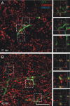
Gephyrin, a postsynaptic marker of GABAergic synapses, is apposed to labeled spines. A, B, Gephyrin (red) puncta are adjacent and apposed to spine heads on labeled cells (green) at 21 dpi (A) and 42 dpi (B), labeling the postsynaptic mitral/tufted dendrite receiving the synapse from the GFP-labeled spines. Images on the left are maximum projections of confocal images, and images on the right are higher-magnification single optical planes of the boxed areas on the left, showing the z dimension. Scale bar, 10 μm.
Figure 5.
Adult-generated granule cells express GluR2/3, a postsynaptic marker in the EPL. GluR2/3 (red) puncta colocalize in labeled dendrites and spines in the EPL by 21 dpi. The image on the left is maximum projection of a confocal image, and images on the right are higher magnification, single optical plane, of the boxed areas on the left, showing the z dimension. Scale bar, 10 μm.
Figure 6.
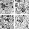
Electron micrographs of synapses in the EPL. A, A dendrodendritic synapse in the EPL between a mitral cell dendrite and a granule cell spine, unlabeled, for reference. The mitral to granule synapse is defined by a collection of small spherical vesicles closely apposed to the presynaptic membrane of the mitral cell secondary dendrite, and an asymmetrically thick membrane specialization in the granule cell spine head. The reciprocal granule to mitral synapse is defined by the elliptical cluster of vesicles in the spine head and symmetrical thickenings in the presynaptic and postsynaptic membranes. B, C, Examples of mitral to granule excitatory synapses on labeled spines at 42 dpi. Virally labeled cells were marked by GFP immunohistochemistry and DAB to form an electron dense product, so spines of new granule cells are darkly stained. D, An example of a bidirectional dendrodendritic synapse on the same spine head. On the right, the mitral to granule synapse can be seen, and on the left, the granule to mitral inhibitory synapse. Arrows indicate the direction of the synapse. Scale bar, 1 μm. Md, Mitral cell dendrite; Gr, granule cell spine.
Figure 7.
Synapses are first found in the GCL. A, PSD-95 puncta (red) are present on the cell body and basal dendrites at 10 dpi. The image is from a single optical plane. B, In the GCL, cell bodies also express GluR2/3 (red) by 10 dpi. At this point, expression is low and very punctuate. C, At 21 dpi, GluR2/3 expression has increased, so the cell body is surrounded by puncta. Basal dendrites also have GluR2/3 puncta (arrows). Images are single optical planes; the _z_-dimension is shown to the right and below. Scale bars, 10 μm.
Figure 8.
Electron micrographs of synapses in the GCL at 10 dpi. A, Examples of typical axodendritic synapses onto unlabeled dendrites. Clusters of vesicles in the axon terminal are accompanied by asymmetric membrane thickening and the presence of a synaptic cleft. B, An immature (developing) synapse onto the basal dendrite of a labeled granule cell. The immature synapse is defined by a small cluster of vesicles and a very small membrane thickening. C, A synapse that appears more morphologically mature with a well defined asymmetric postsynaptic thickening in the labeled basal dendrite. D, A single axon terminal forms asymmetrical synapses onto both a labeled and an unlabeled basal dendrite. Arrows indicate direction of synapses. Scale bar, 1 μm. a, Axon terminal; d, basal dendrite.
Figure 9.
Cholinergic fibers contact new cells in the GCL. A, ChAT fibers terminate in the GL and GCL. B, An example of a cholinergic fiber contacting a new cell 14 dpi. The image is a projection of a confocal stack. Scale bar: (in B) A, 32 μm; B, 10 μm.
Similar articles
- Complementary postsynaptic activity patterns elicited in olfactory bulb by stimulation of mitral/tufted and centrifugal fiber inputs to granule cells.
Laaris N, Puche A, Ennis M. Laaris N, et al. J Neurophysiol. 2007 Jan;97(1):296-306. doi: 10.1152/jn.00823.2006. Epub 2006 Oct 11. J Neurophysiol. 2007. PMID: 17035366 Free PMC article. - Plasticity of dendrodendritic microcircuits following mitral cell loss in the olfactory bulb of the murine mutant Purkinje cell degeneration.
Greer CA, Halász N. Greer CA, et al. J Comp Neurol. 1987 Feb 8;256(2):284-98. doi: 10.1002/cne.902560208. J Comp Neurol. 1987. PMID: 3558882 - Dendrodendritic synapses in the mouse olfactory bulb external plexiform layer.
Bartel DL, Rela L, Hsieh L, Greer CA. Bartel DL, et al. J Comp Neurol. 2015 Jun 1;523(8):1145-61. doi: 10.1002/cne.23714. Epub 2015 Feb 17. J Comp Neurol. 2015. PMID: 25420934 Free PMC article. - synaptic organization of the glomerulus in the main olfactory bulb: compartments of the glomerulus and heterogeneity of the periglomerular cells.
Kosaka K, Kosaka T. Kosaka K, et al. Anat Sci Int. 2005 Jun;80(2):80-90. doi: 10.1111/j.1447-073x.2005.00092.x. Anat Sci Int. 2005. PMID: 15960313 Review. - Computing with dendrodendritic synapses in the olfactory bulb.
Urban NN, Arevian AC. Urban NN, et al. Ann N Y Acad Sci. 2009 Jul;1170:264-9. doi: 10.1111/j.1749-6632.2009.03899.x. Ann N Y Acad Sci. 2009. PMID: 19686145 Review.
Cited by
- GluN2B-containing NMDA receptors promote wiring of adult-born neurons into olfactory bulb circuits.
Kelsch W, Li Z, Eliava M, Goengrich C, Monyer H. Kelsch W, et al. J Neurosci. 2012 Sep 5;32(36):12603-11. doi: 10.1523/JNEUROSCI.1459-12.2012. J Neurosci. 2012. PMID: 22956849 Free PMC article. - Continuous neural plasticity in the olfactory intrabulbar circuitry.
Cummings DM, Belluscio L. Cummings DM, et al. J Neurosci. 2010 Jul 7;30(27):9172-80. doi: 10.1523/JNEUROSCI.1717-10.2010. J Neurosci. 2010. PMID: 20610751 Free PMC article. - Microglial depletion disrupts normal functional development of adult-born neurons in the olfactory bulb.
Wallace J, Lord J, Dissing-Olesen L, Stevens B, Murthy VN. Wallace J, et al. Elife. 2020 Mar 9;9:e50531. doi: 10.7554/eLife.50531. Elife. 2020. PMID: 32150529 Free PMC article. - Electrical responses of three classes of granule cells of the olfactory bulb to synaptic inputs in different dendritic locations.
Simões-de-Souza FM, Antunes G, Roque AC. Simões-de-Souza FM, et al. Front Comput Neurosci. 2014 Oct 13;8:128. doi: 10.3389/fncom.2014.00128. eCollection 2014. Front Comput Neurosci. 2014. PMID: 25360108 Free PMC article. - Distributed organization of a brain microcircuit analyzed by three-dimensional modeling: the olfactory bulb.
Migliore M, Cavarretta F, Hines ML, Shepherd GM. Migliore M, et al. Front Comput Neurosci. 2014 Apr 29;8:50. doi: 10.3389/fncom.2014.00050. eCollection 2014. Front Comput Neurosci. 2014. PMID: 24808855 Free PMC article.
References
- Alvarez VA, Sabatini BL. Anatomical and physiological plasticity of dendritic spines. Annu Rev Neurosci. 2007;30:79–97. - PubMed
- Cameron HA, Kaliszewski CK, Greer CA. Organization of mitochondria in olfactory bulb granule cell dendritic spines. Synapse. 1991;8:107–118. - PubMed
- Carlen M, Cassidy RM, Brismar H, Smith GA, Enquist LW, Frisen J. Functional integration of adult-born neurons. Curr Biol. 2002;12:606–608. - PubMed
Publication types
MeSH terms
Grants and funding
- T32 GM007205/GM/NIGMS NIH HHS/United States
- DC006291/DC/NIDCD NIH HHS/United States
- GM07205/GM/NIGMS NIH HHS/United States
- F32 DC000210/DC/NIDCD NIH HHS/United States
- DC006972/DC/NIDCD NIH HHS/United States
- AG028054/AG/NIA NIH HHS/United States
- R01 DC006972/DC/NIDCD NIH HHS/United States
- DC00210/DC/NIDCD NIH HHS/United States
- P01 AG028054/AG/NIA NIH HHS/United States
- R01 DC006291/DC/NIDCD NIH HHS/United States
- R01 DC000210/DC/NIDCD NIH HHS/United States
LinkOut - more resources
Full Text Sources
Miscellaneous


