Structural basis for complement factor H linked age-related macular degeneration - PubMed (original) (raw)
. 2007 Oct 1;204(10):2277-83.
doi: 10.1084/jem.20071069. Epub 2007 Sep 24.
Steven Johnson, Pietro Roversi, Andrew P Herbert, Bärbel S Blaum, Jess Tyrrell, Thomas A Jowitt, Simon J Clark, Edward Tarelli, Dusan Uhrín, Paul N Barlow, Robert B Sim, Anthony J Day, Susan M Lea
Affiliations
- PMID: 17893204
- PMCID: PMC2118454
- DOI: 10.1084/jem.20071069
Structural basis for complement factor H linked age-related macular degeneration
Beverly E Prosser et al. J Exp Med. 2007.
Abstract
Nearly 50 million people worldwide suffer from age-related macular degeneration (AMD), which causes severe loss of central vision. A single-nucleotide polymorphism in the gene for the complement regulator factor H (FH), which causes a Tyr-to-His substitution at position 402, is linked to approximately 50% of attributable risks for AMD. We present the crystal structure of the region of FH containing the polymorphic amino acid His402 in complex with an analogue of the glycosaminoglycans (GAGs) that localize the complement regulator on the cell surface. The structure demonstrates direct coordination of ligand by the disease-associated polymorphic residue, providing a molecular explanation of the genetic observation. This glycan-binding site occupies the center of an extended interaction groove on the regulator's surface, implying multivalent binding of sulfated GAGs. This finding is confirmed by structure-based site-directed mutagenesis, nuclear magnetic resonance-monitored binding experiments performed for both H402 and Y402 variants with this and another model GAG, and analysis of an extended GAG-FH complex.
Figures
Figure 1.
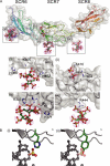
The structure of FH modules 6–8 reveals multiple sulfated sugar-binding sites. (A) The overall structure of FH678402H is shown in cartoon representation colored from blue at the N terminus (residue 320) to red at the C terminus (residue 506). Electron density for the bound SOS is presented as an FO–FC map contoured at 2 σ, which was generated before any modeling of the sugar was attempted. All figures were generated using PyMOL (25). SOS-binding sites are labeled (i–iv) and enlarged views are shown below the main panel. SOS is represented as a ball and stick colored by atom type (C, green; O, red; N, blue; S, yellow). Amino acid sidechains that contact the sugar are shown in stick representation, whereas the rest of the protein molecule is shown as a ribbon. Intermolecular hydrogen bonds are shown as dashed orange lines. The polymorphic residue associated with AMD (His402) is highlighted. (B) Model illustrating the proposed ability of the Tyr/His polymorphism at position 402 to differentially bind sulfated sugars. Position of Tyr402 was taken from the NMR structure of FH7402Y (Protein Data Bank ID, 2JGX).
Figure 2.
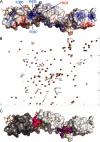
Characterization of GAG-binding sites. (A) Electrostatic surface potential (plotted at ± 5 kT/e) of FH678402H reveals a positively charged groove that extends between the three major SOS-binding sites. The residues lining the groove that have previously associated with polyanion binding, Arg387, Lys388, and Lys405 (16, 18), are highlighted in blue. The polymorphic residue associated with AMD (His402) is highlighted in red (–5). (B; 1H, 15N) HSQC spectrum for free FH78402Y (50 μM; black) and in the presence of SOS. Protein/ligand ratios are 1:0.6 (red), 1:1.2 (orange), 1:2.5 (green), 1:5 (blue), and 1:10 (purple). The y axis corresponds to 15N chemical shifts, and the x axis corresponds to 1H chemical shifts, both in parts per million. Conditions were 20 mM potassium phosphate, pH 7.4, 298 K. (C) Chemical shift perturbations mapped on the surface of FH678402H. SCR6 is shown in dark gray, as it was not present in the constructs used for these experiments. The sidechains of residues that exhibit the largest combined 15N and 1H chemical shift perturbations are highlighted in pink (FH7) and purple (FH78). The data presented in B and C are also shown in Fig. S4 with the molecule rotated in other views. Fig. S4 is available at
http://www.jem.org/cgi/content/full/jem.20071069/DC1
.
Figure 3.
Proposed model for FH interactions with sulfate-rich domains of GAGs on the cell surface. (A) Analysis of binding of different fragments of FH402H to a heparin-HiTrap column confirms the binding site in SCR6 and reveals pH dependency of binding by SCR6 and 7. This supports the involvement of histidine sidechains in glycan coordination, as revealed by the structure. (B) Mutation of sidechains seen to coordinate SOS in SCR6 alter the affinity of FH67402H for a heparin-HiTrap column. (C and D) Model for simultaneous FH binding to C3b (light blue spheres; PDB code, 2I07 [reference 26]) and GAGs (red/green spheres; PDB code, 1HPN [reference 27]) on the retinal epithelium. FH modules for which structural data is available are represented as cartoons and semitransparent surfaces: SCR5 (28), SCR6–8 (PDB code 2UWN; this study), SCR15/16 (Protein Data Bank ID, 1HFH [reference 29]), SCR19/20 (PDB code 2G7I/2BZM [references 30, 31]). Mutational data (dark blue spheres; for review see [reference 32]) mapped onto the structure of C3b suggest FH coils around C3b like a snake.
Similar articles
- Structure shows that a glycosaminoglycan and protein recognition site in factor H is perturbed by age-related macular degeneration-linked single nucleotide polymorphism.
Herbert AP, Deakin JA, Schmidt CQ, Blaum BS, Egan C, Ferreira VP, Pangburn MK, Lyon M, Uhrín D, Barlow PN. Herbert AP, et al. J Biol Chem. 2007 Jun 29;282(26):18960-8. doi: 10.1074/jbc.M609636200. Epub 2007 Mar 13. J Biol Chem. 2007. PMID: 17360715 - Molecular Mechanisms of Macular Degeneration Associated with the Complement Factor H Y402H Mutation.
Harrison RES, Morikis D. Harrison RES, et al. Biophys J. 2019 Jan 22;116(2):215-226. doi: 10.1016/j.bpj.2018.12.007. Epub 2018 Dec 14. Biophys J. 2019. PMID: 30616835 Free PMC article. - Functional and structural implications of the complement factor H Y402H polymorphism associated with age-related macular degeneration.
Ormsby RJ, Ranganathan S, Tong JC, Griggs KM, Dimasi DP, Hewitt AW, Burdon KP, Craig JE, Hoh J, Gordon DL. Ormsby RJ, et al. Invest Ophthalmol Vis Sci. 2008 May;49(5):1763-70. doi: 10.1167/iovs.07-1297. Epub 2008 Feb 8. Invest Ophthalmol Vis Sci. 2008. PMID: 18263814 - Multiple interactions of complement Factor H with its ligands in solution: a progress report.
Perkins SJ, Nan R, Okemefuna AI, Li K, Khan S, Miller A. Perkins SJ, et al. Adv Exp Med Biol. 2010;703:25-47. doi: 10.1007/978-1-4419-5635-4_3. Adv Exp Med Biol. 2010. PMID: 20711705 Review. - Complement factor H and age-related macular degeneration: the role of glycosaminoglycan recognition in disease pathology.
Clark SJ, Bishop PN, Day AJ. Clark SJ, et al. Biochem Soc Trans. 2010 Oct;38(5):1342-8. doi: 10.1042/BST0381342. Biochem Soc Trans. 2010. PMID: 20863311 Review.
Cited by
- Structural analysis of the C-terminal region (modules 18-20) of complement regulator factor H (FH).
Morgan HP, Mertens HD, Guariento M, Schmidt CQ, Soares DC, Svergun DI, Herbert AP, Barlow PN, Hannan JP. Morgan HP, et al. PLoS One. 2012;7(2):e32187. doi: 10.1371/journal.pone.0032187. Epub 2012 Feb 28. PLoS One. 2012. PMID: 22389686 Free PMC article. - Protection of host cells by complement regulators.
Schmidt CQ, Lambris JD, Ricklin D. Schmidt CQ, et al. Immunol Rev. 2016 Nov;274(1):152-171. doi: 10.1111/imr.12475. Immunol Rev. 2016. PMID: 27782321 Free PMC article. Review. - The proteoglycan glycomatrix: a sugar microenvironment essential for complement regulation.
Clark SJ, Bishop PN, Day AJ. Clark SJ, et al. Front Immunol. 2013 Nov 26;4:412. doi: 10.3389/fimmu.2013.00412. Front Immunol. 2013. PMID: 24324472 Free PMC article. Review. No abstract available. - New functional and structural insights from updated mutational databases for complement factor H, Factor I, membrane cofactor protein and C3.
Rodriguez E, Rallapalli PM, Osborne AJ, Perkins SJ. Rodriguez E, et al. Biosci Rep. 2014 Oct 22;34(5):e00146. doi: 10.1042/BSR20140117. Biosci Rep. 2014. PMID: 25188723 Free PMC article. - Identification of factor H-like protein 1 as the predominant complement regulator in Bruch's membrane: implications for age-related macular degeneration.
Clark SJ, Schmidt CQ, White AM, Hakobyan S, Morgan BP, Bishop PN. Clark SJ, et al. J Immunol. 2014 Nov 15;193(10):4962-70. doi: 10.4049/jimmunol.1401613. Epub 2014 Oct 10. J Immunol. 2014. PMID: 25305316 Free PMC article.
References
- Friedman, D.S., B.J. O'Colmain, B. Munoz, S.C. Tomany, C. McCarty, P.T. de Jong, B. Nemesure, P. Mitchell, and J. Kempen. 2004. Prevalence of age-related macular degeneration in the United States. Arch. Ophthalmol. 122:564–572. - PubMed
- Haines, J.L., M.A. Hauser, S. Schmidt, W.K. Scott, L.M. Olson, P. Gallins, K.L. Spencer, S.Y. Kwan, M. Noureddine, J.R. Gilbert, et al. 2005. Complement factor H variant increases the risk of age-related macular degeneration. Science. 308:419–421. - PubMed
- Edwards, A.O., R. Ritter III, K.J. Abel, A. Manning, C. Panhuysen, and L.A. Farrer. 2005. Complement factor H polymorphism and age-related macular degeneration. Science. 308:421–424. - PubMed
- Hageman, G.S., D.H. Anderson, L.V. Johnson, L.S. Hancox, A.J. Taiber, L.I. Hardisty, J.L. Hageman, H.A. Stockman, J.D. Borchardt, K.M. Gehrs, et al. 2005. A common haplotype in the complement regulatory gene factor H (HF1/CFH) predisposes individuals to age-related macular degeneration. Proc. Natl. Acad. Sci. USA. 102:7227–7232. - PMC - PubMed
Publication types
MeSH terms
Substances
Grants and funding
- G0001089/MRC_/Medical Research Council/United Kingdom
- 078780/z/05/z/WT_/Wellcome Trust/United Kingdom
- G0400389/MRC_/Medical Research Council/United Kingdom
- MC_U138274352/MRC_/Medical Research Council/United Kingdom
- WT_/Wellcome Trust/United Kingdom
- G0400775/MRC_/Medical Research Council/United Kingdom
- 078780/WT_/Wellcome Trust/United Kingdom
- 075415/z/04/z/WT_/Wellcome Trust/United Kingdom
LinkOut - more resources
Full Text Sources
Other Literature Sources
Medical
Molecular Biology Databases
Miscellaneous


