MRI patterns of atrophy associated with progression to AD in amnestic mild cognitive impairment - PubMed (original) (raw)
MRI patterns of atrophy associated with progression to AD in amnestic mild cognitive impairment
J L Whitwell et al. Neurology. 2008.
Abstract
Objective: To compare the patterns of gray matter loss in subjects with amnestic mild cognitive impairment (aMCI) who progress to Alzheimer disease (AD) within a fixed clinical follow-up time vs those who remain stable.
Methods: Twenty-one subjects with aMCI were identified from the Mayo Clinic Alzheimer's research program who remained clinically stable for their entire observed clinical course (aMCI-S), where the minimum required follow-up time from MRI to last follow-up assessment was 3 years. These subjects were age- and gender-matched to 42 aMCI subjects who progressed to AD within 18 months of the MRI (aMCI-P). Each subject was then age- and gender-matched to a control subject. Voxel-based morphometry (VBM) was used to assess patterns of gray matter atrophy in the aMCI-P and aMCI-S groups compared to the control group, and compared to each other.
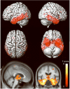
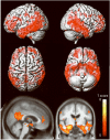
Results: The aMCI-P group showed bilateral loss affecting the medial and inferior temporal lobe, temporoparietal association neocortex, and frontal lobes, compared to controls. The aMCI-S group showed no regions of gray matter loss when compared to controls. When the aMCI-P and aMCI-S groups were compared directly, the aMCI-P group showed greater loss in the medial and inferior temporal lobes, the temporoparietal neocortex, posterior cingulate, precuneus, anterior cingulate, and frontal lobes than the aMCI-S group.
Conclusions: The regions of loss observed in subjects with amnestic mild cognitive impairment (aMCI) who progressed to Alzheimer disease (AD) within 18 months of the MRI are typical of subjects with AD. The lack of gray matter loss in subjects with aMCI who remained clinically stable for their entire observed clinical course is consistent with the notion that patterns of atrophy on MRI at baseline map well onto the subsequent clinical course.
Figures
Figure 1
Flow chart illustrating the subject selection process.
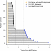
Figure 2
Schematic plot showing the time from baseline MRI to either progress to a diagnosis of AD, or the end of clinical follow-up, for all aMCI subjects. Subjects 1–21 were classified as the aMCI-S subjects that do not progress to AD over their entire follow-up, and subjects 22–63 were classified as the aMCI-P subjects since they progressed within 18 months of the MRI scan. The gray vertical lines indicate the 18 month and three year time-points.
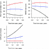
Figure 3
Plots showing the change in MMSE, CDR sum of boxes and AVLT sum of learning over trials 1–5 over three years from the time of the MRI in the aMCI-P (black), aMCI-S (blue) and control subjects (red).
Figure 4
Patterns of grey matter loss identified in the aMCI-P group compared to controls (corrected for multiple comparisons, p<0.05). The patterns of cortical atrophy are shown on a 3D surface render (top). In addition the results are shown on a sagittal and coronal slice through the customized template, selected to highlight changes in the cingulate cortex and the medial temporal lobes (bottom). L = left; R = right
Figure 5
Regions that show greater grey matter loss in the aMCI-P group compared to the aMCI-S group (corrected for multiple comparisons, p<0.05). The patterns of cortical atrophy are shown on a 3D surface render (top). In addition the results are shown on a sagittal and coronal slice through the customized template, selected to highlight changes in the cingulate cortex and the medial temporal lobes (bottom). L = left; R = right
Comment in
- Is amnestic mild cognitive impairment always AD?
Jagust W. Jagust W. Neurology. 2008 Feb 12;70(7):502-3. doi: 10.1212/01.wnl.0000299190.17488.b3. Neurology. 2008. PMID: 18268243 No abstract available.
Similar articles
- 3D maps from multiple MRI illustrate changing atrophy patterns as subjects progress from mild cognitive impairment to Alzheimer's disease.
Whitwell JL, Przybelski SA, Weigand SD, Knopman DS, Boeve BF, Petersen RC, Jack CR Jr. Whitwell JL, et al. Brain. 2007 Jul;130(Pt 7):1777-86. doi: 10.1093/brain/awm112. Epub 2007 May 28. Brain. 2007. PMID: 17533169 Free PMC article. - Patterns of Grey Matter Atrophy at Different Stages of Parkinson's and Alzheimer's Diseases and Relation to Cognition.
Kunst J, Marecek R, Klobusiakova P, Balazova Z, Anderkova L, Nemcova-Elfmarkova N, Rektorova I. Kunst J, et al. Brain Topogr. 2019 Jan;32(1):142-160. doi: 10.1007/s10548-018-0675-2. Epub 2018 Sep 11. Brain Topogr. 2019. PMID: 30206799 - Atrophy rates accelerate in amnestic mild cognitive impairment.
Jack CR Jr, Weigand SD, Shiung MM, Przybelski SA, O'Brien PC, Gunter JL, Knopman DS, Boeve BF, Smith GE, Petersen RC. Jack CR Jr, et al. Neurology. 2008 May 6;70(19 Pt 2):1740-52. doi: 10.1212/01.wnl.0000281688.77598.35. Epub 2007 Nov 21. Neurology. 2008. PMID: 18032747 Free PMC article. - Structural magnetic resonance imaging for the early diagnosis of dementia due to Alzheimer's disease in people with mild cognitive impairment.
Lombardi G, Crescioli G, Cavedo E, Lucenteforte E, Casazza G, Bellatorre AG, Lista C, Costantino G, Frisoni G, Virgili G, Filippini G. Lombardi G, et al. Cochrane Database Syst Rev. 2020 Mar 2;3(3):CD009628. doi: 10.1002/14651858.CD009628.pub2. Cochrane Database Syst Rev. 2020. PMID: 32119112 Free PMC article. - Gray Matter Atrophy in Amnestic Mild Cognitive Impairment: A Voxel-Based Meta-Analysis.
Zhang J, Liu Y, Lan K, Huang X, He Y, Yang F, Li J, Hu Q, Xu J, Yu H. Zhang J, et al. Front Aging Neurosci. 2021 Mar 31;13:627919. doi: 10.3389/fnagi.2021.627919. eCollection 2021. Front Aging Neurosci. 2021. PMID: 33867968 Free PMC article.
Cited by
- Comparison of regional gray matter atrophy, white matter alteration, and glucose metabolism as a predictor of the conversion to Alzheimer's disease in mild cognitive impairment.
Sohn BK, Yi D, Seo EH, Choe YM, Kim JW, Kim SG, Choi HJ, Byun MS, Jhoo JH, Woo JI, Lee DY. Sohn BK, et al. J Korean Med Sci. 2015 Jun;30(6):779-87. doi: 10.3346/jkms.2015.30.6.779. Epub 2015 May 13. J Korean Med Sci. 2015. PMID: 26028932 Free PMC article. - Cerebrospinal Fluid Markers of Neurodegeneration and Rates of Brain Atrophy in Early Alzheimer Disease.
Tarawneh R, Head D, Allison S, Buckles V, Fagan AM, Ladenson JH, Morris JC, Holtzman DM. Tarawneh R, et al. JAMA Neurol. 2015 Jun;72(6):656-65. doi: 10.1001/jamaneurol.2015.0202. JAMA Neurol. 2015. PMID: 25867677 Free PMC article. - Regional rates of neocortical atrophy from normal aging to early Alzheimer disease.
McDonald CR, McEvoy LK, Gharapetian L, Fennema-Notestine C, Hagler DJ Jr, Holland D, Koyama A, Brewer JB, Dale AM; Alzheimer's Disease Neuroimaging Initiative. McDonald CR, et al. Neurology. 2009 Aug 11;73(6):457-65. doi: 10.1212/WNL.0b013e3181b16431. Neurology. 2009. PMID: 19667321 Free PMC article. - Mapping cerebral atrophic trajectory from amnestic mild cognitive impairment to Alzheimer's disease.
Wei X, Du X, Xie Y, Suo X, He X, Ding H, Zhang Y, Ji Y, Chai C, Liang M, Yu C, Liu Y, Qin W; Alzheimer’s Disease Neuroimaging Initiative. Wei X, et al. Cereb Cortex. 2023 Feb 7;33(4):1310-1327. doi: 10.1093/cercor/bhac137. Cereb Cortex. 2023. PMID: 35368064 Free PMC article. - Theta band high definition transcranial alternating current stimulation, but not transcranial direct current stimulation, improves associative memory performance.
Lang S, Gan LS, Alrazi T, Monchi O. Lang S, et al. Sci Rep. 2019 Jun 12;9(1):8562. doi: 10.1038/s41598-019-44680-8. Sci Rep. 2019. PMID: 31189985 Free PMC article. Clinical Trial.
References
- Petersen RC, Smith GE, Waring SC, Ivnik RJ, Tangalos EG, Kokmen E. Mild cognitive impairment: clinical characterization and outcome. Arch Neurol. 1999;56:303–308. - PubMed
- Petersen RC. Mild cognitive impairment as a diagnostic entity. J Intern Med. 2004;256:183–194. - PubMed
- Jicha GA, Parisi JE, Dickson DW, et al. Neuropathologic outcome of mild cognitive impairment following progression to clinical dementia. Arch Neurol. 2006;63:674–681. - PubMed
- Ganguli M, Dodge HH, Shen C, DeKosky ST. Mild cognitive impairment, amnestic type: an epidemiologic study. Neurology. 2004;63:115–121. - PubMed
Publication types
MeSH terms
Grants and funding
- P50 AG16574/AG/NIA NIH HHS/United States
- R01 AG11378/AG/NIA NIH HHS/United States
- P50 AG016574/AG/NIA NIH HHS/United States
- R01 AG011378/AG/NIA NIH HHS/United States
- U01 AG06786/AG/NIA NIH HHS/United States
- U01 AG006786/AG/NIA NIH HHS/United States
LinkOut - more resources
Full Text Sources
Other Literature Sources
Medical


