Sialic acid receptor detection in the human respiratory tract: evidence for widespread distribution of potential binding sites for human and avian influenza viruses - PubMed (original) (raw)
Sialic acid receptor detection in the human respiratory tract: evidence for widespread distribution of potential binding sites for human and avian influenza viruses
John M Nicholls et al. Respir Res. 2007.
Abstract
Background: Influenza virus binds to cell receptors via sialic acid (SA) linked glycoproteins. They recognize SA on host cells through their haemagglutinins (H). The distribution of SA on cell surfaces is one determinant of host tropism and understanding its expression on human cells and tissues is important for understanding influenza pathogenesis. The objective of this study therefore was to optimize the detection of alpha2,3-linked and alpha2,6-linked SA by lectin histochemistry by investigating the binding of Sambucus nigra agglutinin (SNA) for SAalpha2,6Gal and Maackia amurensis agglutinin (MAA) for SAalpha2,3Gal in the respiratory tract of normal adults and children.
Methods: We used fluorescent and biotinylated SNA and MAA from different suppliers on archived and prospectively collected biopsy and autopsy specimens from the nasopharynx, trachea, bronchus and lungs of fetuses, infants and adults. We compared different methods of unmasking for tissue sections to determine if these would affect lectin binding. Using serial sections we then compared the lectin binding of MAA from different suppliers.
Results: We found that unmasking using microwave treatment in citrate buffer produced increased lectin binding to the ciliated and glandular epithelium of the respiratory tract. In addition we found that there were differences in tissue distribution of the alpha2,3 linked SA when 2 different isoforms of MAA (MAA1 and MAA2) lectin were used. MAA1 had widespread binding throughout the upper and lower respiratory tract and showed more binding to the respiratory epithelium of children than in adults. By comparison, MAA2 binding was mainly restricted to the alveolar epithelial cells of the lung with weak binding to goblet cells. SNA binding was detected in bronchial and alveolar epithelial cells and binding of this lectin was stronger to the paediatric epithelium compared to adult epithelium. Furthermore, the MAA lectins from 2 suppliers (Roche and EY Labs) tended to only bind in a pattern similar to MAA1 (Vector Labs) and produced a different binding pattern to MAA2 from Vector Labs.
Conclusion: The lectin binding pattern of MAA may vary depending on the supplier and the different isoforms of MAA show a different tissue distribution in the respiratory tract. This finding is important if conclusions about the potential binding sites of SAalpha2,3 binding viruses, such as influenza or human parainfluenza are to be made.
Figures
Figure 1
Effect of different retrieval techniques on lectin binding. Single labelling of paediatric respiratory mucosa by FITC- conjugated Sambucus nigra agglutinin (SNA) (A-F) and Maackia amurensis agglutinin (MAA) (G-J) using different methods of retrieval. No antigen retrieval, (A) and (F), Citrate buffer (B) and (G), EDTA (C) and (H), Pronase (D) and (I), and Trypsin (E) and (J). Stain intensity was graded as strong (++) and a weaker pattern as +. In the absence of unmasking techniques there was minimal to weak (-/+) SNA binding and weak (+) MAA binding in the basal epithelium and epithelial cells of the bronchial mucosa of paediatric tissues. All forms of retrieval enhanced the lectin staining of the surface epithelial cells and mucus containing cells for both SNA and MAA. Examination with dual FITC/Rhodamine filter. Magnification × 200.
Figure 2
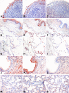
Lectin binding to upper and lower respiratory tract. Tissue distribution of Sambucus nigra agglutinin (SNA) for SAα2,6, Maackia amurensis agglutinin 1 (MAA1), and Maackia amurensis agglutinin 2 (MAA2) for SAα2,3 binding in the adult and paediatric respiratory tract. Serial sections of nasopharynx (A-C), adult bronchus (D-F), adult lung (G-I), paediatric bronchus (J-L), and paediatric lung (M-O) are shown and stained with SNA (A,D,G,J,M), MAA1 (B,E,H,K,N) and MAA2 (C,F,I,L,O). The adult nasopharynx shows SNA and MAA1 binding in the epithelium but no MAA2 binding. A similar pattern is also present in the adult bronchus and in addition the pneumocytes show MAA1 and MAA2 binding (E,F). Alveolar macrophages (G-I) demonstrate minimal SNA and no MAA2 binding but are positive for MAA1. The paediatric bronchus shows a greater binding of the epithelium with MAA1 (K) than the adult (E). The pneumocytes (M) also show more SNA binding than the adult (G). Staining using HRP conjugated SNA and biotin conjugated MAA1 and MAA2. (A-F) and (J-L) at 200 × magnification and (G-I) and (M-O) at 400 × magnification.
Figure 3
Lectin binding to bronchial epithelium using different detection methods. Serial sections for comparison of detection methods for lectin binding of Sambucus nigra agglutinin (SNA) to SAα2,6 and Maackia amurensis (MAA) agglutinin for binding to SAα2,3 in adult bronchial epithelium. Double fluoresecence for FITC labelled SNA and TRITC labelled MAA shows a heterogeneous pattern (A), with more binding to the basal epithelium with MAA (B) than SNA (C). HRP labelling of MAA (D) and SNA (E) shows a similar pattern of binding to the fluorescent labelled lectin. 200 × magnification.
Figure 4
Comparison of MAA binding using MAA from different suppliers. Serial sections of adult lung tissue for comparison of lectin binding of Maackia amurensis (MAA) for SAα2,3Gal. Biotin conjugated MAA1(also known as MAL) from Vector Laboratories (A) and (E), Biotin conjugated MAA2 (also known as MAH) from Vector Laboratories (B) and (F), Digoxigenin conjugated MAA from Roche (C) and (G) and HRP conjugated MAA from EY Laboratories (D) and (H). Orange arrows indicate alveolar macrophages, Green arrows indicate alveolar pneumocytes and blue arrows indicate bronchiolar epithelium. Haematoxylin counterstain 200 × magnification.
Similar articles
- Expression and distribution of sialic acid influenza virus receptors in wild birds.
França M, Stallknecht DE, Howerth EW. França M, et al. Avian Pathol. 2013 Feb;42(1):60-71. doi: 10.1080/03079457.2012.759176. Avian Pathol. 2013. PMID: 23391183 Free PMC article. - Distribution of sialic acid receptors and influenza A virus of avian and swine origin in experimentally infected pigs.
Trebbien R, Larsen LE, Viuff BM. Trebbien R, et al. Virol J. 2011 Sep 8;8:434. doi: 10.1186/1743-422X-8-434. Virol J. 2011. PMID: 21902821 Free PMC article. - Comparative distribution of human and avian type sialic acid influenza receptors in the pig.
Nelli RK, Kuchipudi SV, White GA, Perez BB, Dunham SP, Chang KC. Nelli RK, et al. BMC Vet Res. 2010 Jan 27;6:4. doi: 10.1186/1746-6148-6-4. BMC Vet Res. 2010. PMID: 20105300 Free PMC article. - Tissue and host tropism of influenza viruses: importance of quantitative analysis.
Zhang H. Zhang H. Sci China C Life Sci. 2009 Dec;52(12):1101-10. doi: 10.1007/s11427-009-0161-x. Epub 2009 Dec 17. Sci China C Life Sci. 2009. PMID: 20016966 Review. - Adaptation of influenza viruses to human airway receptors.
Thompson AJ, Paulson JC. Thompson AJ, et al. J Biol Chem. 2021 Jan-Jun;296:100017. doi: 10.1074/jbc.REV120.013309. Epub 2020 Nov 22. J Biol Chem. 2021. PMID: 33144323 Free PMC article. Review.
Cited by
- Influenza B Virus Receptor Specificity: Closing the Gap between Binding and Tropism.
Page CK, Tompkins SM. Page CK, et al. Viruses. 2024 Aug 24;16(9):1356. doi: 10.3390/v16091356. Viruses. 2024. PMID: 39339833 Free PMC article. Review. - A comprehensive review of influenza B virus, its biological and clinical aspects.
Ashraf MA, Raza MA, Amjad MN, Ud Din G, Yue L, Shen B, Chen L, Dong W, Xu H, Hu Y. Ashraf MA, et al. Front Microbiol. 2024 Sep 4;15:1467029. doi: 10.3389/fmicb.2024.1467029. eCollection 2024. Front Microbiol. 2024. PMID: 39296301 Free PMC article. Review. - Establishment of a humanized ST6GAL1 mouse model for influenza research.
Chao L, Feng H, Qian G, Limin L, Ziwei L, Shuangshuang L, Xiaoyan L, Yuechao H, Mengjie Y, Yingze Z, Jun L, Xuancheng L, Shuguang D. Chao L, et al. Animal Model Exp Med. 2024 Jun;7(3):337-346. doi: 10.1002/ame2.12449. Epub 2024 Jun 11. Animal Model Exp Med. 2024. PMID: 38859745 Free PMC article. - Establishment of Swine Primary Nasal, Tracheal, and Bronchial Epithelial Cell Culture Models for the Study of Influenza Virus Infection.
Krunkosky M, Krunkosky TM, Meliopoulos V, Kyriakis CS, Schultz-Cherry S, Tompkins SM. Krunkosky M, et al. J Virol Methods. 2024 Jun;327:114943. doi: 10.1016/j.jviromet.2024.114943. Epub 2024 Apr 26. J Virol Methods. 2024. PMID: 38679164 - Two Receptor Binding Strategy of SARS-CoV-2 Is Mediated by Both the N-Terminal and Receptor-Binding Spike Domain.
Monti M, Milanetti E, Frans MT, Miotto M, Di Rienzo L, Baranov MV, Gosti G, Somavarapu AK, Nagaraj M, Golbek TW, Rossing E, Moons SJ, Boltje TJ, van den Bogaart G, Weidner T, Otzen DE, Tartaglia GG, Ruocco G, Roeters SJ. Monti M, et al. J Phys Chem B. 2024 Jan 18;128(2):451-464. doi: 10.1021/acs.jpcb.3c06258. Epub 2024 Jan 8. J Phys Chem B. 2024. PMID: 38190651 Free PMC article.
References
- Lamblin G, Lhermitte M, Klein A, Roussel P, Van Halbeek H, Vliegenthart JF. Biochem Soc Trans. 1984/08/01. 4. Vol. 12. 1984. Carbohydrate chains from human bronchial mucus glycoproteins: a wide spectrum of oligosaccharide structures; pp. 599–600. - PubMed
- Baum LG, Paulson JC. Acta Histochem Suppl. 1990/01/01. Vol. 40. 1990. Sialyloligosaccharides of the respiratory epithelium in the selection of human influenza virus receptor specificity; pp. 35–38. - PubMed
- Delmotte P, Degroote S, Merten MD, Van Seuningen I, Bernigaud A, Figarella C, Roussel P, Perini JM. Glycoconj J. 2002/06/27. 6. Vol. 18. 2001. Influence of TNFalpha on the sialylation of mucins produced by a transformed cell line MM-39 derived from human tracheal gland cells; pp. 487–497. - DOI - PubMed
Publication types
MeSH terms
Substances
LinkOut - more resources
Full Text Sources
Other Literature Sources
Medical
Research Materials


