iNOS activity is critical for the clearance of Burkholderia mallei from infected RAW 264.7 murine macrophages - PubMed (original) (raw)
iNOS activity is critical for the clearance of Burkholderia mallei from infected RAW 264.7 murine macrophages
Paul J Brett et al. Cell Microbiol. 2008 Feb.
Abstract
Burkholderia mallei is a facultative intracellular pathogen that can cause fatal disease in animals and humans. To better understand the role of phagocytic cells in the control of infections caused by this organism, studies were initiated to examine the interactions of B. mallei with RAW 264.7 murine macrophages. Utilizing modified kanamycin-protection assays, B. mallei was shown to survive and replicate in RAW 264.7 cells infected at multiplicities of infection (moi) of < or = 1. In contrast, the organism was efficiently cleared by the macrophages when infected at an moi of 10. Interestingly, studies demonstrated that the monolayers only produced high levels of TNF-alpha, IL-6, IL-10, GM-CSF, RANTES and IFN-beta when infected at an moi of 10. In addition, nitric oxide assays and inducible nitric oxide synthase (iNOS) immunoblot analyses revealed a strong correlation between iNOS activity and clearance of B. mallei from RAW 264.7 cells. Furthermore, treatment of activated macrophages with the iNOS inhibitor, aminoguanidine, inhibited clearance of B. mallei from infected monolayers. Based upon these results, it appears that moi significantly influence the outcome of interactions between B. mallei and murine macrophages and that iNOS activity is critical for the clearance of B. mallei from activated RAW 264.7 cells.
Figures
Fig. 1
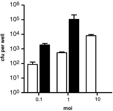
Survival characteristics of B. mallei in RAW 264.7 cells. Monolayers were infected with B. mallei at moi ranging from 0.1 to 10. Uptake (white bars) and intracellular survival (black bars) were quantified at 3 and 24 h post infection respectively. Values represent the means ± SD of three independent experiments.
Fig. 3
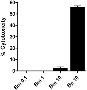
Cellular integrity of RAW 264.7 cells infected with B. mallei and B. pseudomallei. Monolayers were infected with B. mallei at moi 0.1 (Bm 0.1), moi 1 (Bm 1), moi 10 (Bm 10) and B. pseudomallei at moi 10 (Bp 10). Per cent cytotoxicity was determined by assaying for LDH release in culture supernatants at 24 h post infection. Values represent the means ± SD of three independent experiments assayed in duplicate.
Fig. 2
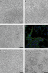
Microscopic analysis of RAW 264.7 cells infected with B. mallei and B. pseudomallei. Monolayers were fixed and examined at 24 h post infection. Light micrographs: (A) mock infected, (B) B. mallei moi 0.1, (C) B. mallei moi 1, (E) B. mallei moi 10, and (F) B. pseudomallei moi 10. Black scale bars = 1 mm. D. Confocal micrograph of RAW 264.7 cells infected with B. mallei at an moi of 1. Bacteria were stained red with rabbit anti-Burkholderia thailandensis polyclonal sera and anti-rabbit IgG Alexa Fluor® 568, actin was stained green with Alexa Fluor® 488 phalloidin, and nuclei were stained blue with DRAQ5™. White scale bar = 20 μm. Micrographs are representative of at least three independent experiments.
Fig. 4
B. mallei is a weak activator of RAW 264.7 cells. Culture supernatants from monolayers infected with B. mallei (open circles) or E. coli (filled circles) at moi ranging from 0.1 to 10 were harvested at (A–E) 6 h or (F–J) 18 h post infection and assayed for the production of (A and F) TNF-α, (B and G) IL-6, (C and H) IL-10, (D and I) GM-CSF, and (E and J) RANTES. Values represent the means ± SD of three independent experiments assayed in duplicate.
Fig. 5
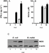
Production of IFN-β and NO by B. mallei- and _E. coli_-infected RAW 264.7 cells. Culture supernatants from monolayers infected with B. mallei (white bars) or E. coli (black bars) at moi of 1 and 10 were harvested (A) 6 h and (B) 24 h post infection and assayed for the production of IFN-β and NO respectively. Supernatants from mock-infected monolayers were included as negative controls. Values represent the means ± SD of three independent experiments assayed in duplicate or triplicate. C. Cell lysates prepared from _E. coli_-infected and _B. mallei_-infected monolayers harvested 24 h post infection were assayed for the presence of iNOS by immunoblot analysis. β-actin was included as a loading control. Results are representative of three independent experiments.
Fig. 6
Clearance of B. mallei from activated RAW 264.7 macrophages correlates with iNOS activity. Monolayers were infected with B. mallei at an moi of 10. A. Bacterial loads were determined at 3, 6, 12, 18 and 24 h post infection. Values represent the means ± SD of three independent experiments. Culture supernatants harvested at 3, 6, 12, 18 and 24 h from mock-infected (open circles) and _B. mallei_-infected (filled circles) monolayers were assayed for the production of (B) TNF-α, (C) RANTES, and (D) NO. Values represent the means ± SD of three independent experiments assayed in duplicate or triplicate. E. Cell lysates prepared from _B. mallei_-infected monolayers harvested at 3, 6, 12, 18 and 24 h post infection were assayed for the presence of iNOS by immunoblot analysis. β-actin was included as a loading control. Results are representative of three independent experiments.
Fig. 7
Inhibition of iNOS activity facilitates survival of B. mallei in activated RAW 264.7 cells. Monolayers infected with B. mallei (Bm) at an moi of 10 were incubated in the absence or presence of AG (200 μg ml−1) at 1 h post infection. A. Uptake (white bars) and intracellular survival (black bars) were determined at 3 and 24 h post infection respectively. Values represent the means ± SD of three independent experiments. Culture supernatants harvested at 24 h post infection from mock-infected and _B. mallei_-infected monolayers were assayed for the production of (B) TNF-α, (C) RANTES and (D) NO. Values represent the means ± SD of three independent experiments assayed in duplicate or triplicate. E. Cell lysates prepared from mock-infected and _B. mallei_-infected monolayers harvested at 24 h post infection were assayed for the presence of iNOS by immunoblot analysis. β-actin was included as a loading control. Results are representative of three independent experiments.
Fig. 8
E. coli LPS stimulates the clearance of B. mallei from RAW 264.7 cells infected at an moi of 1. Infected monolayers were incubated with either AG (200 μg ml−1), LPS (100 ng ml−1), or AG (200 μg ml−1) and LPS (100 ng ml−1) at 1 h post infection. A. Uptake (white bars) and intracellular survival (black bars) were determined at 3 and 24 h post infection respectively. Values represent the means ± SD of three independent experiments. B. Culture supernatants harvested 24 h post infection from mock-infected and _B. mallei_-infected monolayers were assayed for the production of NO. Values represent the means ± SD of three independent experiments assayed in triplicate.
Similar articles
- Burkholderia mallei cluster 1 type VI secretion mutants exhibit growth and actin polymerization defects in RAW 264.7 murine macrophages.
Burtnick MN, DeShazer D, Nair V, Gherardini FC, Brett PJ. Burtnick MN, et al. Infect Immun. 2010 Jan;78(1):88-99. doi: 10.1128/IAI.00985-09. Epub 2009 Nov 2. Infect Immun. 2010. PMID: 19884331 Free PMC article. - Burkholderia pseudomallei interferes with inducible nitric oxide synthase (iNOS) production: a possible mechanism of evading macrophage killing.
Utaisincharoen P, Tangthawornchaikul N, Kespichayawattana W, Chaisuriya P, Sirisinha S. Utaisincharoen P, et al. Microbiol Immunol. 2001;45(4):307-13. doi: 10.1111/j.1348-0421.2001.tb02623.x. Microbiol Immunol. 2001. PMID: 11386421 - Induction of iNOS expression and antimicrobial activity by interferon (IFN)-beta is distinct from IFN-gamma in Burkholderia pseudomallei-infected mouse macrophages.
Utaisincharoen P, Anuntagool N, Arjcharoen S, Limposuwan K, Chaisuriya P, Sirisinha S. Utaisincharoen P, et al. Clin Exp Immunol. 2004 May;136(2):277-83. doi: 10.1111/j.1365-2249.2004.02445.x. Clin Exp Immunol. 2004. PMID: 15086391 Free PMC article. - TNF-alpha controls intracellular mycobacterial growth by both inducible nitric oxide synthase-dependent and inducible nitric oxide synthase-independent pathways.
Bekker LG, Freeman S, Murray PJ, Ryffel B, Kaplan G. Bekker LG, et al. J Immunol. 2001 Jun 1;166(11):6728-34. doi: 10.4049/jimmunol.166.11.6728. J Immunol. 2001. PMID: 11359829 - Innate immune response to Burkholderia mallei.
Saikh KU, Mott TM. Saikh KU, et al. Curr Opin Infect Dis. 2017 Jun;30(3):297-302. doi: 10.1097/QCO.0000000000000362. Curr Opin Infect Dis. 2017. PMID: 28177960 Free PMC article. Review.
Cited by
- Burkholderia mallei cellular interactions in a respiratory cell model.
Whitlock GC, Valbuena GA, Popov VL, Judy BM, Estes DM, Torres AG. Whitlock GC, et al. J Med Microbiol. 2009 May;58(Pt 5):554-562. doi: 10.1099/jmm.0.007724-0. J Med Microbiol. 2009. PMID: 19369515 Free PMC article. - Burkholderia mallei and Burkholderia pseudomallei stimulate differential inflammatory responses from human alveolar type II cells (ATII) and macrophages.
Lu R, Popov V, Patel J, Eaves-Pyles T. Lu R, et al. Front Cell Infect Microbiol. 2012 Dec 28;2:165. doi: 10.3389/fcimb.2012.00165. eCollection 2012. Front Cell Infect Microbiol. 2012. PMID: 23293773 Free PMC article. - Lifestyle-induced metabolic inflexibility and accelerated ageing syndrome: insulin resistance, friend or foe?
Nunn AV, Bell JD, Guy GW. Nunn AV, et al. Nutr Metab (Lond). 2009 Apr 16;6:16. doi: 10.1186/1743-7075-6-16. Nutr Metab (Lond). 2009. PMID: 19371409 Free PMC article. - Phenotypic Characterization of a Novel Virulence-Factor Deletion Strain of Burkholderia mallei That Provides Partial Protection against Inhalational Glanders in Mice.
Bozue JA, Chaudhury S, Amemiya K, Chua J, Cote CK, Toothman RG, Dankmeyer JL, Klimko CP, Wilhelmsen CL, Raymond JW, Zavaljevski N, Reifman J, Wallqvist A. Bozue JA, et al. Front Cell Infect Microbiol. 2016 Feb 26;6:21. doi: 10.3389/fcimb.2016.00021. eCollection 2016. Front Cell Infect Microbiol. 2016. PMID: 26955620 Free PMC article. - Characterization of in vitro phenotypes of Burkholderia pseudomallei and Burkholderia mallei strains potentially associated with persistent infection in mice.
Bernhards RC, Cote CK, Amemiya K, Waag DM, Klimko CP, Worsham PL, Welkos SL. Bernhards RC, et al. Arch Microbiol. 2017 Mar;199(2):277-301. doi: 10.1007/s00203-016-1303-8. Epub 2016 Oct 13. Arch Microbiol. 2017. PMID: 27738703 Free PMC article.
References
- Bartlett JG. Glanders. In: Gorbach SL, Bartlet JG, Blacknow NR, editors. Infectious Diseases. Lippincott: Williams & Wilkins; 2005. pp. 1463–1464.
- Barton GM, Medzhitov R. Toll-like receptor signaling pathways. Science. 2003;300:1524–1525. - PubMed
- Boehm U, Klamp T, Groot M, Howard JC. Cellular responses to interferon-gamma. Annu Rev Immunol. 1997;15:749–795. - PubMed
- Breitbach K, Rottner K, Klocke S, Rohde M, Jenzora A, Wehland J, Steinmetz I. Actin-based motility of Burkholderia pseudomallei involves the Arp 2/3 complex, but not N-WASP and Ena/VASP proteins. Cell Microbiol. 2003;5:385–393. - PubMed
Publication types
MeSH terms
Substances
LinkOut - more resources
Full Text Sources


