Structural extremes in a cretaceous dinosaur - PubMed (original) (raw)
Structural extremes in a cretaceous dinosaur
Paul C Sereno et al. PLoS One. 2007.
Abstract
Fossils of the Early Cretaceous dinosaur, Nigersaurus taqueti, document for the first time the cranial anatomy of a rebbachisaurid sauropod. Its extreme adaptations for herbivory at ground-level challenge current hypotheses regarding feeding function and feeding strategy among diplodocoids, the larger clade of sauropods that includes Nigersaurus. We used high resolution computed tomography, stereolithography, and standard molding and casting techniques to reassemble the extremely fragile skull. Computed tomography also allowed us to render the first endocast for a sauropod preserving portions of the olfactory bulbs, cerebrum and inner ear, the latter permitting us to establish habitual head posture. To elucidate evidence of tooth wear and tooth replacement rate, we used photographic-casting techniques and crown thin sections, respectively. To reconstruct its 9-meter postcranial skeleton, we combined and size-adjusted multiple partial skeletons. Finally, we used maximum parsimony algorithms on character data to obtain the best estimate of phylogenetic relationships among diplodocoid sauropods. Nigersaurus taqueti shows extreme adaptations for a dinosaurian herbivore including a skull of extremely light construction, tooth batteries located at the distal end of the jaws, tooth replacement as fast as one per month, an expanded muzzle that faces directly toward the ground, and hollow presacral vertebral centra with more air sac space than bone by volume. A cranial endocast provides the first reasonably complete view of a sauropod brain including its small olfactory bulbs and cerebrum. Skeletal and dental evidence suggests that Nigersaurus was a ground-level herbivore that gathered and sliced relatively soft vegetation, the culmination of a low-browsing feeding strategy first established among diplodocoids during the Jurassic.
Conflict of interest statement
Competing Interests: The authors have declared that no competing interests exist.
Figures
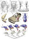
Figure 1. Skull of Nigersaurus taqueti and head posture in sauropodomorphs.
(A)-Lateral view of skull (MNN GAD512). (B)-Anterodorsal view of cranium. (C)-Anterior view of lower jaws. (D)-Premaxillary and dentary tooth series (blue) reconstructed from µCT scans. (E)-Skull reconstruction in anterolateral and dorsal view cut at mid length between muzzle and occipital units (cross-section in red) with the adductor mandibulae muscle shown between the quadrate and surangular. (F)-Endocast in dorsal view showing the cerebrum and olfactory bulbs. (G)-Endocast (above) and transparent skulls with endocasts in place (below) based on µCT scans of the basal sauropodomorph Massospondylus carinatus (BP 1/4779), the basal neosauropod Camarasaurus lentus (CM 11338), the diplodocid Diplodocus longus (CM 11161), and the prototype skull of the rebbachisaurid Nigersaurus taqueti. Endocasts and skulls are oriented with the lateral semicircular canal held horizontal; the angle measurement indicates degrees from the horizontal for a line from the jaw joint to the tip of the upper teeth. Endocasts show brain space and dural sinuses (blue), nerve openings (yellow), inner ear (pink), and internal carotid artery (red). Phylogenetic diagram at bottom shows the relationships of these four sauropodomorphs, with increasing ventral deflection of the snout toward the condition seen in Nigersaurus. Scale bar equals 2 cm in F. Abbreviations: 1–5, fenestrae 1–5; a, angular; amm, adductor mandibulae muscle; antfe, antorbital fenestra; ar, articular; ce, cerebrum; cp, coronoid process; d, dentary; d1, 34, dentary tooth 1, 34; ds, dural sinus; emf, external mandibular fenestra; en, external nares; f, frontal; fo, foramen; j, jugal; m, maxilla; nf, narial fossa; olb, olfactory bulb; olt, olfactory tract; pm, premaxilla; po, postorbital; popr, paroccipital process; q, quadrate; qj, quadratojugal; sa, surangular, saf, surangular foramen; sq, squamosal; vc, vascular canal.
Figure 2. Crown form, wear pattern, and microstructure in Nigersaurus taqueti.
Wear facets and surface detail is from a worn crown (MNN GAD513), and enamel and dentine microstructure is from left premaxillary teeth in cross-section (MNN GAD514). (A)-Crown in lingual (interior) view showing low-angle wear facet. Magnified views of a cast of the lingual wear facet showing (B)-wear striations in the dentine and (C)-the edge of the facet. (D)-Crown in labial (exterior) view showing high-angle wear facet. Magnified views of a cast of the labial facet showing (E)-coarse scratches on the dentine and (F)-fine scratches on the edge of the facet. (G)-Transverse thin section of two successive premaxillary crowns showing thickened labial enamel and circumferential incremental lines of von Ebner in the dentine. (H)-Magnified view of the older (left) crown showing approximately 60 incremental lines of von Ebner. Scale bar in A and D equals 5 mm; scale bar below F equals 0.5 mm in B, E and F and 1.11 mm in C. Abbreviations: de, dentine; en, enamel; fe, facet edge; g, gouge; s, scratch.
Figure 3. Skeleton of Nigersaurus taqueti.
Skeletal reconstruction is based mainly on four specimens (MNN GAD513, GAD 515-518). (A)-Skeletal silhouette showing preserved bones. (B)-Fifth cervical vertebra in lateral view. (C)-Eighth dorsal vertebra in lateral view with two cross-sections from a µCT scan. (D)-Probable eighth caudal vertebra in lateral view with anterior view of the neural spine. (E)-Caudal vertebra (ca. CA37) with low neural spine. (F)-Distal caudal vertebra (ca. CA47) with biconvex centrum and rudimentary neural arch. Human silhouette equals 1.68 meters (5 feet 6 inches). Upper scale bar equals 10 cm for B-E; lower scale bar equals 5 cm for F. Abbreviations: C, cervical vertebra; CA, caudal vertebra; ce, centrum; D, dorsal vertebra; di, diapophysis; ep, epipophysis; ns, neural spine; pa, parapophysis; pl, pleurocoel; poz, postzygapophysis; prz, prezygapophysis; przepl, prezygapophyseal-epipophyseal lamina; r, rib; se, septum; sp, spine.
Figure 4. Calibrated phylogeny of diplodocoid sauropods.
The diagram is based on strict consensus of five minimum-length trees using 13 ingroup taxa and 102 unordered characters (CI = 0.76; RI = 0.78) (Text S5). Scaled icons represent a diplodocid (Apatosaurus) , dicraeosaurid (Dicraeosaurus) , and a rebbachisaurid (Nigersaurus). Geographic distributions include Laurasian diplodocoids (western North America—Apatasaurus, Diplodocus, Suuwassea; Europe—Histriasaurus, Spanish rebbachisaurid) and Gondwanan diplodocoids (South America—Cathartesaura, Limaysaurus, Zapalasaurus; Africa—Rebbachisaurus, Nigersaurus). Temporal boundaries based on a recent timescale . Color scheme: Laurasia (orange); Gondwana (blue); North America (solid orange); Europe (striped orange); South America (blue); Africa (striped blue).
Similar articles
- Inferences of diplodocoid (Sauropoda: Dinosauria) feeding behavior from snout shape and microwear analyses.
Whitlock JA. Whitlock JA. PLoS One. 2011 Apr 6;6(4):e18304. doi: 10.1371/journal.pone.0018304. PLoS One. 2011. PMID: 21494685 Free PMC article. - Neurovascular anatomy of dwarf dinosaur implies precociality in sauropods.
Schade M, Knötschke N, Hörnig MK, Paetzel C, Stumpf S. Schade M, et al. Elife. 2022 Dec 20;11:e82190. doi: 10.7554/eLife.82190. Elife. 2022. PMID: 36537069 Free PMC article. - First complete sauropod dinosaur skull from the Cretaceous of the Americas and the evolution of sauropod dentition.
Chure D, Britt BB, Whitlock JA, Wilson JA. Chure D, et al. Naturwissenschaften. 2010 Apr;97(4):379-91. doi: 10.1007/s00114-010-0650-6. Epub 2010 Feb 24. Naturwissenschaften. 2010. PMID: 20179896 Free PMC article. - Rethinking the nature of fibrolamellar bone: an integrative biological revision of sauropod plexiform bone formation.
Stein K, Prondvai E. Stein K, et al. Biol Rev Camb Philos Soc. 2014 Feb;89(1):24-47. doi: 10.1111/brv.12041. Epub 2013 May 6. Biol Rev Camb Philos Soc. 2014. PMID: 23647662 Review. - Dinosaur biomechanics.
Alexander RM. Alexander RM. Proc Biol Sci. 2006 Aug 7;273(1596):1849-55. doi: 10.1098/rspb.2006.3532. Proc Biol Sci. 2006. PMID: 16822743 Free PMC article. Review.
Cited by
- Photographic Atlas and three-dimensional reconstruction of the holotype skull of Euhelopus zdanskyi with description of additional cranial elements.
Poropat SF, Kear BP. Poropat SF, et al. PLoS One. 2013 Nov 21;8(11):e79932. doi: 10.1371/journal.pone.0079932. eCollection 2013. PLoS One. 2013. PMID: 24278222 Free PMC article. - A possible brachiosaurid (Dinosauria, Sauropoda) from the mid-Cretaceous of northeastern China.
Liao CC, Moore A, Jin C, Yang TR, Shibata M, Jin F, Wang B, Jin D, Guo Y, Xu X. Liao CC, et al. PeerJ. 2021 Aug 20;9:e11957. doi: 10.7717/peerj.11957. eCollection 2021. PeerJ. 2021. PMID: 34484987 Free PMC article. - Reconstructing the past: methods and techniques for the digital restoration of fossils.
Lautenschlager S. Lautenschlager S. R Soc Open Sci. 2016 Oct 12;3(10):160342. doi: 10.1098/rsos.160342. eCollection 2016 Oct. R Soc Open Sci. 2016. PMID: 27853548 Free PMC article. - The endocranial anatomy of Buriolestes schultzi (Dinosauria: Saurischia) and the early evolution of brain tissues in sauropodomorph dinosaurs.
Müller RT, Ferreira JD, Pretto FA, Bronzati M, Kerber L. Müller RT, et al. J Anat. 2021 Apr;238(4):809-827. doi: 10.1111/joa.13350. Epub 2020 Nov 2. J Anat. 2021. PMID: 33137855 Free PMC article. - Sampling impacts the assessment of tooth growth and replacement rates in archosaurs: implications for paleontological studies.
Kosch JCD, Zanno LE. Kosch JCD, et al. PeerJ. 2020 Sep 18;8:e9918. doi: 10.7717/peerj.9918. eCollection 2020. PeerJ. 2020. PMID: 32999766 Free PMC article.
References
- Lavocat R. Sure les dinosauriens du Continental Intercalaire des Kem-Kem de la Daoura. Comptes Rendus de la Dix-Neuviéme Session, Congrès Géologique International, Alger. 1954;21 (1952):65–68.
- Naish D, Martill DM. Saurischian dinosaurs 1: Sauropods. In: Martill DM, Naish D, editors. Dinosaurs of the Isle of Wight. London: The Palaeontological Association; 2001. pp. 185–241.
- Dalla Vecchia FM. Remains of Sauropoda (Reptilia, Saurischia) in the Lower Cretaceous (upper Hauterivianl/lower Baremian) limestones of SW Istria (Croatia). Geol Croatica. 1998;51:105–134.
- Pereda-Suberbiola X, Torcida F, Izquierdo LA, Huerta P, Montero D, et al. First rebbachisaurid dinosaur (Sauropoda, Diplodocoidea) from the Early Cretaceous of Spain: paleobiological implications. Bull Soc Géol France. 2003;174:471–479.
- Calvo JO, Salgado L. Rebbachisaurus tessonei sp. nov. a new Sauropoda from the Albian-Cenomanian of Argentina; new evidence on the origin of Diplodocidae. Gaia. 1995;11:13–33.
Publication types
MeSH terms
LinkOut - more resources
Full Text Sources


