Amino acid substitutions in the S2 subunit of mouse hepatitis virus variant V51 encode determinants of host range expansion - PubMed (original) (raw)
Amino acid substitutions in the S2 subunit of mouse hepatitis virus variant V51 encode determinants of host range expansion
Willie C McRoy et al. J Virol. 2008 Feb.
Abstract
We previously described mouse hepatitis virus (MHV) variant V51 derived from a persistent infection of murine DBT cells with an expanded host range (R. S. Baric, E. Sullivan, L. Hensley, B. Yount, and W. Chen, J. Virol. 73:638-649, 1999). Sequencing of the V51 spike gene, the mediator of virus entry, revealed 13 amino acid substitutions relative to the originating MHV A59 strain. Seven substitutions were located in the amino-terminal S1 cleavage subunit, and six were located in the carboxy-terminal S2 cleavage subunit. Using targeted RNA recombination, we constructed a panel of recombinant viruses to map the mediators of host range to the six substitutions in S2, with a subgroup of four changes of particular interest. This subgroup maps to two previously identified domains within S2, a putative fusion peptide and a heptad repeat, both conserved features of class I fusion proteins. In addition to an altered host range, V51 displayed altered utilization of CEACAM1a, the high-affinity receptor for A59. Interestingly, a recombinant with S1 from A59 and S2 from V51 was severely debilitated in its ability to productively infect cells via CEACAM1a, while the inverse recombinant was not. This result suggests that the S2 substitutions exert powerful effects on the fusion trigger that normally passes from S1 to S2. These novel findings play against the existing data that suggest that MHV host range determinants are located in the S1 subunit, which harbors the receptor binding domain, or involve coordinating changes in both S1 and S2. Mounting evidence also suggests that the class I fusion mechanism may possess some innate plasticity that regulates viral host range.
Figures
FIG. 1.
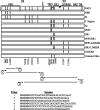
Spike protein structure map and cloning strategy. The panel of 12 chimeric S genes produced for this study is depicted, with the approximate locations of substitutions present in V51 relative to the A59 strain indicated as vertical black bars (numerical locations are indicated along the bottom of the figure). The 1,324-residue spike protein is typically posttranslationally cleaved between residues 717 and 718 into S1 and S2 subunits (cleavage site indicated with black arrow). The locations of characterized functional domains are indicated with black bars. The amino-terminal 330 residues of S1 comprise the RBD. S2 includes the putative fusion peptide PEP3 (residues 929 to 944), HR1 (residues 947 to 1048), a linker region (residues 1049 to 1214), HR2 (residues 1215 to 1262), and a transmembrane domain (TM) (residues 1264 to 1286). The four principal PCR products generated to facilitate chimeric S gene construction are indicated flanked by the corresponding primers. Primer 1 is the forward primer for both products in the S1 region, while primer 2 is the reverse primer for both products in the S2 region. Primer sequences are provided, and the locations of engineered No See'm sites are marked with open arrows. Randomized nucleotides (N′s in the sequences) were incorporated during primer synthesis to increase BsmbI restriction digest yields. Naturally occurring AlwNI (open triangle) and MluI (filled triangle) restriction sites within S2 were used in conjunction with the engineered No See'm sites to subdivide the substitutions present in this region.
FIG. 2.
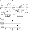
Mapping of host range determinants on human HepG2 cells. Monolayers of HepG2 cells were infected at an MOI of 5, and virus titers at indicated time points were determined via plaque assay with murine DBT cells. (A and B) Representative growth curves reported as PFU per milliliter. Data points are identified by the key provided on the far right. (C) Average titers of recombinants across multiple growth curves. The black squares indicate the average titers, while the arms signify the maximum and minimum titers recorded per recombinant across a minimum of four growth curves.
FIG. 3.
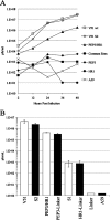
Mapping of host range determinants within the S2 subunit. Monolayers of HepG2 cells were infected at an MOI of 5, and virus titers at indicated time points were determined via plaque assay with murine DBT cells. (A) Representative growth curve dissecting the contributions of substitutions in the PEP3 and HR1 domains on host range. (B) Titers for HepG2 cells at 48-h postinfection, focusing on S2 substitutions in combinations. Infections per recombinant were performed in triplicate, with error bars (standard deviations) indicated.
FIG. 4.
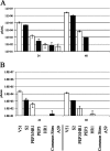
Recombinant virus replication in CHO and MCF7 cells. Monolayers of cells were infected at an MOI of 5 in triplicate, and titers at 24 and 48 h were determined by plaque assay with DBT cells. Infections per recombinant were performed in triplicate, with error bars (standard deviations) indicated. The bar labels at the bottom apply to both panels. (A) Hamster CHO cells. (B) Human MCF7 cells.
FIG. 5.
Immunofluorescence analysis of HepG2 cells. Monolayers of HepG2 cells infected at an MOI of 5 were fixed and stained with anti-N protein monoclonal antibody. Photos are representative fields for each recombinant.
FIG. 6.
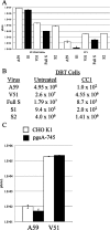
Receptor utilization by recombinant viruses. Monolayers of ST-CEACAM1a, DBT, K1, or pgsA-745 cells were infected at an MOI of 5, and at 24 h postinfection, titers were determined via plaque assay on DBT cells. Infections were performed in triplicate, with error bars (standard deviations) indicated. (A) ST-CEACAM1a cells were untreated or pretreated with blocking antibody CC1. (B) DBT cells were untreated or pretreated with CC1. (C) HS-expressing CHO cells or nonexpressing pgsA-745 cells were infected with A59 or V51.
Similar articles
- Cooperative involvement of the S1 and S2 subunits of the murine coronavirus spike protein in receptor binding and extended host range.
de Haan CA, Te Lintelo E, Li Z, Raaben M, Wurdinger T, Bosch BJ, Rottier PJ. de Haan CA, et al. J Virol. 2006 Nov;80(22):10909-18. doi: 10.1128/JVI.00950-06. Epub 2006 Sep 6. J Virol. 2006. PMID: 16956938 Free PMC article. - The N-terminal region of the murine coronavirus spike glycoprotein is associated with the extended host range of viruses from persistently infected murine cells.
Schickli JH, Thackray LB, Sawicki SG, Holmes KV. Schickli JH, et al. J Virol. 2004 Sep;78(17):9073-83. doi: 10.1128/JVI.78.17.9073-9083.2004. J Virol. 2004. PMID: 15308703 Free PMC article. - Amino acid substitutions and an insertion in the spike glycoprotein extend the host range of the murine coronavirus MHV-A59.
Thackray LB, Holmes KV. Thackray LB, et al. Virology. 2004 Jul 1;324(2):510-24. doi: 10.1016/j.virol.2004.04.005. Virology. 2004. PMID: 15207636 Free PMC article. - The spike glycoprotein of murine coronavirus MHV-JHM mediates receptor-independent infection and spread in the central nervous systems of Ceacam1a-/- Mice.
Miura TA, Travanty EA, Oko L, Bielefeldt-Ohmann H, Weiss SR, Beauchemin N, Holmes KV. Miura TA, et al. J Virol. 2008 Jan;82(2):755-63. doi: 10.1128/JVI.01851-07. Epub 2007 Nov 14. J Virol. 2008. PMID: 18003729 Free PMC article. - A 12-amino acid stretch in the hypervariable region of the spike protein S1 subunit is critical for cell fusion activity of mouse hepatitis virus.
Tsai CW, Chang SC, Chang MF. Tsai CW, et al. J Biol Chem. 1999 Sep 10;274(37):26085-90. doi: 10.1074/jbc.274.37.26085. J Biol Chem. 1999. PMID: 10473557
Cited by
- Identification of the Fusion Peptide-Containing Region in Betacoronavirus Spike Glycoproteins.
Ou X, Zheng W, Shan Y, Mu Z, Dominguez SR, Holmes KV, Qian Z. Ou X, et al. J Virol. 2016 May 27;90(12):5586-5600. doi: 10.1128/JVI.00015-16. Print 2016 Jun 15. J Virol. 2016. PMID: 27030273 Free PMC article. - Evolutionary Arms Race between Virus and Host Drives Genetic Diversity in Bat Severe Acute Respiratory Syndrome-Related Coronavirus Spike Genes.
Guo H, Hu BJ, Yang XL, Zeng LP, Li B, Ouyang S, Shi ZL. Guo H, et al. J Virol. 2020 Sep 29;94(20):e00902-20. doi: 10.1128/JVI.00902-20. Print 2020 Sep 29. J Virol. 2020. PMID: 32699095 Free PMC article. - Coronavirus Spike Protein and Tropism Changes.
Hulswit RJ, de Haan CA, Bosch BJ. Hulswit RJ, et al. Adv Virus Res. 2016;96:29-57. doi: 10.1016/bs.aivir.2016.08.004. Epub 2016 Sep 13. Adv Virus Res. 2016. PMID: 27712627 Free PMC article. Review. - First detection of bovine coronavirus in Yak (Bos grunniens) and a bovine coronavirus genome with a recombinant HE gene.
He Q, Guo Z, Zhang B, Yue H, Tang C. He Q, et al. J Gen Virol. 2019 May;100(5):793-803. doi: 10.1099/jgv.0.001254. Epub 2019 Apr 1. J Gen Virol. 2019. PMID: 30932810 Free PMC article. - Contributions of the S2 spike ectodomain to attachment and host range of infectious bronchitis virus.
Promkuntod N, Wickramasinghe IN, de Vrieze G, Gröne A, Verheije MH. Promkuntod N, et al. Virus Res. 2013 Nov 6;177(2):127-37. doi: 10.1016/j.virusres.2013.09.006. Epub 2013 Sep 13. Virus Res. 2013. PMID: 24041648 Free PMC article.
References
- Beauchemin, N., T. Chen, G. Draber, G. Dveksler, P. Gold, S. Gray-Owen, F. Grunert, S. Hammarstrom, K. V. Holmes, A. Karlson, M. Kuroki, S. H. Lin, L. Lucka, S. M. Najjar, M. Neumaier, B. Obrink, J. E. Shively, K. M. Skubitz, C. P. Stanners, P. Thomas, J. A. Thompson, M. Virji, S. von Kleist, C. Wagener, S. Watt, and W. Zimmerman. 1999. Redefined nomenclature for members of the carcinoembryonic antigen family. Exp. Cell Res. 252243-249. - PubMed
Publication types
MeSH terms
Substances
LinkOut - more resources
Full Text Sources
