Fibrinogen-like protein 1, a hepatocyte derived protein is an acute phase reactant - PubMed (original) (raw)
Fibrinogen-like protein 1, a hepatocyte derived protein is an acute phase reactant
Zhilin Liu et al. Biochem Biophys Res Commun. 2008.
Abstract
Fibrinogen-like protein 1 (FGL1) is a hepatocyte derived protein that is upregulated in regenerating rodent livers following partial hepatectomy. It has been implicated as a mitogen for liver cell proliferation. In this study, we show that recombinant human IL-6 induces FGL1 expression in Hep G2 cells in a pattern similar to those of acute phase reactants. Following induction of acute inflammation in rats by subcutaneous injection of turpentine oil, serum FGL1 levels are also enhanced. Although, a recent report suggests that FGL1 associates almost exclusively with the fibrin matrix, we report here that approximately 20% of the total plasma FGL1 remains free. The enhancement of FGL1 levels in vitro by IL-6 and its induction after turpentine oil injection suggest that it is an acute phase reactant. Its presence in bound and free forms in the blood also implies biological roles that extend beyond the proposed autocrine effect it has on hepatocytes during regeneration.
Figures
Figure 1
IL-6 induces expression of FGL1 in Hep G2 cells: A) Relative mRNA level of FGL1 in Hep G2 cells treated with 50 ng/ml BSA (control), 50 ng/ml of rhIL-6 (IL-6), 1 μM dexamethasone (Dex) and rhIL-6 in the presence of 1 μM dexamethasone for 48 hours. IL-6 treatment results in a 3 fold increase in FGL1 message (compare bar 2 to 1). Dexamethasone alone results in small but reproducible increase in FGL1 levels (bar 3) but augments the effects of IL-6 (compare bar 4 to 2). mRNA levels were normalized to GAPDH. B) Concentration dependence of IL-6 effect on Hep G2 cells. Dose response of Hep G2 cells treated with 0 ng/ml, 5 ng/ml, 15 ng/ml, 30 ng/ml and 50 ng/ml of IL-6 shows that concentration of 5ng/ml result in maximal induction of FGL1 mRNA. Samples were normalized to GAPDH. C) Representative dose response of Hep G2 cells treated with IL-6 at concentrations of 1 to 104 pg/ml. Cells treated with equivalent amounts of BSA were used as control. Fold change in mRNA was determined by dividing the relative level of FGL1 mRNA in IL-6 treated cells by that of FGL1 mRNA in BSA treated cells. FGL1 levels begin to rise 100 pg/ml (graph) and are more profound as expected at 10 ng/ml. β-actin mRNA level was used for normalization. (Bottom, panel) Western blots of cell culture supernatant shows the presence of accumulation of FGL1 protein with increasing concentrations of IL-6 (top). There is no effect of BSA addition on FGl1 protein level (bottom). D) Time dependent effect of IL-6 on FGL1 levels. Representative experiment shows the quantitation of FGL1 mRNA and protein levels following treatment with 10 ng/ml of IL-6. mRNA induction begins at 12 hours and peaks 48 hours after treatment (graph), FGl1 protein accumulates in the culture media as a function of time (bottom, gel). mRNA levels were normalized to GAPDH.
Figure 2
FGL1 is an acute phase reactant. A) mRNA levels of SAA1 (top), FGA (middle) and FGL1 (bottom), 24 hours after treatment with varying doses of IL-6. The pattern of induction is similar for all three genes, with increase in mRNA notable beginning at 100 ng/ml. B) Fold inductions of all three genes at 24 and 48 hours shows that SAA1 levels are at least an order of magnitude higher than those of FGA and FGL1. At 48 hours SAA1 is induced 180 fold compared with 14 fold for FGA and 10 fold for FGL1. mRNA levels were normalized to GAPDH.
Figure 3
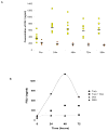
Subcutaneous Turpentine oil injection results in enhancement of serum levels of FGL1 in rats. ELISA assays of FGL1 serum levels at various time points following the subcutaneous injection of turpentine oil (circles) or PBS (triangles). Stimulation of inflammation at an extrahepatic site results in enhanced levels of FGL1. Horizontal bars represent the mean B) Representative experiment on the effect of dexamethasone on turpentine induced FGL1 expression. Injection of dexamethasone dampens effect of turpentine. Compare squares to circles.
Figure 4
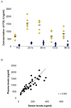
FGL1 is present in the plasma and serum. A) Detection of FGL1 levels in the plasma (circles) and serum (triangles) show that approximately 20% of FGL1 remains in the serum after blood coagulation. The horizontal bars represent the mean. A plot of plasma FGL1 against serum FGL1 shows a near linear correlation with a co-efficient of 0.84. Approximately 20% of FGL1 is in the serum at all times.
Similar articles
- FGL1 and FGL2: emerging regulators of liver health and disease.
Chen J, Wu L, Li Y. Chen J, et al. Biomark Res. 2024 May 31;12(1):53. doi: 10.1186/s40364-024-00601-0. Biomark Res. 2024. PMID: 38816776 Free PMC article. Review. - Fibrinogen-Like Protein 1 Modulates Sorafenib Resistance in Human Hepatocellular Carcinoma Cells.
Son Y, Shin NR, Kim SH, Park SC, Lee HJ. Son Y, et al. Int J Mol Sci. 2021 May 19;22(10):5330. doi: 10.3390/ijms22105330. Int J Mol Sci. 2021. PMID: 34069373 Free PMC article. - CD14 is an acute-phase protein.
Bas S, Gauthier BR, Spenato U, Stingelin S, Gabay C. Bas S, et al. J Immunol. 2004 Apr 1;172(7):4470-9. doi: 10.4049/jimmunol.172.7.4470. J Immunol. 2004. PMID: 15034063 - Targeted disruption of fibrinogen like protein-1 accelerates hepatocellular carcinoma development.
Nayeb-Hashemi H, Desai A, Demchev V, Bronson RT, Hornick JL, Cohen DE, Ukomadu C. Nayeb-Hashemi H, et al. Biochem Biophys Res Commun. 2015 Sep 18;465(2):167-73. doi: 10.1016/j.bbrc.2015.07.078. Epub 2015 Jul 28. Biochem Biophys Res Commun. 2015. PMID: 26225745 Free PMC article. - Role of Fibrinogen-like Protein 1 in Tumor Recurrence Following Hepatectomy.
Shafieizadeh Z, Shafieizadeh Z, Davoudi M, Afrisham R, Miao X. Shafieizadeh Z, et al. J Clin Transl Hepatol. 2024 Apr 28;12(4):406-415. doi: 10.14218/JCTH.2023.00397. Epub 2024 Mar 25. J Clin Transl Hepatol. 2024. PMID: 38638375 Free PMC article. Review.
Cited by
- Perspectives on immune checkpoint ligands: expression, regulation, and clinical implications.
Moon J, Oh YM, Ha SJ. Moon J, et al. BMB Rep. 2021 Aug;54(8):403-412. doi: 10.5483/BMBRep.2021.54.8.054. BMB Rep. 2021. PMID: 34078531 Free PMC article. Review. - Fibrinogen-like Protein 1 as a Predictive Marker for the Incidence of Severe Acute Pancreatitis and Infectious Pancreatic Necrosis.
Sui Y, Zhao Z, Zhang Y, Zhang T, Li G, Liu L, Tan H, Sun B, Li L. Sui Y, et al. Medicina (Kaunas). 2022 Nov 29;58(12):1753. doi: 10.3390/medicina58121753. Medicina (Kaunas). 2022. PMID: 36556955 Free PMC article. - FGL1 and FGL2: emerging regulators of liver health and disease.
Chen J, Wu L, Li Y. Chen J, et al. Biomark Res. 2024 May 31;12(1):53. doi: 10.1186/s40364-024-00601-0. Biomark Res. 2024. PMID: 38816776 Free PMC article. Review. - The FGL-1/LAG-3 Axis is Associated With Disease Course in Alcohol-associated Hepatitis: A Preliminary Report.
Pedersen L, Eriksen LL, Brix FH, Vilstrup H, Deleuran B, Sandahl TD, Støy S. Pedersen L, et al. J Clin Exp Hepatol. 2025 Jan-Feb;15(1):102424. doi: 10.1016/j.jceh.2024.102424. Epub 2024 Oct 10. J Clin Exp Hepatol. 2025. PMID: 39553834 - Protective effect of mesenchymal stem cell-conditioned medium on hepatic cell apoptosis after acute liver injury.
Xagorari A, Siotou E, Yiangou M, Tsolaki E, Bougiouklis D, Sakkas L, Fassas A, Anagnostopoulos A. Xagorari A, et al. Int J Clin Exp Pathol. 2013 Apr 15;6(5):831-40. Print 2013. Int J Clin Exp Pathol. 2013. PMID: 23638214 Free PMC article. Retracted.
References
- Kushner I. The acute phase response: an overview. Methods Enzymol. 1988;163:373–83. - PubMed
- Koj A. Biological functions of acute-phase proteins. In: Gordon AH, Koj A, editors. The acute phase response to injury and infection. Elsheiver Science; New York: 1985. pp. 145–160.
- Gertz MA, Kyle RA. Secondary systemic amyloidosis: response and survival in 64 patients. Medicine (Baltimore) 1991;70:246–56. - PubMed
- Nemeth E, Valore EV, Territo M, Schiller G, Lichtenstein A, Ganz T. Hepcidin, a putative mediator of anemia of inflammation, is a type II acute-phase protein. Blood. 2003;101:2461–3. - PubMed
Publication types
MeSH terms
Substances
LinkOut - more resources
Full Text Sources
Other Literature Sources
Molecular Biology Databases
Research Materials
Miscellaneous


