The Pfam protein families database - PubMed (original) (raw)
. 2008 Jan;36(Database issue):D281-8.
doi: 10.1093/nar/gkm960. Epub 2007 Nov 26.
Affiliations
- PMID: 18039703
- PMCID: PMC2238907
- DOI: 10.1093/nar/gkm960
The Pfam protein families database
Robert D Finn et al. Nucleic Acids Res. 2008 Jan.
Abstract
Pfam is a comprehensive collection of protein domains and families, represented as multiple sequence alignments and as profile hidden Markov models. The current release of Pfam (22.0) contains 9318 protein families. Pfam is now based not only on the UniProtKB sequence database, but also on NCBI GenPept and on sequences from selected metagenomics projects. Pfam is available on the web from the consortium members using a new, consistent and improved website design in the UK (http://pfam.sanger.ac.uk/), the USA (http://pfam.janelia.org/) and Sweden (http://pfam.sbc.su.se/), as well as from mirror sites in France (http://pfam.jouy.inra.fr/) and South Korea (http://pfam.ccbb.re.kr/).
Figures
Figure 1.
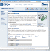
The Pfam family page from the new website. This page shows the summary information for the Piwi domain. The tabs on the left allow users to browse the different types of associated information and beneath the tabs is the ‘jump to’ box, a tool which can direct the user to the page for any other entry in the site, given any type of accession or identifier. The panel at the top right gives a summary of the number of protein architectures, sequences, interactions, species and structures available. The same page layout and navigational tools are common to all of the different types of data in the Pfam website.
Figure 2.
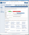
The Pfam protein sequence page, showing the DAS annotation viewer. Various tracks can be selected using the check boxes beneath the annotation view, allowing the user to view features derived from any of the listed DAS sources. For instance, in the example displayed, the membrane topology calculated by Phobius (18) can be viewed alongside the Pfam domain annotations and those from a variety of different domain databases. The figure shows a tool-tip for one feature, which gives the details of the annotation and, in this case, highlights the link to further information.
Figure 3.
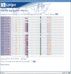
The DAS-based Pfam sequence alignment viewer. A portion of the Piwi domain full alignment is shown with residue conservation highlighted in a ClustalX (19) style colour scheme.
Similar articles
- Pfam: clans, web tools and services.
Finn RD, Mistry J, Schuster-Böckler B, Griffiths-Jones S, Hollich V, Lassmann T, Moxon S, Marshall M, Khanna A, Durbin R, Eddy SR, Sonnhammer EL, Bateman A. Finn RD, et al. Nucleic Acids Res. 2006 Jan 1;34(Database issue):D247-51. doi: 10.1093/nar/gkj149. Nucleic Acids Res. 2006. PMID: 16381856 Free PMC article. - The Pfam protein families database.
Bateman A, Birney E, Cerruti L, Durbin R, Etwiller L, Eddy SR, Griffiths-Jones S, Howe KL, Marshall M, Sonnhammer EL. Bateman A, et al. Nucleic Acids Res. 2002 Jan 1;30(1):276-80. doi: 10.1093/nar/30.1.276. Nucleic Acids Res. 2002. PMID: 11752314 Free PMC article. - The Pfam protein families database.
Bateman A, Coin L, Durbin R, Finn RD, Hollich V, Griffiths-Jones S, Khanna A, Marshall M, Moxon S, Sonnhammer EL, Studholme DJ, Yeats C, Eddy SR. Bateman A, et al. Nucleic Acids Res. 2004 Jan 1;32(Database issue):D138-41. doi: 10.1093/nar/gkh121. Nucleic Acids Res. 2004. PMID: 14681378 Free PMC article. - The Pfam protein families database.
Punta M, Coggill PC, Eberhardt RY, Mistry J, Tate J, Boursnell C, Pang N, Forslund K, Ceric G, Clements J, Heger A, Holm L, Sonnhammer EL, Eddy SR, Bateman A, Finn RD. Punta M, et al. Nucleic Acids Res. 2012 Jan;40(Database issue):D290-301. doi: 10.1093/nar/gkr1065. Epub 2011 Nov 29. Nucleic Acids Res. 2012. PMID: 22127870 Free PMC article. - Pfam 10 years on: 10,000 families and still growing.
Sammut SJ, Finn RD, Bateman A. Sammut SJ, et al. Brief Bioinform. 2008 May;9(3):210-9. doi: 10.1093/bib/bbn010. Epub 2008 Mar 15. Brief Bioinform. 2008. PMID: 18344544 Review.
Cited by
- MYB transcription factors, their regulation and interactions with non-coding RNAs during drought stress in Brassica juncea.
Balhara R, Verma D, Kaur R, Singh K. Balhara R, et al. BMC Plant Biol. 2024 Oct 24;24(1):999. doi: 10.1186/s12870-024-05736-8. BMC Plant Biol. 2024. PMID: 39448923 Free PMC article. - Comparative genomic analyses of the genus Robertmurraya and proposal of the novel species Robertmurraya mangrovi sp. nov., isolated from mangrove soil.
Li M, Hu X, Ni T, Ni Y, Xue D, Li F. Li M, et al. Antonie Van Leeuwenhoek. 2024 Oct 23;118(1):22. doi: 10.1007/s10482-024-02032-1. Antonie Van Leeuwenhoek. 2024. PMID: 39441363 - Telomere-to-Telomere Haplotype-Resolved Genomes of Agrocybe chaxingu Reveals Unique Genetic Features and Developmental Insights.
Chen X, Wei Y, Meng G, Wang M, Peng X, Dai J, Dong C, Huo G. Chen X, et al. J Fungi (Basel). 2024 Aug 25;10(9):602. doi: 10.3390/jof10090602. J Fungi (Basel). 2024. PMID: 39330362 Free PMC article. - Chromosome-level genome assembly of cotton thrips Thrips tabaci (Thysanoptera: Thripidae).
Gao Y, Ji J, Xu C, Wang L, Zhang K, Li D, Wang X, Xin M, Hua H, Chen L, Gao X, Zhu X, Cui J, Luo J. Gao Y, et al. Sci Data. 2024 Sep 16;11(1):1003. doi: 10.1038/s41597-024-03737-8. Sci Data. 2024. PMID: 39294155 Free PMC article. - Creating and leveraging bespoke large-scale knowledge graphs for comparative genomics and multi-omics drug discovery with SocialGene.
Clark CM, Kwan JC. Clark CM, et al. bioRxiv [Preprint]. 2024 Aug 19:2024.08.16.608329. doi: 10.1101/2024.08.16.608329. bioRxiv. 2024. PMID: 39229008 Free PMC article. Preprint.
References
- Sonnhammer ELL, Eddy SR, Durbin R. Pfam: a comprehensive database of protein domain families based on seed alignments. Proteins. 1997;28:405–420. - PubMed
- Bairoch A, Apweiler R, Wu CH, Barker WC, Boeckmann B, Ferro S, Gasteiger E, Huang H, Lopez R, et al. The Universal Protein Resource (UniProt) Nucleic Acids Res. 2007;35:D193–D197. - PubMed
Publication types
MeSH terms
Substances
Grants and funding
- WT_/Wellcome Trust/United Kingdom
- 087656/WT_/Wellcome Trust/United Kingdom
- BB/F010435/1/BB_/Biotechnology and Biological Sciences Research Council/United Kingdom
- G0100305/MRC_/Medical Research Council/United Kingdom
LinkOut - more resources
Full Text Sources
Other Literature Sources


