The all-trans-retinal dimer series of lipofuscin pigments in retinal pigment epithelial cells in a recessive Stargardt disease model - PubMed (original) (raw)
The all-trans-retinal dimer series of lipofuscin pigments in retinal pigment epithelial cells in a recessive Stargardt disease model
So R Kim et al. Proc Natl Acad Sci U S A. 2007.
Abstract
The bis-retinoid pigments that accumulate in retinal pigment epithelial cells as lipofuscin are associated with inherited and age-related retinal disease. In addition to A2E and related cis isomers, we previously showed that condensation of two molecules of all-trans-retinal leads to the formation of a protonated Schiff base conjugate, all-trans-retinal dimer-phosphatidylethanolamine. Here we report the characterization of the related pigments, all-trans-retinal dimer-ethanolamine and unconjugated all-trans-retinal dimer, in human and mouse retinal pigment epithelium. In eyecups of Abcr(-/-) mice, a model of recessive Stargardt macular degeneration, all-trans-retinal dimer-phosphatidylethanolamine was increased relative to wild type and was more abundant than A2E. Total pigment of the all-trans-retinal dimer series (sum of all-trans-retinal dimer-phosphatidylethanolamine, all-trans-retinal dimer-ethanolamine, and all-trans-retinal dimer) increased with age in Abcr(-/-) mice and was modulated by amino acid variants in Rpe65. In in vitro assays, enzyme-mediated hydrolysis of all-trans-retinal dimer-phosphatidylethanolamine generated all-trans-retinal dimer-ethanolamine, and protonation/deprotonation of the Schiff base nitrogen of all-trans-retinal dimer-ethanolamine was pH-dependent. Unconjugated all-trans-retinal dimer was a more efficient generator of singlet oxygen than A2E, and the all-trans-retinal dimer series was more reactive with singlet oxygen than was A2E. By analyzing chromatographic properties and UV-visible spectra together with mass spectrometry, mono- and bis-oxygenated all-trans-retinal dimer photoproducts were detected in Abcr(-/-) mice. The latter findings are significant to an understanding of the adverse effects of retinal pigment epithelial cell lipofuscin.
Conflict of interest statement
The authors declare no conflict of interest.
Figures
Fig. 1.
Structures of the atRAL dimer series of RPE lipofuscin pigments, atRAL dimer, atRAL dimer-E, and atRAL dimer-PE.
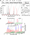
Fig. 2.
Two- and three-dimensional display of HPLC chromatograms demonstrating the detection of atRAL-derived lipofuscin chromophores in extracts of eyecups from _Abcr_−/− mice. Mice were homozygous for Rpe65 Leu-450 (11 months old; pooled sample of eight eyecups). (A Left Lower) HPLC chromatograms obtained when injectant was eyecup extract (black) and a mixture of three standards (red): atRAL dimer-phosphatidylethanolamine, atRAL dimer-E, and unconjugated atRAL dimer. C18 column, 430-nm monitoring, and a gradient of acetonitrile and water (acetonitrile:water = 85:15 → 100:0 for 15 min; 0.8 ml/min) with 0.1% of TFA was used for mobile phase. (A Left Upper) UV-visible absorbance spectra corresponding to A2E, isoA2E, atRALdi-E, atRALdi-PE, and atRAL dimer (atRALdi). (A Right) Monitoring at 510 nm with chromatogram expanded between retention times of 14–18 min (A Right Upper) and monitoring at 510 nm with chromatogram expanded between 14 and 28 min (A Right Lower). (B) Three-dimensional display of retention time, wavelength (270–600 nm), and photodiode array absorbance data obtained by HPLC for the chromatogram in A.
Fig. 3.
HPLC quantitation of RPE lipofuscin pigments in eyecups of _Abcr_−/− and Abcr+/+ mice and in _Abcr_−/− mice genotyped for Rpe65 Leu450Met. Levels of A2E and isoA2E (measured separately and summed, A2E/isoA2E), atRALdi-PE, and total atRAL dimer series (atRAL dimer-phosphatidylethanolamine, atRAL dimer-E, and atRAL dimer measured separately and summed) plotted as a function of age. Detection was at 430 nm (A2E, atRALdi) or 500 nm (atRAL di-PE, atRALdi-E). Mice were homozygous for Rpe65 Leu-450 (L/L) or Rpe65 Met-450 (M/M). Each data point is from a single sample, with two to six eyes per sample.
Fig. 4.
Singlet oxygen generation and quenching. (A and B) Phosphorescence of singlet oxygen generated from A2E, atRALdi-E, and atRALdi. (A) A2E, atRALdi, and atRALdi-E (50 μM in CCl4) were irradiated at 430 nm, and the generation of singlet oxygen was detected by its phosphorescence in the near infrared region. atRAL di-E was irradiated in the presence of TFA (atRALdi-E + TFA) or the absence of TFA (atRALdi-E) to test for the effect of protonation of the Schiff base nitrogen on the singlet oxygen yield. (B) Effect of protonation of atRALdi-E and illumination wavelength on the generation of singlet oxygen. atRALdi-E was excited at either 430 nm or 500 nm in the presence or absence of TFA. (C and D) Comparison of tendency of A2E and atRAL dimer-E (atRALdi-E) to quench singlet oxygen. A2E (C) and atRALdi-E (D) were oxidized by singlet oxygen generated from the endoperoxide of 1,4-dimethylnaphthalene. FAB-MS spectra are shown. A2E and atRALdi-E are detected as molecular ion peaks at m/z ratios of 592 and 594, respectively.
Fig. 5.
Detection of photooxidized products of the RPE lipofuscin pigment atRAL dimer (atRALdi) in extracts of _Abcr_−/− mouse eyecups. (Lower) Chromatographic overlay of HPLC profiles generated with samples of atRALdi plus endoperoxide of 1,4-dimethylnaphthalene (red), atRALdi plus MCPBA (blue), or extract of _Abcr_−/− posterior eyecups (Rpe65 Leu-450; age, 12 months). (Upper) UV-visible absorbance of atRALdi and oxidized products of atRALdi. bP-atRALdi, bisperoxy-atRALdi; bF-atRALdi, bisfurano-atRALdi; mP-atRALdi, monoperoxy-atRALdi; mF-atRALdi, monofurano-atRALdi. a, _cis_-isomers of A2E; b, atRAL dimer-E; c, atRAL dimer-PE.
Similar articles
- Novel lipofuscin bisretinoids prominent in human retina and in a model of recessive Stargardt disease.
Wu Y, Fishkin NE, Pande A, Pande J, Sparrow JR. Wu Y, et al. J Biol Chem. 2009 Jul 24;284(30):20155-66. doi: 10.1074/jbc.M109.021345. Epub 2009 May 28. J Biol Chem. 2009. PMID: 19478335 Free PMC article. - Biosynthesis of a major lipofuscin fluorophore in mice and humans with ABCR-mediated retinal and macular degeneration.
Mata NL, Weng J, Travis GH. Mata NL, et al. Proc Natl Acad Sci U S A. 2000 Jun 20;97(13):7154-9. doi: 10.1073/pnas.130110497. Proc Natl Acad Sci U S A. 2000. PMID: 10852960 Free PMC article. - Treatment with isotretinoin inhibits lipofuscin accumulation in a mouse model of recessive Stargardt's macular degeneration.
Radu RA, Mata NL, Nusinowitz S, Liu X, Sieving PA, Travis GH. Radu RA, et al. Proc Natl Acad Sci U S A. 2003 Apr 15;100(8):4742-7. doi: 10.1073/pnas.0737855100. Epub 2003 Apr 1. Proc Natl Acad Sci U S A. 2003. PMID: 12671074 Free PMC article. - A2E, a byproduct of the visual cycle.
Sparrow JR, Fishkin N, Zhou J, Cai B, Jang YP, Krane S, Itagaki Y, Nakanishi K. Sparrow JR, et al. Vision Res. 2003 Dec;43(28):2983-90. doi: 10.1016/s0042-6989(03)00475-9. Vision Res. 2003. PMID: 14611934 Review. - Lipofuscin and macular degeneration.
Wolf G. Wolf G. Nutr Rev. 2003 Oct;61(10):342-6. doi: 10.1301/nr.2003.oct.342-346. Nutr Rev. 2003. PMID: 14604266 Review.
Cited by
- Primary versus Secondary Elevations in Fundus Autofluorescence.
Parmann R, Tsang SH, Sparrow JR. Parmann R, et al. Int J Mol Sci. 2023 Aug 2;24(15):12327. doi: 10.3390/ijms241512327. Int J Mol Sci. 2023. PMID: 37569703 Free PMC article. Review. - The role of the photoreceptor ABC transporter ABCA4 in lipid transport and Stargardt macular degeneration.
Molday RS, Zhong M, Quazi F. Molday RS, et al. Biochim Biophys Acta. 2009 Jul;1791(7):573-83. doi: 10.1016/j.bbalip.2009.02.004. Epub 2009 Feb 20. Biochim Biophys Acta. 2009. PMID: 19230850 Free PMC article. Review. - ATP-binding cassette transporter ABCA4 and chemical isomerization protect photoreceptor cells from the toxic accumulation of excess 11-cis-retinal.
Quazi F, Molday RS. Quazi F, et al. Proc Natl Acad Sci U S A. 2014 Apr 1;111(13):5024-9. doi: 10.1073/pnas.1400780111. Epub 2014 Mar 20. Proc Natl Acad Sci U S A. 2014. PMID: 24707049 Free PMC article. - Retinol dehydrogenase 8 and ATP-binding cassette transporter 4 modulate dark adaptation of M-cones in mammalian retina.
Kolesnikov AV, Maeda A, Tang PH, Imanishi Y, Palczewski K, Kefalov VJ. Kolesnikov AV, et al. J Physiol. 2015 Nov 15;593(22):4923-41. doi: 10.1113/JP271285. Epub 2015 Oct 18. J Physiol. 2015. PMID: 26350353 Free PMC article. - Complement dysregulation in AMD: RPE-Bruch's membrane-choroid.
Sparrow JR, Ueda K, Zhou J. Sparrow JR, et al. Mol Aspects Med. 2012 Aug;33(4):436-45. doi: 10.1016/j.mam.2012.03.007. Epub 2012 Apr 5. Mol Aspects Med. 2012. PMID: 22504022 Free PMC article. Review.
References
- Eldred GE. In: The Retinal Pigment Epithelium: Function and Disease. Marmor MF, Wolfensberger TJ, editors. New York: Oxford Univ Press; 1998. pp. 651–668.
- Radu RA, Han Y, Bui TV, Nusinowitz S, Bok D, Lichter J, Widder K, Travis GH, Mata NL. Invest Ophthalmol Vis Sci. 2005;46:4393–4401. - PubMed
- Maiti P, Kong J, Kim SR, Sparrow JR, Allikmets R, Rando RR. Biochemistry. 2006;45:852–860. - PubMed
- Sakai N, Decatur J, Nakanishi K, Eldred GE. J Am Chem Soc. 1996;118:1559–1560.
Publication types
MeSH terms
Substances
LinkOut - more resources
Full Text Sources
Other Literature Sources
Medical
Molecular Biology Databases
Miscellaneous


