Neural activations at the junction of the inferior frontal sulcus and the inferior precentral sulcus: interindividual variability, reliability, and association with sulcal morphology - PubMed (original) (raw)
Neural activations at the junction of the inferior frontal sulcus and the inferior precentral sulcus: interindividual variability, reliability, and association with sulcal morphology
Jan Derrfuss et al. Hum Brain Mapp. 2009 Jan.
Abstract
The sulcal morphology of the human frontal lobe is highly variable. Although the structural images usually acquired in functional magnetic resonance imaging studies provide information about this interindividual variability, this information is only rarely used to relate structure and function. Here, we investigated the spatial relationship between posterior frontolateral activations in a task-switching paradigm and the junction of the inferior frontal sulcus and the inferior precentral sulcus (inferior frontal junction, IFJ) on an individual-subject basis. Results show that, although variable in terms of stereotaxic coordinates, the posterior frontolateral activations observed in task-switching are consistently and reliably located at the IFJ in the brains of individual participants. The IFJ shares such consistent localization with other nonprimary areas as motion-sensitive area V5/MT and the frontal eye field. Building on tension-based models of morphogenesis, this structure-function correspondence might indicate that the cytoarchitectonic area underlying activations of the IFJ develops at early stages of cortical folding.
(c) 2007 Wiley-Liss, Inc.
Figures
Figure 1
Left frontolateral views of white‐matter segmentations of six subjects with functional imaging maps overlaid in red. Relevant sulci are highlighted by triangles. Note the consistent activation at the junction of the inferior precentral sulcus and the inferior frontal sulcus.
Figure 2
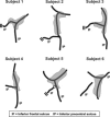
Schematic views of IFJ activations. IFJ activations are shown in grey; black lines indicate the fundi of the inferior precentral and inferior frontal sulci. Note: Double lines indicate truncations of the inferior frontal sulcus. What is marked as inferior frontal sulcus in Subject 4 would be considered as a part of the inferior precentral sulcus following the scheme of Germann et al. [2005]. The circle at the junction of the inferior precentral sulcus and the inferior frontal sulcus in Subject 5 denotes a clear in‐depth separation of these sulci.
Figure 3
Sagittal and axial slices through the IFJ peak for six individuals. The activation peak is marked by a yellow square, the volume of activation by a red outline. On the right, time‐course analyses for the activation peaks are shown.
Figure 4
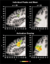
Upper panel: IFJ peak activations of all individuals (yellow squares) overlaid onto an individual brain in Talairach space. Anatomical slices were chosen to correspond to the mean peak location (orange square). Lower panel: Overlap of IFJ activation volumes. The color bar denotes the number of z maps overlapping at a given voxel. There was a maximum overlap of 5 z maps.
Figure 5
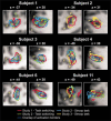
IFJ activations for the six subjects who participated in multiple studies. Volumes of IFJ activations are indicated by colored outlines and are overlaid onto the individual anatomies. For each subject, on the left a sagittal slice is shown and on the right an axial slice. The sulcus abbreviations are explained in Figure 3.
Similar articles
- Involvement of the inferior frontal junction in cognitive control: meta-analyses of switching and Stroop studies.
Derrfuss J, Brass M, Neumann J, von Cramon DY. Derrfuss J, et al. Hum Brain Mapp. 2005 May;25(1):22-34. doi: 10.1002/hbm.20127. Hum Brain Mapp. 2005. PMID: 15846824 Free PMC article. - Functional organization of the left inferior precentral sulcus: dissociating the inferior frontal eye field and the inferior frontal junction.
Derrfuss J, Vogt VL, Fiebach CJ, von Cramon DY, Tittgemeyer M. Derrfuss J, et al. Neuroimage. 2012 Feb 15;59(4):3829-37. doi: 10.1016/j.neuroimage.2011.11.051. Epub 2011 Dec 1. Neuroimage. 2012. PMID: 22155041 - Cognitive control in the posterior frontolateral cortex: evidence from common activations in task coordination, interference control, and working memory.
Derrfuss J, Brass M, von Cramon DY. Derrfuss J, et al. Neuroimage. 2004 Oct;23(2):604-12. doi: 10.1016/j.neuroimage.2004.06.007. Neuroimage. 2004. PMID: 15488410 Clinical Trial. - Three-dimensional cytoarchitectonic analysis of the posterior bank of the human precentral sulcus.
Schmitt O, Modersitzki J, Heldmann S, Wirtz S, Hömke L, Heide W, Kömpf D, Wree A. Schmitt O, et al. Anat Embryol (Berl). 2005 Dec;210(5-6):387-400. doi: 10.1007/s00429-005-0030-8. Anat Embryol (Berl). 2005. PMID: 16177908 - Accurate localization and coactivation profiles of the frontal eye field and inferior frontal junction: an ALE and MACM fMRI meta-analysis.
Bedini M, Olivetti E, Avesani P, Baldauf D. Bedini M, et al. Brain Struct Funct. 2023 May;228(3-4):997-1017. doi: 10.1007/s00429-023-02641-y. Epub 2023 Apr 24. Brain Struct Funct. 2023. PMID: 37093304 Free PMC article.
Cited by
- Morphological patterns and spatial probability maps of the inferior frontal sulcus in the human brain.
Nolan E, Loh KK, Petrides M. Nolan E, et al. Hum Brain Mapp. 2024 Jul 15;45(10):e26759. doi: 10.1002/hbm.26759. Hum Brain Mapp. 2024. PMID: 38989632 Free PMC article. - Brain imaging of a gamified cognitive flexibility task in young and older adults.
Wang P, Guo SJ, Li HJ. Wang P, et al. Brain Imaging Behav. 2024 Aug;18(4):902-912. doi: 10.1007/s11682-024-00883-w. Epub 2024 Apr 17. Brain Imaging Behav. 2024. PMID: 38627304 - An MRI Study of Morphology, Asymmetry, and Sex Differences of Inferior Precentral Sulcus.
Zhao X, Wang Y, Wu X, Liu S. Zhao X, et al. Brain Topogr. 2024 Sep;37(5):748-763. doi: 10.1007/s10548-024-01035-5. Epub 2024 Feb 19. Brain Topogr. 2024. PMID: 38374489 Free PMC article. - On the functional brain networks involved in tool-related action understanding.
Federico G, Osiurak F, Ciccarelli G, Ilardi CR, Cavaliere C, Tramontano L, Alfano V, Migliaccio M, Di Cecca A, Salvatore M, Brandimonte MA. Federico G, et al. Commun Biol. 2023 Nov 14;6(1):1163. doi: 10.1038/s42003-023-05518-2. Commun Biol. 2023. PMID: 37964121 Free PMC article. - A revised perspective on the evolution of the lateral frontal cortex in primates.
Amiez C, Sallet J, Giacometti C, Verstraete C, Gandaux C, Morel-Latour V, Meguerditchian A, Hadj-Bouziane F, Ben Hamed S, Hopkins WD, Procyk E, Wilson CRE, Petrides M. Amiez C, et al. Sci Adv. 2023 May 19;9(20):eadf9445. doi: 10.1126/sciadv.adf9445. Epub 2023 May 19. Sci Adv. 2023. PMID: 37205762 Free PMC article.
References
- Aguirre GK,Zarahn E,D'Esposito M( 1997): Empirical analyses of BOLD fMRI statistics. II. Spatially smoothed data collected under null‐hypothesis and experimental conditions. Neuroimage 5(3): 199–212. - PubMed
- Amunts K,von Cramon DY( 2006): The anatomical segregation of the frontal cortex: What does it mean for function? Cortex 42(4): 525–528. - PubMed
- Amunts K,Schleicher A,Burgel U,Mohlberg H,Uylings HB,Zilles K( 1999): Broca's region revisited: Cytoarchitecture and intersubject variability. J Comp Neurol 412(2): 319–341. - PubMed
- Amunts K,Malikovic A,Mohlberg H,Schormann T,Zilles K( 2000): Brodmann's areas 17 and 18 brought into stereotaxic space‐where and how variable? Neuroimage 11(1): 66–84. - PubMed
Publication types
MeSH terms
LinkOut - more resources
Full Text Sources


