Mechanisms of zoonotic severe acute respiratory syndrome coronavirus host range expansion in human airway epithelium - PubMed (original) (raw)
Mechanisms of zoonotic severe acute respiratory syndrome coronavirus host range expansion in human airway epithelium
Timothy Sheahan et al. J Virol. 2008 Mar.
Abstract
In 2003, severe acute respiratory syndrome coronavirus (SARS-CoV) emerged and caused over 8,000 human cases of infection and more than 700 deaths worldwide. Zoonotic SARS-CoV likely evolved to infect humans by a series of transmission events between humans and animals for sale in China. Using synthetic biology, we engineered the spike protein (S) from a civet strain, SZ16, into our epidemic strain infectious clone, creating the chimeric virus icSZ16-S, which was infectious but yielded progeny viruses incapable of propagating in vitro. After introducing a K479N mutation within the S receptor binding domain (RBD) of SZ16, the recombinant virus (icSZ16-S K479N) replicated in Vero cells but was severely debilitated in growth. The in vitro evolution of icSZ16-S K479N on human airway epithelial (HAE) cells produced two viruses (icSZ16-S K479N D8 and D22) with enhanced growth on HAE cells and on delayed brain tumor cells expressing the SARS-CoV receptor, human angiotensin I converting enzyme 2 (hACE2). The icSZ16-S K479N D8 and D22 virus RBDs contained mutations in ACE2 contact residues, Y442F and L472F, that remodeled S interactions with hACE2. Further, these viruses were neutralized by a human monoclonal antibody (MAb), S230.15, but the parent icSZ16-S K479N strain was eight times more resistant than the mutants. These data suggest that the human adaptation of zoonotic SARS-CoV strains may select for some variants that are highly susceptible to select MAbs that bind to RBDs. The epidemic, icSZ16-S K479N, and icSZ16-S K479N D22 viruses replicate similarly in the BALB/c mouse lung, highlighting the potential use of these zoonotic spike SARS-CoVs to assess vaccine or serotherapy efficacy in vivo.
Figures
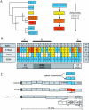
FIG. 1.
Phylogenetic relationships of zoonotic SARS-CoV and construction of zoonotic spike protein chimeras within the SARS-CoV infectious clone. The colors in panels A, B, and C denote the phase of the SARS-CoV epidemic at which the viruses or amino acid residues evolved: blue, animal associated; yellow, early phase; orange, middle phase; and red, late phase. (A) Neighbor joining tree constructed from nucleotide sequences of various SARS-CoV S genes. The numbers represent bootstrap values based on 1,000 bootstrap replicates. (B) Spike protein amino acid differences between Urbani, GD03, and SZ16. Known neutralizing epitopes are denoted A, B, and C. (C) Schematic of infectious clone fragments A to F and the SARS-CoV genes contained therein. Using synthetic biology and site-directed mutagenesis, we reconstructed the SZ16 S that was then inserted into our infectious clone, replacing the epidemic strain S.
FIG. 2.
SARS-CoV SZ16 spike protein chimera icSZ16-S replicates in Vero E6 cells, but infection cannot be passed in culture until a point mutation (K479N) is introduced within the RBD. The expected sizes of the target RT-PCR products are as follows: for 3a, 1,796 bp; E, 949 bp; M, 666 bp; and GAPDH, 235 bp. (A) RT-PCR for the leader containing transcripts detects actively replicating genomic RNA. RT-PCR using RNA extracted from Vero E6 cells transfected with genomic icSZ16-S RNA (passage 0) detects transcripts for the E and M genes of icSZ16-S. (B) The transfer of icSZ16-S supernatants from passage 0 to naive Vero E6 or mink lung epithelial Mv1Lu cells does not result in a productive infection. As a positive control, Vero E6 and Mv1Lu cells were successfully infected with epidemic strain supernatants, as replication was detected by the presence of the leader containing transcripts. (C) In contrast to wild-type icSZ16-S, a point mutation (K479N) in the icSZ16-S spike protein allows for passage of icSZ16-S K479N virus into Vero E6 cells.
FIG. 3.
Growth curve analysis of the mutant virus panel in HAE, Vero E6, DBT-hACE2, and DBT cells. (A) HAE cell cultures were infected with 4.4 × 10⁴ PFU/200 μl of the indicated viruses for 2 h at 37°C. The inoculum was removed, and the apical surfaces were rinsed with DPBS. Apical-surface washes were performed at 0, 6, 12, 24, 36, 48, and 72 hpi. Virus titers were assessed for Vero E6 cells by a standard plaque assay. (B to D) Vero E6, DBT-hACE2, and DBT cells (respectively) were infected with the indicated viruses at an MOI of 0.01 for 1 h at 37°C. The inoculum was removed, cultures were rinsed with DPBS, and growth medium was added. The medium was sampled at 0, 6, 12, 24, and 36 hpi, and virus titer was assessed by plaque assay on Vero E6 cells. The data presented are representative of two separate experiments.
FIG. 4.
Immunofluorescence staining of HAE cell cultures infected with the mutant virus panel. At 72 hpi, mock and infected HAE cell cultures were PFA fixed and paraffin embedded for tissue sectioning. Sections were stained for SARS-CoV N (fluorescein isothiocyanate) and tubulin within cilia (Texas Red) and viewed by fluorescence microscopy. (A) Mock; (B) icSARS; (C) icSZ16-S K479N; (D) icSZ16-S K479N D22.
FIG. 5.
PRNT assay of the mutant panel viruses using human MAb S230.15. One hundred PFU of the indicated virus was incubated at 37°C for 1 h with twofold dilutions of antibody or DPBS in duplicate. After the incubation, the virus-antibody cocktails were used to infect Vero E6 cell monolayers for 1 h, after which cultures were overlaid with growth medium containing agarose. After 48 h, plaques were enumerated. The percentages of neutralization were calculated as follows: 1 − (number of plaques with antibody/number of plaques without antibody) × 100%. Error bars represent the standard errors of the means.
FIG. 6.
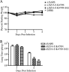
Clinical signs and lung virus titers of 6-week-old BALB/c mice infected with DPBS, icSARS, icSZ16-S K479N, or icSZ16-S K479N D22. (A) Mice were infected with 105 PFU/50 μl intranasally (10 mice per virus), and weight was monitored daily. (B) Lungs were removed on days 2 and 4 postinfection (3 mice per group per day), homogenized, and centrifuged to pellet debris. Supernatants were used in a standard plaque assay to determine lung virus titers (PFU/g).
FIG. 7.
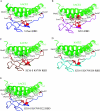
Rosetta Design modeling of “evolved” mutations that enhance spike protein binding to ACE2. Rosetta Design was used to generate structural models of SZ16 and mutant RBDs that were then superimposed onto the existing crystal structure of the SARS Urbani RBD bound to ACE2. (A) Epidemic strain and hACE2 RBD architecture. (B) SZ16 and hACE2 interaction is inhibited by steric clashing, shown as red dots, of K479 of S and residues K31 and H34 of hACE2. (C) Electrostatic repulsion at residue 479 is eradicated, allowing S and ACE2 binding, but local remodeling within the RBD due to hydrogen bonding differences at residue 487 creates cross-reactions whereby residues 442 and 479 of K479N compete with each other for interaction partners H34, K31, and D32 of hACE2. (D) The Y442F mutation of icSZ16-S K479N D8 restores an optimal RBD, allowing for favorable packing to create an architecture similar to that of the wild type. (E) Leucine 472 of the icSZ16-S K479N and icSZ16-S K479N D8 S interacts with L79 and M82 of ACE2. The icSZ16-S K479N D22 L472F mutation is predicted to have hydrophobic interactions with three potential partners, L79, M82, and Y83, of hACE2 that will increase the stability of the binding. Green dots on hACE2 indicate residues which are within 4 angstroms and thus are predicted to interact with the S residues shown in red.
Similar articles
- Pathways of cross-species transmission of synthetically reconstructed zoonotic severe acute respiratory syndrome coronavirus.
Sheahan T, Rockx B, Donaldson E, Corti D, Baric R. Sheahan T, et al. J Virol. 2008 Sep;82(17):8721-32. doi: 10.1128/JVI.00818-08. Epub 2008 Jun 25. J Virol. 2008. PMID: 18579604 Free PMC article. - Synthetic reconstruction of zoonotic and early human severe acute respiratory syndrome coronavirus isolates that produce fatal disease in aged mice.
Rockx B, Sheahan T, Donaldson E, Harkema J, Sims A, Heise M, Pickles R, Cameron M, Kelvin D, Baric R. Rockx B, et al. J Virol. 2007 Jul;81(14):7410-23. doi: 10.1128/JVI.00505-07. Epub 2007 May 16. J Virol. 2007. PMID: 17507479 Free PMC article. - Natural mutations in the receptor binding domain of spike glycoprotein determine the reactivity of cross-neutralization between palm civet coronavirus and severe acute respiratory syndrome coronavirus.
Liu L, Fang Q, Deng F, Wang H, Yi CE, Ba L, Yu W, Lin RD, Li T, Hu Z, Ho DD, Zhang L, Chen Z. Liu L, et al. J Virol. 2007 May;81(9):4694-700. doi: 10.1128/JVI.02389-06. Epub 2007 Feb 21. J Virol. 2007. PMID: 17314167 Free PMC article. - SARS-CoV replication and pathogenesis in an in vitro model of the human conducting airway epithelium.
Sims AC, Burkett SE, Yount B, Pickles RJ. Sims AC, et al. Virus Res. 2008 Apr;133(1):33-44. doi: 10.1016/j.virusres.2007.03.013. Epub 2007 Apr 23. Virus Res. 2008. PMID: 17451829 Free PMC article. Review. - Infection of human airway epithelia by SARS coronavirus is associated with ACE2 expression and localization.
Jia HP, Look DC, Hickey M, Shi L, Pewe L, Netland J, Farzan M, Wohlford-Lenane C, Perlman S, McCray PB Jr. Jia HP, et al. Adv Exp Med Biol. 2006;581:479-84. doi: 10.1007/978-0-387-33012-9_85. Adv Exp Med Biol. 2006. PMID: 17037581 Free PMC article. Review. No abstract available.
Cited by
- Deep mutational scanning of SARS-CoV-2 receptor binding domain reveals constraints on folding and ACE2 binding.
Starr TN, Greaney AJ, Hilton SK, Crawford KHD, Navarro MJ, Bowen JE, Tortorici MA, Walls AC, Veesler D, Bloom JD. Starr TN, et al. bioRxiv [Preprint]. 2020 Jun 17:2020.06.17.157982. doi: 10.1101/2020.06.17.157982. bioRxiv. 2020. PMID: 32587970 Free PMC article. Updated. Preprint. - How the Heart Was Involved in COVID-19 during the First Pandemic Phase: A Review.
Canalella A, Vitale E, Vella F, Senia P, Cannizzaro E, Ledda C, Rapisarda V. Canalella A, et al. Epidemiologia (Basel). 2021 Mar 22;2(1):124-139. doi: 10.3390/epidemiologia2010011. Epidemiologia (Basel). 2021. PMID: 36417195 Free PMC article. Review. - The application of genomics to emerging zoonotic viral diseases.
Haagmans BL, Andeweg AC, Osterhaus AD. Haagmans BL, et al. PLoS Pathog. 2009 Oct;5(10):e1000557. doi: 10.1371/journal.ppat.1000557. Epub 2009 Oct 26. PLoS Pathog. 2009. PMID: 19855817 Free PMC article. Review. - Origin and evolution of pathogenic coronaviruses.
Cui J, Li F, Shi ZL. Cui J, et al. Nat Rev Microbiol. 2019 Mar;17(3):181-192. doi: 10.1038/s41579-018-0118-9. Nat Rev Microbiol. 2019. PMID: 30531947 Free PMC article. Review. - Recombination, reservoirs, and the modular spike: mechanisms of coronavirus cross-species transmission.
Graham RL, Baric RS. Graham RL, et al. J Virol. 2010 Apr;84(7):3134-46. doi: 10.1128/JVI.01394-09. Epub 2009 Nov 11. J Virol. 2010. PMID: 19906932 Free PMC article. Review.
References
- Cello, J., A. V. Paul, and E. Wimmer. 2002. Chemical synthesis of poliovirus cDNA: generation of infectious virus in the absence of natural template. Science 2971016-1018. - PubMed
- Chinese SARS Molecular Epidemiology Consortium. 2004. Molecular evolution of the SARS coronavirus during the course of the SARS epidemic in China. Science 3031666-1669. - PubMed
- Deming, D., T. Sheahan, M. Heise, B. Yount, N. Davis, A. Sims, M. Suthar, J. Harkema, A. Whitmore, R. Pickles, A. West, E. Donaldson, K. Curtis, R. Johnston, and R. Baric. 2006. Vaccine efficacy in senescent mice challenged with recombinant SARS-CoV bearing epidemic and zoonotic spike variants. PLoS Med. 3e525. - PMC - PubMed
Publication types
MeSH terms
Substances
LinkOut - more resources
Full Text Sources
Medical
Miscellaneous


