Coronavirus spike protein inhibits host cell translation by interaction with eIF3f - PubMed (original) (raw)
Coronavirus spike protein inhibits host cell translation by interaction with eIF3f
Han Xiao et al. PLoS One. 2008.
Abstract
In response to viral infection, the expression of numerous host genes, including predominantly a number of proinflammatory cytokines and chemokines, is usually up-regulated at both transcriptional and translational levels. It was noted that in cells infected with coronavirus, transcription and translation of some of these genes were differentially induced. Drastic induction of their expression at the transcriptional level was observed in cells infected with coronavirus. However, induction of the same genes at the translational level was usually found to be minimal to moderate. To investigate the underlying mechanisms, yeast two-hybrid screen was carried out using SARS-CoV proteins as baits, revealing that a subunit of the eukaryotic initiation factor 3 (eIF3), eIF3f, may interact with the N-terminal region of the SARS-CoV spike (S) protein. This interaction was subsequently confirmed by co-immunoprecipitation and immunofluorescent staining. Meanwhile, parallel experiments confirmed that eIF3f could also interact with the S protein of another coronavirus, the avian coronavirus infectious bronchitis virus (IBV). These interactions led to the inhibition of translation of a reporter gene in both in vitro expression system and intact cells. Interestingly, IBV-infected cells stably expressing a Flag-tagged eIF3f showed much higher translation of IL-6 and IL-8, suggesting that the interaction between coronavirus S protein and eIF3f plays a functional role in controlling the expression of host genes, especially genes that are induced during coronavirus infection cycles. This study reveals a novel mechanism exploited by coronavirus to regulate viral pathogenesis.
Conflict of interest statement
Competing Interests: The authors have declared that no competing interests exist.
Figures
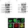
Figure 1. Confirmation of the interaction between SARS-CoV S protein and eIF3f by co-immunoprecipitation and immunofluorescence.
a. Interaction of eIF3f with the N-terminal portion of SARS-CoV S protein (SΔC). HeLa cells expressing the Myc-tagged SΔC (lane 1), the Flag-tagged eIF3f (lane 2) and eIF3f+SΔC (lane 3) were harvested at 24 hours post-transfection and lysed. The total lysates were either detected directly by Western blot with anti-Myc (top panel) and anti-Flag (second panel) antibodies, respectively, or immunoprecipitated with anti-Flag antibody. The precipitates were analyzed by Western blot with anti-Flag (third panel) and anti-Myc (bottom panel) antibodies. b. Interaction of eIF3f with the full-length SARS-CoV S protein. HeLa cells expressing the SARS-CoV S (lane 1), the Flag-tagged eIF3f (lane 2) and eIF3f+S (lane 3), were lysed. The total lysates were either detected directly by Western blot with anti-S (top panel) and anti-Flag (second panel) antibodies, respectively, or immunoprecipitated with anti-S antibodies. The precipitates were analyzed by Western blot with anti-S (third panel) and anti-Flag (bottom panel) antibodies. c. Subcellular localization of eIF3f and SARS-CoV S protein by immunofluorescent staining of HeLa cells expressing eIF3f (panel A), SARS-CoV S protein (panel B) or co-expressing eIF3f and S (panels C, D and E). The subcellular localization of the proteins were examined at 12 hours post-transfection by dual labelling with a mixture of anti-S (rabbit) and anti-Flag (mouse) antibodies, followed by incubating with a mixture of FITC-conjugated anti-mouse and tetramethyl rhodamine isocyanate-conjugated anti-rabbit antisera. Panel C shows the staining profile of eIF3f, panel D shows the staining pattern of SARS-CoV S protein, and panels E shows the overlapping images C and D.
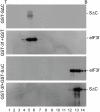
Figure 2. Co-fractionation of SΔC with eIF3f in vitro in sucrose gradients.
Ten micrograms each of the purified GST-SΔC (top panel), GST-3f+GST (second panel) and GST3f+GST-SΔC (third and bottom panels) were loaded onto the top of 15–50% sucrose gradients and centrifuged at 40,000 g for 20 hours. Fourteen fractions were collected from the top to the bottom and analyzed by Western blot with anti-S (top and bottom panels) and anti-eIF3f (second and third panels) antibodies.
Figure 3. Inhibition of protein synthesis by interaction of SARS-CoV S protein with eIF3f.
a. Dose dependent inhibition of luciferase mRNA translation by GST-SΔC in vitro (lanes 1–10) and rescue of the inhibition by addition of the purified GST-3f (lanes 11–14). In vitro transcribed luciferase mRNA (0.5 µg) was translated in vitro in rabbit reticulocyte lysates in the presence of [35S] methionine. Glutathione buffer (lane 1), increasing amounts of GST-SΔC (lanes 2–8), GST (lanes 9–10), purified GST (lane 11), GST-SΔC (lane 12), GST+GST-SΔC (lane 13), or GST-SΔC+GST-3f (lane 14) were added to the translation system as indicated. Polypeptides were separated on SDS-10% polyacrylamide gel and visualized by autoradiography (upper panels). A duplicate of the in vitro translation reactions in the absence of [35S] methionine (replaced by cold-methionine) was used to measure the luciferase activity (lower graph). b. Inhibition of luciferase translation by the full-length SARS-CoV S protein in intact cells. HeLa cells on 6-well plates were infected in duplicate with the recombinant vaccinia virus vTF7-3, and transfected with 1.2 µg of empty vector pKT0 (V) (lanes 1 and 6), 1 µg pKT0/S+0.2 µg pLUC (lanes 2 and 5), and 1 µg pKT0+0.2 µg pLUC (lanes 3 and 4), respectively. Cells were harvested at 24 hours post-transfection, and protein expression was analyzed by Western blot with anti-luciferase and anti-S antibodies. The same membrane was also probed with anti-actin antibodies as a loading control. The luciferase gene expression at the mRNA level in the transfected cells was also analyzed by Northern blot with a Dig-labeled DNA probe corresponding to the 5′-end 550 nucleotides of the luciferase gene (lanes 4–7).
Figure 4. Interaction of the IBV S protein with eIF3f and inhibition of protein synthesis.
a. Confirmation of the interaction between the Flag-tagged eIF3f and the full-length IBV S protein by co-immunoprecipitation. HeLa cells expressing the Flag-tagged eIF3f (lane 1), IBV S (lane 2) and eIF3f+IBV S (lane 3) by using the vaccinia/T7 expression system were harvested at 24 hours post-transfection and lysed. The total cell lysates were either detected directly by Western blot with anti-Flag (top panel) and anti-IBV S (second panel) antibodies or immunoprecipitated with anti-IBV S antibodies. The precipitates were analyzed by Western blot with anti-IBV S (third panel) and anti-Flag (bottom panel) antibodies. b. Interaction of the Flag-tagged eIF3f with the N-terminal region of the IBV S protein (IBV SΔC). HeLa cells expressing the Flag-tagged eIF3f (lane 1), the Myc-tagged IBV SΔC (lane 2) and eIF3f+IBV SΔC (lane 3), were harvested at 24 hours post-transfection and lysed. The total cells lysates were either detected directly by Western blot with anti-Flag (top panel) and anti-Myc (second panel) antibodies, or immunoprecipitated with anti-Flag antibody. The precipitates were analyzed by Western blot with anti-Flag (third panel) or anti-Myc (bottom panel) antibodies. c. Inhibition of luciferase activity by IBV and SARS-CoV S protein. HeLa cells expressing luciferase, luciferase+IBV S and luciferase+SARS-CoV S by using the vaccinia/T7 system were harvested at 24 hours post-transfection, lysed, and the luciferase activities in the total cell lysates were measured. d. Co-immunoprecipitation analysis of the interaction between IBV S protein and the endogenous eIF3f in IBV-infected Vero cells. Mock- and IBV-infected Vero cells were harvested at 24 hours post-infection and lysed. The total cell lysates were either detected directly by Western blot with anti-IBV S (top panel), anti-IBV N (third panel) and anti-actin (bottom panel) antibodies, respectively, or immunoprecipitated with anti-eIF3f antibody. The precipitates were analyzed by Western blot with anti-IBV S (second panel) or N (fourth panel) antibodies.
Figure 5. The relationship between the fusogenicity of S protein and its expression level in cells expressing fusion-competent and fusion-incompetent S protein.
a. Detection of cell-cell fusion by indirect immunofluorescence. Vero cells were infected with the vaccinia/T7 recombinant virus and transfected with S(EP3) and S(p65) constructs. At 6, 8, 10 and 12 hours post-transfecion, respectively, cells were fixed with 4% paraformaldehyde and stained with rabbit anti-IBV S polyclonal antibodies. b. Expression of fusion-competent and fusion-incompetent IBV S protein. Vero cells were infected with vaccinia/T7 recombinant virus and transfected with S(EP3) and S(p65) constructs. Cells were harvested at 6, 8, 10 and 12 hours post-transfection, respectively, and lysates prepared. The viral protein expression was analyzed by Western blot with rabbit anti-IBV S antibodies. The same membrane was also probed with anti-β-tubulin monoclonal antibody as a loading control. c. Quantitative analysis of the expression of fusion-competent and fusion-incompetent IBV S protein. Vero cells were infected with vaccinia/T7 recombinant virus and transfected with S(EP3)-luciferase and S(p65)-luciferase constructs. Cells were harvested at 12 and 14 hours post-transfection, respectively, lysates prepared, and the luciferase activity was determined.
Figure 6. Analysis of IL-6 and IL-8 expression at mRNA and protein levels in IBV-infected cells stably expressing the Flag-tagged eIF-3f.
a. IBV infection of Vero cell clones stably expressing of the Flag-tagged eIF3f. Two stable Vero cell clones with high level expression of the Flag-tagged eIF3f (lanes 2, 4, 6, 8 and 10) and undetectable expression of the Flag-tagged eIF3f (lanes 1, 3, 5, 7 and 9), respectively, were infected with IBV at a multiplicity of infection of 2 and harvested at 0, 8, 12, 16 and 24 hours post-infection. The expression of the Flag-tagged eIF3f was analyzed by Western blot with anti-Flag antibody. The same membrane was also probed with anti-actin antibodies as a loading control. b. Analysis of viral RNA replication and protein synthesis in IBV-infected stable cells clones with high expression of eIF3f (+eIF3f) (lanes 1–5) and undetectable expression of eIF3f (-eIF3f) (lanes 6–10). The two stable Vero cell clones were infected with IBV at a multiplicity of infection of 2 and harvested at 0, 8, 12, 16 and 24 hours post-infection. Ten micrograms of total RNA extracted from one portion of the harvested infected cells were separated on 1.3% agarose gel and transferred to a Hybond N+ membrane. Viral RNAs were probed with a Dig-labeled DNA probe corresponding to the 3′ end 680 nucleotides of the IBV genome. Bands corresponding to the genomic and subgenomic mRNA are indicated by numbers 1–6 on the right. The same membrane was also probed with a Dig-labeled GAPDH probe as loading control. The viral protein expression was analyzed by Western blot of total cell lysates prepared from another portion of the harvested infected cells with anti-IBV S protein antibodies. The same membrane was also analyzed by Western blot with anti-actin antibodies. c. Analysis of IL-8 expression. The total RNA preparations were analyzed by Northern blot with a Dig-labelled IL-8 probe (upper panels). The IL-8 expression at the protein level was analyzed by Western blot analysis with anti-IL-8 antibodies (lower panels). d. Analysis of IL-6 expression. The total RNA preparations were analyzed by Northern blot with a Dig-labelled IL-6 probe (lanes 1–8). The IL-6 expression at the protein level was analyzed by Western blot analysis of culture media (lanes 9 and 10) and total cell lysates (lanes 11–12) harvested 24 h post-infection with anti-IL-6 antibodies. Western blot analysis of the total cell lysates with anti-actin antibodies was included as a loading control (lanes 11 and 12).
Similar articles
- Decreased expression of eukaryotic initiation factor 3f deregulates translation and apoptosis in tumor cells.
Shi J, Kahle A, Hershey JW, Honchak BM, Warneke JA, Leong SP, Nelson MA. Shi J, et al. Oncogene. 2006 Aug 10;25(35):4923-36. doi: 10.1038/sj.onc.1209495. Epub 2006 Mar 13. Oncogene. 2006. PMID: 16532022 - The coronavirus spike protein induces endoplasmic reticulum stress and upregulation of intracellular chemokine mRNA concentrations.
Versteeg GA, van de Nes PS, Bredenbeek PJ, Spaan WJ. Versteeg GA, et al. J Virol. 2007 Oct;81(20):10981-90. doi: 10.1128/JVI.01033-07. Epub 2007 Aug 1. J Virol. 2007. PMID: 17670839 Free PMC article. - Intracellular targeting signals contribute to localization of coronavirus spike proteins near the virus assembly site.
Lontok E, Corse E, Machamer CE. Lontok E, et al. J Virol. 2004 Jun;78(11):5913-22. doi: 10.1128/JVI.78.11.5913-5922.2004. J Virol. 2004. PMID: 15140989 Free PMC article. - Coronavirus spike proteins in viral entry and pathogenesis.
Gallagher TM, Buchmeier MJ. Gallagher TM, et al. Virology. 2001 Jan 20;279(2):371-4. doi: 10.1006/viro.2000.0757. Virology. 2001. PMID: 11162792 Free PMC article. Review. No abstract available. - The translational factor eIF3f: the ambivalent eIF3 subunit.
Marchione R, Leibovitch SA, Lenormand JL. Marchione R, et al. Cell Mol Life Sci. 2013 Oct;70(19):3603-16. doi: 10.1007/s00018-013-1263-y. Epub 2013 Jan 25. Cell Mol Life Sci. 2013. PMID: 23354061 Free PMC article. Review.
Cited by
- SARS-CoV-2 Harnesses Host Translational Shutoff and Autophagy To Optimize Virus Yields: the Role of the Envelope (E) Protein.
Waisner H, Grieshaber B, Saud R, Henke W, Stephens EB, Kalamvoki M. Waisner H, et al. Microbiol Spectr. 2023 Feb 14;11(1):e0370722. doi: 10.1128/spectrum.03707-22. Epub 2023 Jan 9. Microbiol Spectr. 2023. PMID: 36622177 Free PMC article. - Prospective Roles of Tumor Necrosis Factor-Alpha (TNF-α) in COVID-19: Prognosis, Therapeutic and Management.
Mohd Zawawi Z, Kalyanasundram J, Mohd Zain R, Thayan R, Basri DF, Yap WB. Mohd Zawawi Z, et al. Int J Mol Sci. 2023 Mar 24;24(7):6142. doi: 10.3390/ijms24076142. Int J Mol Sci. 2023. PMID: 37047115 Free PMC article. Review. - Translational shutdown and evasion of the innate immune response by SARS-CoV-2 NSP14 protein.
Hsu JC, Laurent-Rolle M, Pawlak JB, Wilen CB, Cresswell P. Hsu JC, et al. Proc Natl Acad Sci U S A. 2021 Jun 15;118(24):e2101161118. doi: 10.1073/pnas.2101161118. Proc Natl Acad Sci U S A. 2021. PMID: 34045361 Free PMC article. - Molecular Regulation of Skeletal Muscle Growth and Organelle Biosynthesis: Practical Recommendations for Exercise Training.
Solsona R, Pavlin L, Bernardi H, Sanchez AM. Solsona R, et al. Int J Mol Sci. 2021 Mar 8;22(5):2741. doi: 10.3390/ijms22052741. Int J Mol Sci. 2021. PMID: 33800501 Free PMC article. Review.
References
- Cebulla CM, Miller DM, Sedmak DD. Viral inhibition of interferon signal transduction. Intervirology. 1999;42:325–330. - PubMed
- Cheung CY, Poon LL, Lau AS, Luk W, Lau YL, et al. Induction of proinflammatory cytokines in human macrophages by influenza A (H5N1) viruses: a mechanism for the unusual severity of human disease? Lancet. 2002;360:1831–1837. - PubMed
- Guidotti LG, Chisari FV. Cytokine-mediated control of viral infections. Virology. 2000;273:221–227. - PubMed
Publication types
MeSH terms
Substances
LinkOut - more resources
Full Text Sources
Molecular Biology Databases
Miscellaneous


