Peripheral injection of human umbilical cord blood stimulates neurogenesis in the aged rat brain - PubMed (original) (raw)
Peripheral injection of human umbilical cord blood stimulates neurogenesis in the aged rat brain
Adam D Bachstetter et al. BMC Neurosci. 2008.
Abstract
Background: Neurogenesis continues to occur throughout life but dramatically decreases with increasing age. This decrease is mostly related to a decline in proliferative activity as a result of an impoverishment of the microenvironment of the aged brain, including a reduction in trophic factors and increased inflammation.
Results: We determined that human umbilical cord blood mononuclear cells (UCBMC) given peripherally, by an intravenous injection, could rejuvenate the proliferative activity of the aged neural stem/progenitor cells. This increase in proliferation lasted for at least 15 days after the delivery of the UCBMC. Along with the increase in proliferation following UCBMC treatment, an increase in neurogenesis was also found in the aged animals. The increase in neurogenesis as a result of UCBMC treatment seemed to be due to a decrease in inflammation, as a decrease in the number of activated microglia was found and this decrease correlated with the increase in neurogenesis.
Conclusion: The results demonstrate that a single intravenous injection of UCBMC in aged rats can significantly improve the microenvironment of the aged hippocampus and rejuvenate the aged neural stem/progenitor cells. Our results raise the possibility of a peripherally administered cell therapy as an effective approach to improve the microenvironment of the aged brain.
Figures
Figure 1
Proliferation is increased in aged rats following UCBMC treatment. To determine if UCBMC could stimulate proliferation of the hippocampal neural progenitor/stem cells rats received two i.p. injections of BrdU (50 mg/kg) and were sacrifice the following day. (A) Quantification of the BrdU immunoreactive cell in the SGZ/GCL in aged rats 2 days after the UCBMC treatment showed that there was a significant (p < 0.005) increase in the number of BrdU immunoreactive cells. (B, C) Photomicrographs of the dentate gyrus of a media-treated rat (B) and a UCBMC-treated rat (C) shows the BrdU staining in those animals sacrificed 2 days after the treatment. (D)The arrow in C points to a cluster of BrdU immunoreactive cells from the UCBMC-treated rat shown in D at higher magnification. (E) To determine how long proliferation might remain elevated injections of BrdU (50 mg/kg) began 14 days after the treatment. Quantification of the BrdU immunoreactive cells determine that the UCBMC-treated group had significantly (p < 0.01) more cells in the SGZ/GCL then the animals that received media alone. (F, G) BrdU staining of the media-treated (F) and the UCBMC-treated (G) animals in the dentate gyrus of the hippocampus 15 days after the treatment. (H) Arrow in G points to cells shown at higher magnification in H. (scale bar for B, C, F, G is 100 μm; scale bar for D, H is 25 μm)
Figure 2
15 days after a UCBMC treatment neurogenesis is increase in aged rats. To determine if UCBMC treatment could stimulate neurogenesis aged F344 rats were sacrificed and immunohistochemical stained for DCX and BrdU. (A) A significant increase (p < 0.05) in the number of DCX+ cells, quantified in the SGZ/GCL, was found in the UCBMC treated rats. (B, C) Photomicrographs show the dentate gyrus demonstrating the DCX immunohistochemistry in the media-treated (B) and UCBMC-treated (C) rats. (D) A higher magnification photomicrograph of area indicated in C shows a number of DCX+ cells showing the different morphologies of the cells. (E) The results obtained with DCX were confirmed by BrdU. BrdU was injected i.p. for five consecutive days after the single injection of UCBMC. 10 days after the last injection of BrdU the animals were sacrificed. Compare to both a media control as well as an human adult peripheral blood (PBMC) control the UCBMC treated animals had significantly more BrdU+ cells (p < 0.01). (F, G, H) Photomicorgaphs of dentate gyrus shows BrdU immunohistochemistry in the media-treated (F), PBMC-treated (G) and UCBMC-treated (H) rats. (I, J) Immunofluorescence was conducted to determine the phenotype of the BrdU+ cells. (I) An example of the cells double labeled with BrdU+/NeuN+ (I; shown in orthogonal projection) and BrdU+/TUJ1+ (J; shown using maximum projection). (scale bar for B, C, F, G, H is 100 μm; scale bar for D is 25 μm)
Figure 3
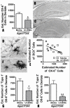
The decrease in microglia activation correlates with neurogenesis. 15 days after the UCBMC treatment a significant reduction (p < 0.05) was found in the number of OX-6+ cells in the dentate gyrus of the aged rats (A). (B, C) Photomicrographs are shown of the hippocampus of media-treated (C) and UCBMC-treated (C) rats. (D) A higher magnification photomicrograph of area indicated by arrow in B. (E) A significant negative correlation (p < 0.01) was found between the number of OX-6+ cells and the amount of neurogenesis as determine by the number of DCX+ cells. (F) The OX-6+ were further characterized based on morphology. The cell on the left represents a typical 'Type 1' cell the cell on the right represents a typical 'Type 2' cell. Both 'Type 1' (p < 0.05; G) and 'Type 2' (p < 0.01; H) OX-6+ cells were significantly reduced in the aged animals following UCBMC treatment, but there was a greater reduction in 'Type 2' cells amounting to a four fold change. (scale bar for B, C is 200 μm; scale bar for D is 25 μm)
Similar articles
- Umbilical cord blood cells regulate the differentiation of endogenous neural stem cells in hypoxic ischemic neonatal rats via the hedgehog signaling pathway.
Wang X, Zhao Y, Wang X. Wang X, et al. Brain Res. 2014 Apr 29;1560:18-26. doi: 10.1016/j.brainres.2014.02.019. Epub 2014 Feb 22. Brain Res. 2014. PMID: 24565927 - Spirulina promotes stem cell genesis and protects against LPS induced declines in neural stem cell proliferation.
Bachstetter AD, Jernberg J, Schlunk A, Vila JL, Hudson C, Cole MJ, Shytle RD, Tan J, Sanberg PR, Sanberg CD, Borlongan C, Kaneko Y, Tajiri N, Gemma C, Bickford PC. Bachstetter AD, et al. PLoS One. 2010 May 5;5(5):e10496. doi: 10.1371/journal.pone.0010496. PLoS One. 2010. PMID: 20463965 Free PMC article. - Umbilical cord blood cells regulate endogenous neural stem cell proliferation via hedgehog signaling in hypoxic ischemic neonatal rats.
Wang XL, Zhao YS, Hu MY, Sun YQ, Chen YX, Bi XH. Wang XL, et al. Brain Res. 2013 Jun 26;1518:26-35. doi: 10.1016/j.brainres.2013.04.038. Epub 2013 Apr 28. Brain Res. 2013. PMID: 23632377 - Therapeutic potential of neurogenesis for prevention and recovery from Alzheimer's disease: allopregnanolone as a proof of concept neurogenic agent.
Brinton RD, Wang JM. Brinton RD, et al. Curr Alzheimer Res. 2006 Jul;3(3):185-90. doi: 10.2174/156720506777632817. Curr Alzheimer Res. 2006. PMID: 16842093 Review. - Neurogenesis in the adult hippocampus.
Ehninger D, Kempermann G. Ehninger D, et al. Cell Tissue Res. 2008 Jan;331(1):243-50. doi: 10.1007/s00441-007-0478-3. Epub 2007 Oct 16. Cell Tissue Res. 2008. PMID: 17938969 Review.
Cited by
- Human umbilical cord blood plasma alleviates age-related olfactory dysfunction by attenuating peripheral TNF-α expression.
Lee BC, Kang I, Lee SE, Lee JY, Shin N, Kim JJ, Choi SW, Kang KS. Lee BC, et al. BMB Rep. 2019 Apr;52(4):259-264. doi: 10.5483/BMBRep.2019.52.4.124. BMB Rep. 2019. PMID: 30293545 Free PMC article. - Human Umbilical Cord Blood Serum-derived α-Secretase: Functional Testing in Alzheimer's Disease Mouse Models.
Habib A, Hou H, Mori T, Tian J, Zeng J, Fan S, Giunta B, Sanberg PR, Sawmiller D, Tan J. Habib A, et al. Cell Transplant. 2018 Mar;27(3):438-455. doi: 10.1177/0963689718759473. Epub 2018 Mar 21. Cell Transplant. 2018. PMID: 29560732 Free PMC article. - Neuroimmunomodulation and Aging.
Gemma C. Gemma C. Aging Dis. 2010 Dec 1;1(3):169-172. Aging Dis. 2010. PMID: 21297896 Free PMC article. - Could cord blood cell therapy reduce preterm brain injury?
Li J, McDonald CA, Fahey MC, Jenkin G, Miller SL. Li J, et al. Front Neurol. 2014 Oct 9;5:200. doi: 10.3389/fneur.2014.00200. eCollection 2014. Front Neurol. 2014. PMID: 25346720 Free PMC article. Review. - Deafferentation enhances neurogenesis in the young and middle aged hippocampus but not in the aged hippocampus.
Shetty AK, Hattiangady B, Rao MS, Shuai B. Shetty AK, et al. Hippocampus. 2011 Jun;21(6):631-46. doi: 10.1002/hipo.20776. Epub 2010 Mar 23. Hippocampus. 2011. PMID: 20333732 Free PMC article.
References
Publication types
MeSH terms
Grants and funding
- R01AG020927/AG/NIA NIH HHS/United States
- P01AG04418/AG/NIA NIH HHS/United States
- R21 AG024165/AG/NIA NIH HHS/United States
- R01 AG020927-05/AG/NIA NIH HHS/United States
- P01 AG004418/AG/NIA NIH HHS/United States
- R01 AG020927/AG/NIA NIH HHS/United States
- R21AG024165/AG/NIA NIH HHS/United States
LinkOut - more resources
Full Text Sources
Other Literature Sources
Medical


