Differential regulation of central nervous system autoimmunity by T(H)1 and T(H)17 cells - PubMed (original) (raw)
Differential regulation of central nervous system autoimmunity by T(H)1 and T(H)17 cells
Ingunn M Stromnes et al. Nat Med. 2008 Mar.
Abstract
Multiple sclerosis is an inflammatory, demyelinating disease of the central nervous system (CNS) characterized by a wide range of clinical signs. The location of lesions in the CNS is variable and is a crucial determinant of clinical outcome. Multiple sclerosis is believed to be mediated by myelin-specific T cells, but the mechanisms that determine where T cells initiate inflammation are unknown. Differences in lesion distribution have been linked to the HLA complex, suggesting that T cell specificity influences sites of inflammation. We demonstrate that T cells that are specific for different myelin epitopes generate populations characterized by different T helper type 17 (T(H)17) to T helper type 1 (T(H)1) ratios depending on the functional avidity of interactions between TCR and peptide-MHC complexes. Notably, the T(H)17:T(H)1 ratio of infiltrating T cells determines where inflammation occurs in the CNS. Myelin-specific T cells infiltrate the meninges throughout the CNS, regardless of the T(H)17:T(H)1 ratio. However, T cell infiltration and inflammation in the brain parenchyma occurs only when T(H)17 cells outnumber T(H)1 cells and trigger a disproportionate increase in interleukin-17 expression in the brain. In contrast, T cells showing a wide range of T(H)17:T(H)1 ratios induce spinal cord parenchymal inflammation. These findings reveal critical differences in the regulation of inflammation in the brain and spinal cord.
Figures
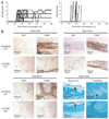
Figure 1
CNS autoimmunity differs in C3H MHC congenic mice. (a) Clinical course of EAE in C3H.SW (left, n = 11) and C3HeB/Fej (right, n = 13) mice. C3H.SW mice were scored according to a classic EAE scale and C3HeB/Fej mice were scored using an atypical EAE scale. Results are representative of two experiments. (b) Immunopathology of CNS tissues from mice killed at onset of EAE. Immunochemically stained CD4+ and F4/80+ cells were detected as brown foci. Pale-staining regions of Luxol-Fast Blue–stained sections demonstrate areas of myelin loss (arrows).
Figure 2
T cell skewing toward a TH17 or TH1 phenotype directs inflammation to the brain or spinal cord. (a) Representative CD4 (red) and F4/80 (green) staining of cerebellum and lumbar spinal cord sections of F1 recipients of epitope-specific T cells (the atypical:classic EAE ratios were rMOG, 3:0; MOG35–55, 0:6; MOG79–90, 2:7; MOG97–114, 11:0). Scale bar, 200 µm. (b) The distribution of inflammation between brain and spinal cord, as quantified by image analysis software, was significantly different for all specificities (P < 0.0001). Error bars indicate s.d. (c) Percentage of F1 recipients showing atypical or classic EAE after transfer of T cells cultured with either peptide alone, peptide and IL-12, or peptide and IL-23 (n = 5–11 recipients per group). Some IL-23–skewed T cell recipients with atypical EAE also showed tail paralysis (3/10, 2/5 and 4/6 for MOG97–114-, MOG79–90- and MOG35–55-specific T cells, respectively). Atypical EAE incidence was significantly higher for non-skewed MOG97–114- compared with MOG79–90- (P = 0.0001) and MOG35–55-specific T cells (P = 0.0003). The difference in disease phenotype induced by IL-23– and IL-12–skewed cells was significant (P = 7.7 × 10⁻¹⁹). (d) Immunohistochemical staining for CD4+ and F4/80+ cells in representative brain and spinal cord sections from IL-12– or IL-23–skewed MOG97–114-specific T cell recipients. Scale bar, 200 µm. (e) Flow cytometric analyses of recipients in c revealed significantly more CD4+, F4/80+, Gr-1+ and CD11c+ cells in the brains of atypical compared with classic EAE mice (P ≤ 0.04). Error bars represent s.e.m.
Figure 3
IL-17 activity triggered by high TH17:TH1 ratios in the brain is required for parenchymal brain inflammation. (a) The numbers of MOG-specific TH17 and TH1 cells in brain and spinal cord of F1 recipients with atypical or classic EAE determined at disease onset by ELISPOT. Each circle represents an individual mouse. *P = 0.005. (b) Epitope-specific TH17 cell numbers in the brains of IL-12– or IL-23–skewed T cell recipients with either classic or atypical EAE, respectively. Differences between atypical versus classic EAE were observed only for MOG79–90 recipients (P = 0.03). (c) The log10 TH17:TH1 ratios in the spleen, brain and spinal cord of F1 recipients of either IL-12– or IL-23–skewed epitope-specific T cells. Correlation of TH17:TH1 ratios >1 with atypical disease and <1 with classic disease was highly significant (P = 5 × 10⁻⁹). (d) Mean clinical course of TH17-biased MOG97–114 T cell recipients receiving either IL-17RA–Fc protein (scores are for classic EAE) or purified mouse IgG (scores are for atypical EAE). Data are pooled from two independent experiments (n = 10 mice per group). Mean clinical scores at day 6 and 7 post-transfer were significantly different (P < 0.008). Because of disease severity, control mice receiving IgG were killed at d 7. (e) The percentage of classic or atypical EAE observed for each group in d. (f) Representative images of brain and spinal cord sections stained with CD4 (red), F4/80 (green) and DAPI (blue). Dashed white line represents boundary between meninges and parenchyma. Scale bars, 5 µm.
Figure 4
TH17:TH1 ratio of epitope-specific T cells is influenced by functional avidity. (a) TH17:TH1 ratio of MOG-epitope specific T cells from spleens of rMOG-primed F1 mice determined by ELISPOT (each circle represents an individual mouse). (b) Representative dose response of rMOG-primed T cells to MOG peptides. MOG97–114-specific T cells showed a significantly higher functional avidity compared with the other specificities (P < 0.05, n = 19 experiments). (c) Proliferation of MBPAc1–11-specific TCR transgenic T cells in response to MBPAc1–11 and MBPAc1–11Met4Lys8 peptides. (d) TH17:TH1 ratios of MBPAc1–11Met4Lys8- and MBPAc1–11-specific T cells isolated from spleens of B10.PL mice immunized with either MBPAc1–11 or MBPAc1–11Met4Lys8 were determined by ELISPOT. *P < 0.05. Error bars represent s.d.
Similar articles
- Relapsing-remitting central nervous system autoimmunity mediated by GFAP-specific CD8 T cells.
Sasaki K, Bean A, Shah S, Schutten E, Huseby PG, Peters B, Shen ZT, Vanguri V, Liggitt D, Huseby ES. Sasaki K, et al. J Immunol. 2014 Apr 1;192(7):3029-42. doi: 10.4049/jimmunol.1302911. Epub 2014 Mar 3. J Immunol. 2014. PMID: 24591371 Free PMC article. - Experimental allergic encephalomyelitis. T cell trafficking to the central nervous system in a resistant Thy-1 congenic mouse strain.
Skundric DS, Huston K, Shaw M, Tse HY, Raine CS. Skundric DS, et al. Lab Invest. 1994 Nov;71(5):671-9. Lab Invest. 1994. PMID: 7526038 - Myelin-specific CD8+ T cells exacerbate brain inflammation in CNS autoimmunity.
Wagner CA, Roqué PJ, Mileur TR, Liggitt D, Goverman JM. Wagner CA, et al. J Clin Invest. 2020 Jan 2;130(1):203-213. doi: 10.1172/JCI132531. J Clin Invest. 2020. PMID: 31573979 Free PMC article. - Interplay between pathogenic Th17 and regulatory T cells.
Oukka M. Oukka M. Ann Rheum Dis. 2007 Nov;66 Suppl 3(Suppl 3):iii87-90. doi: 10.1136/ard.2007.078527. Ann Rheum Dis. 2007. PMID: 17934104 Free PMC article. Review. - Multiple sclerosis: a complicated picture of autoimmunity.
McFarland HF, Martin R. McFarland HF, et al. Nat Immunol. 2007 Sep;8(9):913-9. doi: 10.1038/ni1507. Nat Immunol. 2007. PMID: 17712344 Review.
Cited by
- All-trans-retinoic acid ameliorates experimental allergic encephalomyelitis by affecting dendritic cell and monocyte development.
Zhan XX, Liu Y, Yang JF, Wang GY, Mu L, Zhang TS, Xie XL, Wang JH, Liu YM, Kong QF, Li HL, Sun B. Zhan XX, et al. Immunology. 2013 Apr;138(4):333-45. doi: 10.1111/imm.12040. Immunology. 2013. PMID: 23181351 Free PMC article. - Animal models of Multiple Sclerosis.
Procaccini C, De Rosa V, Pucino V, Formisano L, Matarese G. Procaccini C, et al. Eur J Pharmacol. 2015 Jul 15;759:182-91. doi: 10.1016/j.ejphar.2015.03.042. Epub 2015 Mar 27. Eur J Pharmacol. 2015. PMID: 25823807 Free PMC article. Review. - Ferroptosis promotes T-cell activation-induced neurodegeneration in multiple sclerosis.
Luoqian J, Yang W, Ding X, Tuo QZ, Xiang Z, Zheng Z, Guo YJ, Li L, Guan P, Ayton S, Dong B, Zhang H, Hu H, Lei P. Luoqian J, et al. Cell Mol Immunol. 2022 Aug;19(8):913-924. doi: 10.1038/s41423-022-00883-0. Epub 2022 Jun 8. Cell Mol Immunol. 2022. PMID: 35676325 Free PMC article. - Genome-wide association study of severity in multiple sclerosis.
International Multiple Sclerosis Genetics Consortium. International Multiple Sclerosis Genetics Consortium. Genes Immun. 2011 Dec;12(8):615-25. doi: 10.1038/gene.2011.34. Epub 2011 Jun 9. Genes Immun. 2011. PMID: 21654844 Free PMC article. - Exacerbated egg-induced immunopathology in murine Schistosoma mansoni infection is primarily mediated by IL-17 and restrained by IFN-γ.
Rutitzky LI, Stadecker MJ. Rutitzky LI, et al. Eur J Immunol. 2011 Sep;41(9):2677-87. doi: 10.1002/eji.201041327. Epub 2011 Aug 12. Eur J Immunol. 2011. PMID: 21660933 Free PMC article.
References
- Sospedra M, Martin R. Immunology of multiple sclerosis. Annu. Rev. Immunol. 2005;23:683–747. - PubMed
- Fukazawa T, et al. Both the HLA-CPB1 and -DRB1 alleles correlate with risk for multiple sclerosis in Japanese: clinical phenotypes and gender as important factors. Tissue Antigens. 2000;55:199–205. - PubMed
- Raine C. The lesion in multiple sclerosis and chronic relapsing experimental allergic encephalomyelitis: a structural comparison. In: Raine CS, McFarland HF, Tourtellotte WW, editors. Multiple Sclerosis: Clinical and Pathogenetic Basis. London: Chapman and Hall; 1997. pp. 243–286.
Publication types
MeSH terms
Substances
Grants and funding
- AI072737/AI/NIAID NIH HHS/United States
- R01 AI072737/AI/NIAID NIH HHS/United States
- R01 AI072737-11/AI/NIAID NIH HHS/United States
- T32 CA009537/CA/NCI NIH HHS/United States
- T32-CA009537/CA/NCI NIH HHS/United States
LinkOut - more resources
Full Text Sources
Other Literature Sources
Research Materials


