A biodegradable and biocompatible gecko-inspired tissue adhesive - PubMed (original) (raw)
. 2008 Feb 19;105(7):2307-12.
doi: 10.1073/pnas.0712117105. Epub 2008 Feb 19.
Lino Ferreira, Cathryn Sundback, Jason W Nichol, Edwin P Chan, David J D Carter, Chris J Bettinger, Siamrut Patanavanich, Loice Chignozha, Eli Ben-Joseph, Alex Galakatos, Howard Pryor, Irina Pomerantseva, Peter T Masiakos, William Faquin, Andreas Zumbuehl, Seungpyo Hong, Jeffrey Borenstein, Joseph Vacanti, Robert Langer, Jeffrey M Karp
Affiliations
- PMID: 18287082
- PMCID: PMC2268132
- DOI: 10.1073/pnas.0712117105
A biodegradable and biocompatible gecko-inspired tissue adhesive
Alborz Mahdavi et al. Proc Natl Acad Sci U S A. 2008.
Abstract
There is a significant medical need for tough biodegradable polymer adhesives that can adapt to or recover from various mechanical deformations while remaining strongly attached to the underlying tissue. We approached this problem by using a polymer poly(glycerol-co-sebacate acrylate) and modifying the surface to mimic the nanotopography of gecko feet, which allows attachment to vertical surfaces. Translation of existing gecko-inspired adhesives for medical applications is complex, as multiple parameters must be optimized, including: biocompatibility, biodegradation, strong adhesive tissue bonding, as well as compliance and conformability to tissue surfaces. Ideally these adhesives would also have the ability to deliver drugs or growth factors to promote healing. As a first demonstration, we have created a gecko-inspired tissue adhesive from a biocompatible and biodegradable elastomer combined with a thin tissue-reactive biocompatible surface coating. Tissue adhesion was optimized by varying dimensions of the nanoscale pillars, including the ratio of tip diameter to pitch and the ratio of tip diameter to base diameter. Coating these nanomolded pillars of biodegradable elastomers with a thin layer of oxidized dextran significantly increased the interfacial adhesion strength on porcine intestine tissue in vitro and in the rat abdominal subfascial in vivo environment. This gecko-inspired medical adhesive may have potential applications for sealing wounds and for replacement or augmentation of sutures or staples.
Conflict of interest statement
The authors declare no conflict of interest.
Figures
Fig. 1.
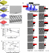
Development of biodegradable synthetic gecko patterns. (a) Nanomolding of the PGSA prepolymer is accomplished by photocuring the prepolymer under UV light followed by removal of the pattern and subsequent spin coating of DXTA on the surface of the pillars. SEM demonstrated excellent pattern transfer and fidelity. (b) Gecko patterns having different pillar size and center to center pitch were developed as illustrated by the SEM images. Pillar dimensions were measured by using optical profilometry as represented by the bar graphs, with red representing the height of pillars; black, the center to center pitch; light gray, diameter of pillar base; and dark gray, diameter of the tip. (Small and large scale bars, 1 and 10 μm, respectively.) (c) Adhesion trend of the longest pillar heights (2.4 μm) shows adhesion of nanopattern with respect to flat polymer as a function of ratio of tip diameter to pitch. _R_2 value of linear fit is 0.99. (d) Adhesion trend of the patterns is plotted as a function of ratio of tip diameter to base diameter of pillars. _R_2 values of linear fit for the low- and high-pitch patterns are 0.96 and 0.99, respectively.
Fig. 2.
DXTA coating of gecko surfaces. (a) Oxidation of DXT by sodium periodate yielding DXT functionalized with aldehyde groups. (b) FTIR spectra of PGSA network coated with DXTA (1, 2) or DXT (3), before (1) or after being washed with water (2, 3). The higher absorbance at 3,300 cm−1 (normalized by the absorbance at 2,930 cm−1 corresponding to the stretching of C–H bonds) in PGSA nanopatterns coated with DXTA rather than with DXT is indirect evidence that DXTA remained on the surface of the PGSA to a higher extent than DXT, after washing. (c) C1s XPS high-resolution spectra of amine-functionalized glass and amine-functionalized glass coated with DXTA before and after being washed with water. The shift in spectra at positions 286–288 eV corresponding to the carbon–oxygen bond shows the presence of DXTA on the surface after rinsing with water (DXTA +W). Data were normalized to the C–C and C-H spectra peak at 285 eV.
Fig. 3.
DXTA coating of nanopatterned PGSA polymer improves tissue adhesion in vitro. (a–c) Relative adhesion of nanopatterned vs. unpatterned PGSA polymer to porcine tissue slides as a function of DXTA surface coating concentration. A represents PGSA DA = 0.8, B is PGSA DA = 0.3 with 5% PEGDA, and C is PGSA DA = 0.3. Data were normalized to the unpatterned DA = 0.8 PGSA polymer without DXTA coating. (d) Normalized adhesion results of the PGSA DA = 0.3 with 5% PEG DA shows the effect of washing on improving adhesion at various DXTA concentrations. (e) Nanopatterned PGSA polymer after surface spin coating with water as control. (F and G) Nanopatterned PGSA after surface spin coating with 0.05% DXTA solution shows adhesion of neighboring pillar tips. The black arrow indicates how DXTA polymer may cause neighboring pillar tips to stick together. (h) Five percent DXTA completely obstructed the underlying nanopattern. (i) The baseline adhesion and maximum values obtained for each material used.
Fig. 4.
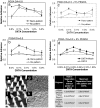
In vivo characterization of synthetic tissue tape. (a) Time-lapsed optical profilometry measurements of pillar dimensions during in vitro degradation in 1 M sodium hydroxide solution (color scheme as in Fig. 1_b_). (b) SEM image of PGSA DA = 0.3 gecko-patterned surface shows presence of pillars after eight days of in vitro degradation under physiological conditions in 1 units/ml of cholesterol esterase enzyme. (Scale bar, 10 μm.) (c) Representative image of 1-cm2 patches of gecko tissue tape, which were used for in vivo experiments. Elasticity of the samples is demonstrated through stretching and bending of the samples using forceps (Inset). (d) Weight-loss measurements after 1 week implantation of samples with different compositions of PGSA and a polyurethane control (PU). (e) Shear adhesion tests were performed on explanted tissue. (f) Within a narrow range, patterned adhesives may exhibit enhanced surface area of contact with tissue when the distance between pillars are sufficiently large and the tip diameter sufficiently low. (g) In vivo adhesion strength of DXTA-coated PGSA DA = 0.8 samples after being implanted for 48 h. (h and i) Tissue response to nanopatterned PGSA disks with DA = 0.8, s.c. implanted in the rat dorsum. Low-magnification photomicrographs of (h) H&E and (i) Masson's trichrome-stained tissue sections immediately adjacent to PGSA implants. PGSA implants formerly occupied open spaces denoted by *. Nanotopography was placed next to muscle tissue (down), and samples were harvested after 1-week implantation. A mild response was observed with a thin inflammatory infiltrate without collagen deposition. (Scale bar, 400 μm.) (j–m) High-magnification photomicrographs of H&E-stained tissue sections immediately adjacent to PGSA implants with (j) DA = 0.3, (k) DA = 0.3 with 5% PEGDA, and (l) DA = 0.8 and (m) unpatterned polyurethane implants. Nanotopography was placed next to muscle tissue (down), and samples were harvested after 1-week implantation. The tissue responses were mild in all PGS implantation but more pronounced in the polyurethane implantation. (Scale bar, 100 μm.)
Similar articles
- Biodegradable and radically polymerized elastomers with enhanced processing capabilities.
Ifkovits JL, Padera RF, Burdick JA. Ifkovits JL, et al. Biomed Mater. 2008 Sep;3(3):034104. doi: 10.1088/1748-6041/3/3/034104. Epub 2008 Aug 8. Biomed Mater. 2008. PMID: 18689916 - A tough biodegradable elastomer.
Wang Y, Ameer GA, Sheppard BJ, Langer R. Wang Y, et al. Nat Biotechnol. 2002 Jun;20(6):602-6. doi: 10.1038/nbt0602-602. Nat Biotechnol. 2002. PMID: 12042865 - Enzymatic and oxidative degradation of poly(polyol sebacate).
Li Y, Thouas GA, Shi H, Chen Q. Li Y, et al. J Biomater Appl. 2014 Apr;28(8):1138-50. doi: 10.1177/0885328213499195. Epub 2013 Jul 31. J Biomater Appl. 2014. PMID: 23904286 - Micro- and Nanostructured Biomaterials for Sutureless Tissue Repair.
Frost SJ, Mawad D, Hook J, Lauto A. Frost SJ, et al. Adv Healthc Mater. 2016 Feb 18;5(4):401-14. doi: 10.1002/adhm.201500589. Epub 2016 Jan 4. Adv Healthc Mater. 2016. PMID: 26725593 Review. - Sticking to the story: outstanding challenges in gecko-inspired adhesives.
Niewiarowski PH, Stark AY, Dhinojwala A. Niewiarowski PH, et al. J Exp Biol. 2016 Apr;219(Pt 7):912-9. doi: 10.1242/jeb.080085. J Exp Biol. 2016. PMID: 27030772 Review.
Cited by
- Repeated origin and loss of adhesive toepads in geckos.
Gamble T, Greenbaum E, Jackman TR, Russell AP, Bauer AM. Gamble T, et al. PLoS One. 2012;7(6):e39429. doi: 10.1371/journal.pone.0039429. Epub 2012 Jun 27. PLoS One. 2012. PMID: 22761794 Free PMC article. - Nanotechnology development in surgical applications: recent trends and developments.
Abaszadeh F, Ashoub MH, Khajouie G, Amiri M. Abaszadeh F, et al. Eur J Med Res. 2023 Nov 24;28(1):537. doi: 10.1186/s40001-023-01429-4. Eur J Med Res. 2023. PMID: 38001554 Free PMC article. Review. - Intelligent Nanomaterials for Wearable and Stretchable Strain Sensor Applications: The Science behind Diverse Mechanisms, Fabrication Methods, and Real-Time Healthcare.
Babu VJ, Anusha M, Sireesha M, Sundarrajan S, Abdul Haroon Rashid SSA, Kumar AS, Ramakrishna S. Babu VJ, et al. Polymers (Basel). 2022 May 30;14(11):2219. doi: 10.3390/polym14112219. Polymers (Basel). 2022. PMID: 35683893 Free PMC article. Review. - Hierarchical architecture of spider attachment setae reconstructed from scanning nanofocus X-ray diffraction data.
Schaber CF, Flenner S, Glisovic A, Krasnov I, Rosenthal M, Stieglitz H, Krywka C, Burghammer M, Müller M, Gorb SN. Schaber CF, et al. J R Soc Interface. 2019 Jan 31;16(150):20180692. doi: 10.1098/rsif.2018.0692. J R Soc Interface. 2019. PMID: 30958170 Free PMC article. - A blood-resistant surgical glue for minimally invasive repair of vessels and heart defects.
Lang N, Pereira MJ, Lee Y, Friehs I, Vasilyev NV, Feins EN, Ablasser K, O'Cearbhaill ED, Xu C, Fabozzo A, Padera R, Wasserman S, Freudenthal F, Ferreira LS, Langer R, Karp JM, del Nido PJ. Lang N, et al. Sci Transl Med. 2014 Jan 8;6(218):218ra6. doi: 10.1126/scitranslmed.3006557. Sci Transl Med. 2014. PMID: 24401941 Free PMC article.
References
- Autumn K, Liang YA, Hsieh ST, Zesch W, Chan WP, et al. Adhesive force of a single gecko foot-hair. Nature. 2000;405:681–685. - PubMed
- Pennisi E. Biomechanics. Geckos climb by the hairs of their toes. Science. 2000;288:1717–1718. - PubMed
Publication types
MeSH terms
Substances
LinkOut - more resources
Full Text Sources
Other Literature Sources
Research Materials


