Human articular chondrocytes produce IL-7 and respond to IL-7 with increased production of matrix metalloproteinase-13 - PubMed (original) (raw)
Human articular chondrocytes produce IL-7 and respond to IL-7 with increased production of matrix metalloproteinase-13
David Long et al. Arthritis Res Ther. 2008.
Abstract
Introduction: Fibronectin fragments have been found in the articular cartilage and synovial fluid of patients with osteoarthritis and rheumatoid arthritis. These matrix fragments can stimulate production of multiple mediators of matrix destruction, including various cytokines and metalloproteinases. The purpose of this study was to discover novel mediators of cartilage destruction using fibronectin fragments as a stimulus.
Methods: Human articular cartilage was obtained from tissue donors and from osteoarthritic cartilage removed at the time of knee replacement surgery. Enzymatically isolated chondrocytes in serum-free cultures were stimulated overnight with the 110 kDa alpha5beta1 integrin-binding fibronectin fragment or with IL-1, IL-6, or IL-7. Cytokines and matrix metalloproteinases released into the media were detected using antibody arrays and quantified by ELISA. IL-7 receptor expression was evaluated by flow cytometry, immunocytochemical staining, and PCR.
Results: IL-7 was found to be produced by chondrocytes treated with fibronectin fragments. Compared with cells isolated from normal young adult human articular cartilage, increased IL-7 production was noted in cells isolated from older adult tissue donors and from osteoarthritic cartilage. Chondrocyte IL-7 production was also stimulated by combined treatment with the catabolic cytokines IL-1 and IL-6. Chondrocytes were found to express IL-7 receptors and to respond to IL-7 stimulation with increased production of matrix metalloproteinase-13 and with proteoglycan release from cartilage explants.
Conclusion: These novel findings indicate that IL-7 may contribute to cartilage destruction in joint diseases, including osteoarthritis.
Figures
Figure 1
Chondrocytes produce IL-7 in response to stimulation with fibronectin fragments. Human articular chondrocytes obtained from normal articular cartilage and cultured in serum-free media were treated overnight with 500 nmol/l of the 110 kDa fibronectin fragment (FN-f). Media was collected and analyzed for cytokine production using (a) an inflammation antibody array or (b) an IL-7 ELISA. Results are representative of three experiments for each result with different donor cells used in each experiment. The IL-7 spots on the array are shown in the red circles. (Other spots that were shown to change after fibronectin fragment stimulation included IL-6, soluble IL-6 receptor [sIL-6R], interferon-inducible protein [IP]-10, and monocyte chemotactic protein [MCP]-1.)
Figure 2
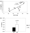
Effects of age and OA on chondrocyte production of IL-7. Media was collected 48 hours after changing to serum-free conditions in chondrocyte cultures established from (a) nonarthritic cartilage from 10 donors of different ages or from (b) cartilage from age-matched nonarthritic (n = 7) and osteoarthritic cartilage (n = 5). IL-7 was measured in the media using ELISA. The relationship of age to IL-7 levels was analyzed by Spearman correlation. The numbers in parentheses above the data points in panel a are the Collin's scores for the donor samples. OA, osteoarthritis.
Figure 3
Chondrocyte expression of IL-7 receptors. (a) Chondrocytes isolated from normal cartilage (n = 1) were incubated with a fluorescently labeled recombinant IL-7 to demonstrate binding of IL-7 to the cell surface. Labeled cells were examined by flow cytometry. The peak that is shaded purple with the black line shows cells stained with IL-7, the peak with the pink line shows blocking antibody negative control, and the peak with the green line shows cells stained with the biotin negative control. (b) Chondrocytes isolated from normal cartilage were incubated with a fluorescently labeled recombinant IL-7 as above. Labeled cells were examined by confocal microscopy. IL-7 staining is shown in green. Top left is the green channel, top right is differential intermittent contrast, and bottom left is the merged image. Chondrocytes from eight different donors showed similar results. (c) Pooled RNA isolated from 10 different sets of cultured chondrocytes was subjected to reverse transcription and real-time PCR with an IL-7 receptor primer set. An amplification plot is shown to demonstrate positive signal. Amplified chondrocyte cDNA in triplicate is shown with the blue lines. Negative control with no reverse transcription of RNA before real-time PCR is shown with a red line. Negative control with no cDNA is shown with the black line.
Figure 4
Chondrocytes respond to IL-7 stimulation with increased PYK2 phosphorylation and production of MMP-13. (a) Chondrocytes isolated from normal adult cartilage were stimulated with 10 ng/mL recombinant IL-7 and lysates were made at indicated time points for immunoblotting with an antibody to phosphorylated proline-rich tyrosine kinase (PYK)2 (Tyr402). The blot was then stripped and probed with total PYK2 antibody to confirm equal loading. (b) Densitometric scanning of the blot shown in panel a. (c) Medium was collected from serum-free chondrocyte cultures after overnight stimulation with 10 ng/ml recombinant IL-7 and examined for the presence of multiple matrix metalloproteinase (MMP) family members using an MMP antibody array. MMP-13 spots are shown in circles. (d,e) Media was collected from serum-free chondrocyte cultures after overnight stimulation with 10 ng/ml recombinant IL-7 or IL-1β, or the two together, and examined for the presence of MMP-13 using a commercially available ELISA. Results are the mean of seven experiments.
Figure 5
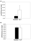
IL-7 causes proteoglycan release, but not nitric oxide production, in cartilage explants. Cartilage explants were stimulated for 72 hours with 10 ng/ml recombinant human IL-7 before media collection. (a) Medium was analyed for sulfated glycosaminoglycan (sGAG) using the dimethylmethylene blue assay and normalized for the wet weight of the tissue. (b) Total nitrite was measured in the media as a marker for nitric oxide production using commercially available colorimetric nitrate/nitrite assay kit. Results represent four experiments.
Figure 6
Role for IL-1 and IL-6 in stimulation of IL-7 production by chondrocytes. (a) Chondrocytes were pretreated with either an IL-6 neutralizing antibody or the IL-1 receptor antagonist, or the combination of the two inhibitors, and then subsequently stimulated with fibronectin fragment. After overnight stimulation media samples were collected and used for an inflammation antibody array. IL-7 spots are shown in red circles. (b) Chondrocytes were stimulated with either IL-1β (10 ng/ml) or IL-6/soluble IL-6 receptor (10 ng/ml and 20 ng/ml) or the combination of cytokines. Medium was collected and subsequently analyzed with an IL-7 ELISA.
Comment in
- Role of interleukin-7 in degenerative and inflammatory joint diseases.
van Roon JA, Lafeber FP. van Roon JA, et al. Arthritis Res Ther. 2008;10(2):107. doi: 10.1186/ar2395. Epub 2008 Apr 18. Arthritis Res Ther. 2008. PMID: 18466642 Free PMC article.
Similar articles
- Articular chondrocytes express the receptor for advanced glycation end products: Potential role in osteoarthritis.
Loeser RF, Yammani RR, Carlson CS, Chen H, Cole A, Im HJ, Bursch LS, Yan SD. Loeser RF, et al. Arthritis Rheum. 2005 Aug;52(8):2376-85. doi: 10.1002/art.21199. Arthritis Rheum. 2005. PMID: 16052547 Free PMC article. - Genetic abrogation of the fibronectin-α5β1 integrin interaction in articular cartilage aggravates osteoarthritis in mice.
Almonte-Becerril M, Gimeno-LLuch I, Villarroya O, Benito-Jardón M, Kouri JB, Costell M. Almonte-Becerril M, et al. PLoS One. 2018 Jun 5;13(6):e0198559. doi: 10.1371/journal.pone.0198559. eCollection 2018. PLoS One. 2018. PMID: 29870552 Free PMC article. - Integrins and chondrocyte-matrix interactions in articular cartilage.
Loeser RF. Loeser RF. Matrix Biol. 2014 Oct;39:11-6. doi: 10.1016/j.matbio.2014.08.007. Epub 2014 Aug 25. Matrix Biol. 2014. PMID: 25169886 Free PMC article. Review. - Role of integrins and their ligands in osteoarthritic cartilage.
Tian J, Zhang FJ, Lei GH. Tian J, et al. Rheumatol Int. 2015 May;35(5):787-98. doi: 10.1007/s00296-014-3137-5. Epub 2014 Sep 27. Rheumatol Int. 2015. PMID: 25261047 Review.
Cited by
- Fibronectin in tissue regeneration: timely disassembly of the scaffold is necessary to complete the build.
Stoffels JM, Zhao C, Baron W. Stoffels JM, et al. Cell Mol Life Sci. 2013 Nov;70(22):4243-53. doi: 10.1007/s00018-013-1350-0. Epub 2013 Jun 12. Cell Mol Life Sci. 2013. PMID: 23756580 Free PMC article. Review. - Long Intergenic Noncoding RNAs Mediate the Human Chondrocyte Inflammatory Response and Are Differentially Expressed in Osteoarthritis Cartilage.
Pearson MJ, Philp AM, Heward JA, Roux BT, Walsh DA, Davis ET, Lindsay MA, Jones SW. Pearson MJ, et al. Arthritis Rheumatol. 2016 Apr;68(4):845-56. doi: 10.1002/art.39520. Arthritis Rheumatol. 2016. PMID: 27023358 Free PMC article. - Interleukin 7 receptor gene polymorphisms and haplotypes are associated with susceptibility to IgA nephropathy in Korean children.
Hahn WH, Suh JS, Park HJ, Cho BS. Hahn WH, et al. Exp Ther Med. 2011 Nov;2(6):1121-1126. doi: 10.3892/etm.2011.322. Epub 2011 Jul 21. Exp Ther Med. 2011. PMID: 22977631 Free PMC article. - Analysing the role of endogenous matrix molecules in the development of osteoarthritis.
Sofat N. Sofat N. Int J Exp Pathol. 2009 Oct;90(5):463-79. doi: 10.1111/j.1365-2613.2009.00676.x. Int J Exp Pathol. 2009. PMID: 19765101 Free PMC article. Review. - Interleukin-6 is elevated in synovial fluid of patients with focal cartilage defects and stimulates cartilage matrix production in an in vitro regeneration model.
Tsuchida AI, Beekhuizen M, Rutgers M, van Osch GJ, Bekkers JE, Bot AG, Geurts B, Dhert WJ, Saris DB, Creemers LB. Tsuchida AI, et al. Arthritis Res Ther. 2012 Dec 3;14(6):R262. doi: 10.1186/ar4107. Arthritis Res Ther. 2012. PMID: 23206933 Free PMC article.
References
- Nietfeld JJ, Wilbrink B, Helle M, van Roy JL, den Otter W, Swaak AJ, Huber-Bruning O. Interleukin-1-induced interleukin-6 is required for the inhibition of proteoglycan synthesis by interleukin-1 in human articular cartilage. Arthritis Rheum. 1990;33:1695–1701. doi: 10.1002/art.1780331113. - DOI - PubMed
Publication types
MeSH terms
Substances
Grants and funding
- AG16697/AG/NIA NIH HHS/United States
- R01 AG016697/AG/NIA NIH HHS/United States
- R37 AR049003/AR/NIAMS NIH HHS/United States
- R01 AR049003/AR/NIAMS NIH HHS/United States
- AR49003/AR/NIAMS NIH HHS/United States
LinkOut - more resources
Full Text Sources
Other Literature Sources


