Border formation in a Bmp gradient reduced to single dissociated cells - PubMed (original) (raw)
Border formation in a Bmp gradient reduced to single dissociated cells
Jia Sheng Hu et al. Proc Natl Acad Sci U S A. 2008.
Abstract
Conversions of signaling gradients into sharp "all-or-none" borders are fundamental to tissue and organismal development. However, whether such conversions can be meaningfully reduced to dissociated cells in culture has been uncertain. Here we describe ultrasensitivity, the phenomenon equivalent to an all-or-none response, in dissociated neural precursor cells (NPCs) exposed to bone morphogenetic protein 4 (Bmp4). NPC ultrasensitivity is evident at the population and single-cell levels based on Msx1 induction, a well known Bmp target response, and occurs in the context of gene expression changes consistent with Bmp4 activity as a morphogen. Dissociated NPCs also display immediate early kinetics and irreversibility for Msx1 induction after brief Bmp4 exposure, which are attractive features for initial border formation. Relevance to border formation in vivo is provided by Bmp4 gain-of-function studies in explants and evidence for single-cell ultrasensitivity in normal and mutant Bmp gradient contexts in the developing forebrain. Together, these studies demonstrate relatively simple, robust, and reducible cell-intrinsic properties that contribute to developmental border formation within a signaling gradient.
Conflict of interest statement
The authors declare no conflict of interest.
Figures
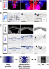
Fig. 1.
Confirmations of graded Bmp activity and NPC threshold conversion in dorsal forebrain explants. (A) Coronal schematics of the E10.5 telencephalon (light blue) and Msx1-expressing dorsal midline (dark blue). (B) Graded Bmp activity (pSmad1/5/8) and three thresholded Msx1 readouts at the E10.5 dorsal midline. Msx2 is more broadly expressed than Msx1 in this region (50), accounting for the larger expression domain detected with the Msx1/2 antibody. Right highlights expression in neural tissue for clarity. (Scale bar: 0.1 mm.) (C) Models of Msx1 threshold conversion with and without exogenous Bmp4 point sources (beads), which predict dose-, distance-, and orientation-dependent ectopic Msx1 induction. The endogenous Bmp gradient profile is based on previous measurements (10). (D) Explant and bead schematic. (E and F) Ectopic Msx1 mRNA and nlacZ inductions (arrowheads) around Bmp4-soaked, but not BSA-soaked, blue Affigel (E) or clear heparin acrylic beads (F) in E10.5 explants cultured for 2 days. Findings confirm predictions described in C and establish Bmp4 and cortical NPC sufficiencies for threshold conversion in explants. [Scale bars: 1 mm (low power) and 0.2 mm (high power).] c, cortex; d, diencephalon; dm, dorsal midline; ee, epidermal ectoderm; l, lateral ganglionic eminence; m, medial ganglionic eminence; me, mesenchyme.
Fig. 2.
NPC ultrasensitivity and irreversibility in dissociated cultures at the population level. (A and B) Schematic Michaelis–Menten, ultrasensitive, bistable, and irreversible response curves. (C–F) Hill plots of real-time qRT-PCR data from E10.5 Msx1-nlacZ (D), E12.5 Msx1-nlacZ (E), and E12.5 wild-type (F) cortical NPC cultures. Native and nlacZ-containing Msx1 transcript inductions display significant ultrasensitivity in response to Bmp4 (nH = 2.4–3.8), whereas Tgif induction is non-ultrasensitive (nH = 0.3 using data points up to 128 or 256 ng/ml Bmp4). Tgif up-regulations at the two highest Bmp4 doses were significant by t test (P < 0.05). (G) Real-time qRT-PCR data from E10.5 Msx1-nlacZ cells for other midline genes. Unlike endogenous Msx1 transcripts, Ttr is up-regulated at intermediate Bmp4 concentrations, whereas both Ttr and Lmx1a display suppression at higher Bmp4 concentrations associated with maximal Msx1 induction, consistent with a morphogen effect. Ttr up-regulations at 8 and 16 ng/ml Bmp4 were significant by t test (P < 0.05). (H–J) Induction kinetics, wild-type cortical NPCs, 50 ng/ml Bmp4. Msx1 up-regulation is detected by 15 min, is statistically significant by 2 h, and is stable for at least 5 days in the continuous presence of Bmp4. (K and L) Washout studies, wild-type cortical NPCs, 50 ng/ml Bmp4. Maximal Msx1 induction requires only brief Bmp4 exposure (no more than 15 min) and does not require Bmp4 thereafter, thus displaying irreversibility. Error bars show standard errors. t tests: *, P < 0.05; **, P < 0.01; ***, P < 0.001.
Fig. 3.
NPC ultrasensitivity in dissociated cultures at the single-cell level. (A) Schematics of graded and ultrasensitive responses at the single-cell level. (B) Culture schematic. (C) Double label immunocytochemistry of E12.5 wild-type cortical NPCs. Msx1/2 induction (red) is ultrasensitive compared with pSmad (green), which is graded and more homogeneous. Similar graded pSmad findings were seen in four independent cultures. (Scale bar: 0.05 mm.) (D) Percentage of X-Gal-positive cells in E12.5 Msx1-nlacZ cortical NPCs (
SI Fig. 6
). Msx1-nlacZ induction occurs equally well in cells that are completely isolated compared with those in contact with other cells. Errors bars show standard errors from two independent cultures.
Fig. 4.
Evidence for single-cell ultrasensitivity in intact tissues. (A) Msx1/2-positive NPCs (arrows) surrounded by negative cells near the E10.5 cortex–midline border. [Scale bars: 0.1 mm (low power) and 0.05 mm (high power).] (B) Scattered Msx1-nlacZ-positive NPCs (arrows) amid negative cells surrounding a Bmp4-soaked heparin acrylic bead in E10.5 explants cultured for 2 days. B Right Inset is inverted and thresholded for clarity. Dashed lines, beads; arrowhead, endogenous Msx1-nlacZ-expressing dorsal midline domain. [Scale bars: 0.2 mm (low power) and 0.05 mm (high power).] (C) Scattered Msx1 transcript-positive NPCs (arrowheads) amid negative cells in the E10.5 dorsal midline region after roof plate ablation. The pSmad1/5/8 gradient, which is reduced and flattened compared with normal (10), remains continuous across the cortex–midline border. C Right highlights neural expression for clarity. [Scale bars: 0.1 mm (low power) and 0.05 mm (high power).] (D) Model using NPC-intrinsic ultrasensitivity and irreversibility to initially convert the Bmp gradient into a crude Msx1 border, which is then refined by additional mechanisms into a sharp cortex–midline boundary. cx, cortex; dm, dorsal midline.
Similar articles
- A BMP-FGF morphogen toggle switch drives the ultrasensitive expression of multiple genes in the developing forebrain.
Srinivasan S, Hu JS, Currle DS, Fung ES, Hayes WB, Lander AD, Monuki ES. Srinivasan S, et al. PLoS Comput Biol. 2014 Feb 13;10(2):e1003463. doi: 10.1371/journal.pcbi.1003463. eCollection 2014 Feb. PLoS Comput Biol. 2014. PMID: 24550718 Free PMC article. - Bone morphogenetic proteins (BMPs) as regulators of dorsal forebrain development.
Furuta Y, Piston DW, Hogan BL. Furuta Y, et al. Development. 1997 Jun;124(11):2203-12. doi: 10.1242/dev.124.11.2203. Development. 1997. PMID: 9187146 - Msx1/Bmp4 genetic pathway regulates mammalian alveolar bone formation via induction of Dlx5 and Cbfa1.
Zhang Z, Song Y, Zhang X, Tang J, Chen J, Chen Y. Zhang Z, et al. Mech Dev. 2003 Dec;120(12):1469-79. doi: 10.1016/j.mod.2003.09.002. Mech Dev. 2003. PMID: 14654219 - Bone morphogenetic proteins.
Chen D, Zhao M, Mundy GR. Chen D, et al. Growth Factors. 2004 Dec;22(4):233-41. doi: 10.1080/08977190412331279890. Growth Factors. 2004. PMID: 15621726 Review.
Cited by
- Responses of Epibranchial Placodes to Disruptions of the FGF and BMP Signaling Pathways in Embryonic Mice.
Washausen S, Knabe W. Washausen S, et al. Front Cell Dev Biol. 2021 Sep 13;9:712522. doi: 10.3389/fcell.2021.712522. eCollection 2021. Front Cell Dev Biol. 2021. PMID: 34589483 Free PMC article. - Dynamic assignment and maintenance of positional identity in the ventral neural tube by the morphogen sonic hedgehog.
Dessaud E, Ribes V, Balaskas N, Yang LL, Pierani A, Kicheva A, Novitch BG, Briscoe J, Sasai N. Dessaud E, et al. PLoS Biol. 2010 Jun 1;8(6):e1000382. doi: 10.1371/journal.pbio.1000382. PLoS Biol. 2010. PMID: 20532235 Free PMC article. - Endothelial Msx1 transduces hemodynamic changes into an arteriogenic remodeling response.
Vandersmissen I, Craps S, Depypere M, Coppiello G, van Gastel N, Maes F, Carmeliet G, Schrooten J, Jones EA, Umans L, Devlieger R, Koole M, Gheysens O, Zwijsen A, Aranguren XL, Luttun A. Vandersmissen I, et al. J Cell Biol. 2015 Sep 28;210(7):1239-56. doi: 10.1083/jcb.201502003. Epub 2015 Sep 21. J Cell Biol. 2015. PMID: 26391659 Free PMC article. - A BMP-FGF morphogen toggle switch drives the ultrasensitive expression of multiple genes in the developing forebrain.
Srinivasan S, Hu JS, Currle DS, Fung ES, Hayes WB, Lander AD, Monuki ES. Srinivasan S, et al. PLoS Comput Biol. 2014 Feb 13;10(2):e1003463. doi: 10.1371/journal.pcbi.1003463. eCollection 2014 Feb. PLoS Comput Biol. 2014. PMID: 24550718 Free PMC article. - Genetics and signaling mechanisms of orofacial clefts.
Reynolds K, Zhang S, Sun B, Garland MA, Ji Y, Zhou CJ. Reynolds K, et al. Birth Defects Res. 2020 Nov;112(19):1588-1634. doi: 10.1002/bdr2.1754. Epub 2020 Jul 15. Birth Defects Res. 2020. PMID: 32666711 Free PMC article. Review.
References
- Shen Q, Wang Y, Dimos JT, Fasan CA, Phoenix TN, Lemischka IR, Ivanova NB, Stifani S, Morrisey EE, Temple S. Nat Neurosci. 2006;9:743–751. - PubMed
- Wilson PA, Lagna G, Suzuki A, Hemmati-Brivanlou A. Development. 1997;124:3177–3184. - PubMed
- Gurdon JB, Standley H, Dyson S, Butler K, Langon T, Ryan K, Stennard F, Shimizu K, Zorn A. Development. 1999;126:5309–5317. - PubMed
- Wolpert L. J Theor Biol. 1969;25:1–47. - PubMed
Publication types
MeSH terms
Substances
LinkOut - more resources
Full Text Sources
Other Literature Sources
Medical
Miscellaneous


