Sustained axenic metabolic activity by the obligate intracellular bacterium Coxiella burnetii - PubMed (original) (raw)
Sustained axenic metabolic activity by the obligate intracellular bacterium Coxiella burnetii
Anders Omsland et al. J Bacteriol. 2008 May.
Abstract
Growth of Coxiella burnetii, the agent of Q fever, is strictly limited to colonization of a viable eukaryotic host cell. Following infection, the pathogen replicates exclusively in an acidified (pH 4.5 to 5) phagolysosome-like parasitophorous vacuole. Axenic (host cell free) buffers have been described that activate C. burnetii metabolism in vitro, but metabolism is short-lived, with bacterial protein synthesis halting after a few hours. Here, we describe a complex axenic medium that supports sustained (>24 h) C. burnetii metabolic activity. As an initial step in medium development, several biological buffers (pH 4.5) were screened for C. burnetii metabolic permissiveness. Based on [(35)S]Cys-Met incorporation, C. burnetii displayed optimal metabolic activity in citrate buffer. To compensate for C. burnetii auxotrophies and other potential metabolic deficiencies, we developed a citrate buffer-based medium termed complex Coxiella medium (CCM) that contains a mixture of three complex nutrient sources (neopeptone, fetal bovine serum, and RPMI cell culture medium). Optimal C. burnetii metabolism occurred in CCM with a high chloride concentration (140 mM) while the concentrations of sodium and potassium had little effect on metabolism. CCM supported prolonged de novo protein and ATP synthesis by C. burnetii (>24 h). Moreover, C. burnetii morphological differentiation was induced in CCM as determined by the transition from small-cell variant to large-cell variant. The sustained in vitro metabolic activity of C. burnetii in CCM provides an important tool to investigate the physiology of this organism including developmental transitions and responses to antimicrobial factors associated with the host cell.
Figures
FIG. 1.
Permissiveness of acidic buffers for C. burnetii metabolism. The metabolic activity of purified C. burnetii in acidic (pH 4.5) buffers was determined by measuring [35S]Cys-Met radiolabel incorporation by bacteria after a 3-h incubation in the indicated buffers without (A and C), or with (B and D) 1 mM glutamate. The level of de novo synthesized protein was determined by scintillation counting (A and B) and SDS-PAGE with autoradiography (C and D). The magnitude of radiolabel incorporation is expressed as the relative increase over incorporation observed in P-25 buffer (pH 7.0) (negative control). Asterisks indicate statistically significant differences (P < 0.05) between the indicated sample and P-25 buffer (pH 7.0). The broken line represents the level of radiolabel incorporation in P-25 buffer (pH 7.0) normalized to 1. Representative autoradiograms are shown. citrate-P, citrate-phosphate; MW, molecular weight (in thousands).
FIG. 2.
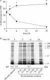
C. burnetii metabolic fitness in supplemented P-25 buffer and CCM. The ability of P-25 buffer supplemented with 5 mM glutamate (broken line) and CCM (solid line) to sustain C. burnetii metabolic fitness was directly compared by examining de novo protein synthesis in labeling buffer following preincubation in these solutions for 2, 6, and 24 h. Following preincubations, C. burnetii was labeled with [35S]Cys-Met in labeling buffer (pH 4.5) for 3 h and then subjected to scintillation counting (A) and SDS-PAGE and autoradiography (B). In panel A, results are expressed as the relative increase in radiolabel incorporation compared to bacteria that were directly labeled in labeling buffer (pH 7.0) (negative control). The 0-h time point represents the relative increase in radiolabel incorporation by C. burnetii cells that were directly labeled in labeling buffer (pH 4.5) without preincubation. Asterisks indicate statistically significant differences (P < 0.05) between CCM and supplemented P-25 buffer. A representative autoradiogram is shown. MW, molecular weight (in thousands).
FIG. 3.
Effect of CCM nutrient supplements on C. burnetii metabolic fitness. The relative impact of CCM nutrient supplements on C. burnetii metabolic fitness was determined by incubating organisms in CSB supplemented with neopeptone (neopep), FBS, or RPMI medium. Bacteria were preincubated in the respective medium formulations for 24 h and then labeled with [35S]Cys-Met in labeling buffer (pH 4.5) for 3 h. De novo protein synthesis by C. burnetii was measured by quantification of radiolabel incorporation by scintillation counting. Results are expressed as the relative increase in radiolabel incorporation compared to the incorporation of bacteria labeled in labeling buffer (pH 7.0) (negative control). Asterisks indicate statistically significant (P < 0.05) differences compared to CSB. The broken line represents the level of radiolabel incorporation in CCM (pH 7.0) normalized to 1.
FIG. 4.
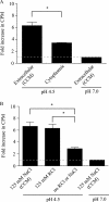
Effect of CCM ion concentrations on C. burnetii metabolic activity. (A) The effect of medium ion levels on C. burnetii metabolic activity was assessed by measuring incorporation of [35S]Cys-Met into C. burnetii de novo synthesized protein following a 3-h incubation in CCM with Cl−, K+, and Na+ concentrations similar to the host cell cytoplasm (26 mM Cl−, 110 mM K+, and 17 mM Na+) or extracellular (140 mM Cl−, 4.3 mM K+, and 190 mM Na+) environment. Incorporation of radiolabel was quantified by scintillation counting and is expressed as the increase relative to the incorporation of C. burnetii incubated in CCM (pH 7.0) with extracellular levels of Cl−, K+, and Na+ (negative control). Asterisks indicate statistically significant differences (P < 0.05) between media with extracellular or intracellular ion levels. The broken line represents the level of radiolabel incorporation in medium with extracellular ion levels at pH 7.0 normalized to 1. (B) The importance of the source of chloride ion on C. burnetii metabolic activity was examined by measuring [35S]Cys-Met incorporation of organisms incubated for 3 h in CCM containing 125 mM KCl or NaCl or in CCM without added KCl or NaCl. Incorporation of radiolabel was quantified by scintillation counting and is expressed as the relative increase over the incorporation of C. burnetii incubated in CCM (pH 7.0) containing 125 mM NaCl (negative control). Asterisks indicate statistically significant differences (P < 0.05) compared to CCM without KCl or NaCl. The broken line represents the level of radiolabel incorporation in medium containing 125 mM NaCl at pH 7.0 normalized to 1.
FIG. 5.
Effects of individual carbon source levels on C. burnetii metabolic activity. The effect of supplementing CCM with efficiently oxidized carbon sources (16) on C. burnetii de novo protein synthesis was measured. Organisms were incubated for 3 h in CCM containing [35S]Cys-Met and supplemented with 1 or 5 mM glutamate, succinate, or pyruvate. Incorporation of radiolabel was quantified by scintillation counting and is expressed as the increase relative to the incorporation of C. burnetii incubated in CCM (pH 7.0) (negative control). Asterisks indicate statistically significant (P < 0.05) differences between radiolabel incorporation by C. burnetii incubated in CCM and in CCM supplemented with a 1 or 5 mM concentration of the carbon source. The broken line represents the level of radiolabel incorporation in CCM (pH 7.0) normalized to 1.
FIG. 6.
Effect of CCM pH on C. burnetii metabolic activity. The pH for optimal C. burnetii metabolism in CCM was determined by incubating bacteria for 3 h in CCM adjusted to different pH values in the presence of [35S]Cys-Met. Radiolabel incorporation was measured by scintillation counting. C. burnetii incorporation of [35S]Cys-Met is expressed as the increase relative to the incorporation of organisms incubated in CCM (pH 7.0) (negative control). Asterisks indicate statistically significant (P < 0.05) differences between the indicated samples. The broken line represents the level of radiolabel incorporation in CCM (pH 7.0) normalized to 1.
FIG. 7.
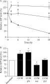
Analysis of C. burnetii ATP pool during incubation in CCM. (A) C. burnetii ATP pools were compared following incubation in CCM (⧫), CSB (▾), or P-25 buffer (▪) for 2, 6, and 24 h. Data are expressed as the percentage of the total ATP pool of the inoculum (time zero). *, statistically significant differences (P < 0.05) between CCM and P-25 buffer; #, statistically significant differences (P < 0.05) between P-25 buffer and CSB. (B) The stability of the C. burnetii ATP pool during incubation in CCM supplemented with an efficiently oxidized carbon source was assessed. ATP was measured following a 3-h incubation in CCM or in CCM supplemented with 5 mM glutamate (glu), succinate (suc), or pyruvate (pyr). Asterisks indicate statistically significant differences between the inoculum and indicated sample. The broken line represents the ATP level of the inoculum normalized to 100%.
FIG. 8.
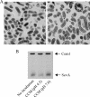
Transition of C. burnetii SCV to LCV during incubation in CCM. Purified C. burnetii SCVs were analyzed by transmission electron microscopy before and after a 24-h incubation in CCM to assess potential morphological transitions. (A) SCVs prior to axenic incubation in CCM showed ultrastructural characteristics of this cell form (left panel). Following SCV incubation in CCM, organisms displayed an ultrastructure more characteristic of the LCV form (e.g., size of >0.5 μm and relaxed chromatin; right panel). Bar, 0.5 μm. (B) Scanning densitometry of an immunoblot probed for ScvA (3.5 kDa), a protein specific to SCVs, showed that the ScvA level decreased by approximately 50% following incubation of SCVs in CCM (pH 4.5) for 24 h. SCVs incubated in CCM (pH 7.0) for 24 h exhibited no decrease in ScvA. The levels of Com1 (27 kDa), a protein equally expressed in SCVs and LCVs, did not change. Data from representative experiments are shown.
Similar articles
- Complementation of Arginine Auxotrophy for Genetic Transformation of Coxiella burnetii by Use of a Defined Axenic Medium.
Sandoz KM, Beare PA, Cockrell DC, Heinzen RA. Sandoz KM, et al. Appl Environ Microbiol. 2016 May 2;82(10):3042-51. doi: 10.1128/AEM.00261-16. Print 2016 May 15. Appl Environ Microbiol. 2016. PMID: 26969695 Free PMC article. - Host cell-free growth of the Q fever bacterium Coxiella burnetii.
Omsland A, Cockrell DC, Howe D, Fischer ER, Virtaneva K, Sturdevant DE, Porcella SF, Heinzen RA. Omsland A, et al. Proc Natl Acad Sci U S A. 2009 Mar 17;106(11):4430-4. doi: 10.1073/pnas.0812074106. Epub 2009 Feb 25. Proc Natl Acad Sci U S A. 2009. PMID: 19246385 Free PMC article. - Axenic growth of Coxiella burnetii.
Omsland A. Omsland A. Adv Exp Med Biol. 2012;984:215-29. doi: 10.1007/978-94-007-4315-1_11. Adv Exp Med Biol. 2012. PMID: 22711634 Review. - The Effect of pH on Antibiotic Efficacy against Coxiella burnetii in Axenic Media.
Smith CB, Evavold C, Kersh GJ. Smith CB, et al. Sci Rep. 2019 Dec 2;9(1):18132. doi: 10.1038/s41598-019-54556-6. Sci Rep. 2019. PMID: 31792307 Free PMC article. - Life on the outside: the rescue of Coxiella burnetii from its host cell.
Omsland A, Heinzen RA. Omsland A, et al. Annu Rev Microbiol. 2011;65:111-28. doi: 10.1146/annurev-micro-090110-102927. Annu Rev Microbiol. 2011. PMID: 21639786 Review.
Cited by
- Host-microbe interaction systems biology: lifecycle transcriptomics and comparative genomics.
Sturdevant DE, Virtaneva K, Martens C, Bozinov D, Ogundare O, Castro N, Kanakabandi K, Beare PA, Omsland A, Carlson JH, Kennedy AD, Heinzen RA, Celli J, Greenberg DE, DeLeo FR, Porcella SF. Sturdevant DE, et al. Future Microbiol. 2010 Feb;5(2):205-19. doi: 10.2217/fmb.09.125. Future Microbiol. 2010. PMID: 20143945 Free PMC article. Review. - Complex Signaling Networks Control Coxiella burnetii.
Thomas DR, Newton HJ. Thomas DR, et al. J Bacteriol. 2023 Mar 21;205(3):e0001323. doi: 10.1128/jb.00013-23. Epub 2023 Feb 27. J Bacteriol. 2023. PMID: 36847508 Free PMC article. - Characterization of the GDP-D-mannose biosynthesis pathway in Coxiella burnetii: the initial steps for GDP-β-D-virenose biosynthesis.
Narasaki CT, Mertens K, Samuel JE. Narasaki CT, et al. PLoS One. 2011;6(10):e25514. doi: 10.1371/journal.pone.0025514. Epub 2011 Oct 31. PLoS One. 2011. PMID: 22065988 Free PMC article. - Complementation of Arginine Auxotrophy for Genetic Transformation of Coxiella burnetii by Use of a Defined Axenic Medium.
Sandoz KM, Beare PA, Cockrell DC, Heinzen RA. Sandoz KM, et al. Appl Environ Microbiol. 2016 May 2;82(10):3042-51. doi: 10.1128/AEM.00261-16. Print 2016 May 15. Appl Environ Microbiol. 2016. PMID: 26969695 Free PMC article. - Q Fever-A Neglected Zoonosis.
Ullah Q, Jamil T, Saqib M, Iqbal M, Neubauer H. Ullah Q, et al. Microorganisms. 2022 Jul 28;10(8):1530. doi: 10.3390/microorganisms10081530. Microorganisms. 2022. PMID: 36013948 Free PMC article. Review.
References
- Appelberg, R. 2006. Macrophage nutriprive antimicrobial mechanisms. J. Leukoc. Biol. 791117-1128. - PubMed
- Chen, S. Y., M. Vodkin, H. A. Thompson, and J. C. Williams. 1990. Isolated Coxiella burnetii synthesizes DNA during acid activation in the absence of host cells. J. Gen. Microbiol. 13689-96. - PubMed
Publication types
MeSH terms
Substances
LinkOut - more resources
Full Text Sources
Other Literature Sources
Research Materials
Miscellaneous


