Effects of estrogen receptor agonists on regulation of the inflammatory response in astrocytes from young adult and middle-aged female rats - PubMed (original) (raw)
Effects of estrogen receptor agonists on regulation of the inflammatory response in astrocytes from young adult and middle-aged female rats
Danielle K Lewis et al. J Neuroimmunol. 2008 Mar.
Abstract
Estrogen has been shown to attenuate the inflammatory response following injury or lipopolysaccharide treatment in several organ systems. Estrogen's actions are transduced through two estrogen receptor sub-types, estrogen receptor (ER) -alpha and estrogen receptor-beta, whose actions may be overlapping or independent of each other. The present study examined the effects of ERalpha- and ERbeta-specific ligands in regulating the inflammatory response in primary astrocyte cultures. Pre-treatment with 17beta-estradiol (ERalpha/ERbeta agonist), HPTE (ERalpha agonist/ERbeta antagonist) and DPN (ERbeta agonist) led to attenuation of IL-1beta, TNFalpha, and MMP-9 in astrocyte media derived from young adult (3-4 mos.) and reproductive senescent female (9-11 mos., acyclic) astrocyte cultures, while pretreatment with PPT (ERalpha agonist) attenuated IL-1beta (but not MMP-9) in both young and senescent-derived astrocyte cultures. Our previous work determined that 17beta-estradiol was unable to attenuate the LPS-induced increase in IL-1beta in olfactory bulb primary microglial cultures derived from either young adult or reproductive senescent females. In young adult-derived microglial cultures, the LPS-induced increase in IL-1beta was not attenuated by pre-treatment with 17beta-estradiol, PPT or HPTE. Interestingly, the ERbeta agonist, DPN significantly decreased IL-1beta following LPS treatment in young adult-derived microglia. Thus while both microglia and astrocytes synthesize and release inflammatory mediators, the present data shows that compounds which bind ERbeta are more effective in attenuating proinflammatory cytokines in both cell types and may therefore be a more effective agent for future therapeutic use.
Figures
Figure 1
Glial fibrillary acidic protein (GFAP) mRNA expression levels increase in the olfactory bulb following an excitotoxic injury. Ovariectomized, placebo-replaced or estrogen-replaced female rats were subjected to a 24 hour NMDA lesion directed to the olfactory bulb. RNA was extracted and subjected to real time PCR. The relative expression of each mRNA was calculated by the comparative CT method using the following formula 2-ΔΔCt [where the amount of the target (GFAP) was normalized to the endogenous reference (Cyclophilin) and relative to a calibrator constant; ABI methods]. Main effect of hormone (a), main effect of injury (b), interaction effect (c) was significant at p≤0.05. OS: ovariectomized, placebo-replaced, sham-lesioned; OL: ovariectomized, NMDA-lesioned; ES: ovariectomized, estrogen-replaced, sham-lesioned; EL: ovariectomized, estrogen-replaced, NMDA-lesioned.
Figure 2
Characterization of primary astrocyte cultures derived from the olfactory bulb of young adult and reproductive senescent female rats. Primary astrocytes cultures were immunoreactive for GFAP (red: a,b) and were counter-stained with the nuclear dye, Hoechst (blue). Primary astrocyte cultures derived from young adult (c) and reproductive senescent females (d) were not immunoreactive for the neuronal marker, microtubule-associated protein-2 (MAP2). A small proportion (5-8%) of cells in these primary cultures derived from young adults (e,g) and reproductive senescent females (f,h) were immunopositive for the microglial markers, ionized-calcium binding adapter molecule 1 (Iba1) and the cell surface glycoprotein Mac-1 (CD11b). Iba-1 and CD11b immunopositive cells and their associated nuclei stained with Hoechst are indicated by white arrows. Immunohistochemistry with the nuclear dye Hoechst is shown in the bottom panel (c-h). Bar: 50 μm.
Figure 3
Gene expression of estrogen receptor alpha (ERα) and estrogen receptor beta (ERβ) in primary astrocyte cultures derived from young adult and reproductive senescent females as determined by RT-PCR. The ERα-specific primers span Exon 4 to Exon 6 while the ERβ-specific primers span from Exon 4 to Exon 5. Two gene-specific products approximately 300 base pairs long (ERα) and 200 bases long (ERβ) were present in both young and senescent astrocyte cultures. Cyclophilin was used as a loading control (cy) for the transcription of each gene. The bands were visualized on a 1.5% agarose gel and verified by sequencing.
Figure 4
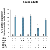
Primary astrocyte cultures derived from young adult and reproductive senescent females express the toll like recepter-4 (TLR-4). TLR4 (a, red) was observed in astrocytes derived from young adult and reproductive senescent females. Cells were counter-stained with the nuclear dye Hoechst (a, blue). Bar: 50 μm. IL-1β protein expression in astrocyte media from young adults (left panel, b) and reproductive senescent females (right panel,b) was determined following a 4-hour pre-treatment with 17β-estradiol (17bE, 20 nM), PPT (2 nM), HPTE (1μM) and DPN (10 nM) followed by a 24 hour treatment with LPS (10 μg/mL) and appropriate agonist. Asterisk (*) indicates significance at p≤0.05 relative to the saline/LPS-treated condition. Zero (0) indicates no detectable levels of expression in saline controls.
Figure 5
TNF-α protein expression in astrocyte media from young adults (left panel) and reproductive senescent females (right panel) was determined following a 4-hour pretreatment with 17β-estradiol (17bE, 20 nM), PPT (2 nM), HPTE (1μM) and DPN (10 nM) followed by a 24 hour treatment with LPS (10 μg/mL) and appropriate agonist. Asterisk (*) indicates significance at p≤0.05 relative to the saline/LPS-treated condition. Zero (0) indicates no detectable levels of expression in saline controls.
Figure 6
MMP-9 activity was measured by zymography in astrocyte media derived from young adults and reproductive senescent females following a 4-hour pretreatment with 17β-estradiol (17bE, 20 nM), PPT (2 nM), HPTE (1μM) and DPN (10 nM) followed by a 24 hour treatment with LPS (10 μg/mL) and appropriate agonist. Asterisk (*) indicates significance at p≤0.05 relative to the saline/LPS-treated condition. Zero (0) indicates no detectable levels of expression in saline controls.
Figure 7
IL-1β protein expression in microglial media from young adults was determined following a 4-hour pretreatment with 17β-estradiol (17bE, 2 nM), PPT (2 nM), HPTE (2μM) and DPN (10 nM) followed by a 24 hour treatment with LPS (10 μg/mL) and appropriate agonist. Asterisk (*) indicates significance at p≤0.05 relative to the saline/LPS-treated condition. Zero (0) indicates no detectable levels of expression in saline controls.
Similar articles
- Estrogen's effects on central and circulating immune cells vary with reproductive age.
Johnson AB, Sohrabji F. Johnson AB, et al. Neurobiol Aging. 2005 Nov-Dec;26(10):1365-74. doi: 10.1016/j.neurobiolaging.2004.12.006. Epub 2005 Feb 17. Neurobiol Aging. 2005. PMID: 16243607 - Temporal expression of IL-1beta protein and mRNA in the brain after systemic LPS injection is affected by age and estrogen.
Johnson AB, Bake S, Lewis DK, Sohrabji F. Johnson AB, et al. J Neuroimmunol. 2006 May;174(1-2):82-91. doi: 10.1016/j.jneuroim.2006.01.019. Epub 2006 Mar 10. J Neuroimmunol. 2006. PMID: 16530273 - Differential effects of estrogen in the injured forebrain of young adult and reproductive senescent animals.
Nordell VL, Scarborough MM, Buchanan AK, Sohrabji F. Nordell VL, et al. Neurobiol Aging. 2003 Sep;24(5):733-43. doi: 10.1016/s0197-4580(02)00193-8. Neurobiol Aging. 2003. PMID: 12885581 - Molecular mechanisms of estrogen action: selective ligands and receptor pharmacology.
Katzenellenbogen BS, Choi I, Delage-Mourroux R, Ediger TR, Martini PG, Montano M, Sun J, Weis K, Katzenellenbogen JA. Katzenellenbogen BS, et al. J Steroid Biochem Mol Biol. 2000 Nov 30;74(5):279-85. doi: 10.1016/s0960-0760(00)00104-7. J Steroid Biochem Mol Biol. 2000. PMID: 11162936 Review. - Functional importance of estrogen receptors in the periodontium.
Nebel D. Nebel D. Swed Dent J Suppl. 2012;(221):11-66. Swed Dent J Suppl. 2012. PMID: 22479908 Review.
Cited by
- Cytotoxicity Of Chalcone Of Eugenia aquea Burm F. Leaves Against T47D Breast Cancer Cell Lines And Its Prediction As An Estrogen Receptor Antagonist Based On Pharmacophore-Molecular Dynamics Simulation.
Muchtaridi M, Yusuf M, Syahidah HN, Subarnas A, Zamri A, Bryant SD, Langer T. Muchtaridi M, et al. Adv Appl Bioinform Chem. 2019 Nov 6;12:33-43. doi: 10.2147/AABC.S217205. eCollection 2019. Adv Appl Bioinform Chem. 2019. PMID: 31807030 Free PMC article. - Ovariectomy and subsequent treatment with estrogen receptor agonists tune the innate immune system of the hippocampus in middle-aged female rats.
Sárvári M, Kalló I, Hrabovszky E, Solymosi N, Liposits Z. Sárvári M, et al. PLoS One. 2014 Feb 13;9(2):e88540. doi: 10.1371/journal.pone.0088540. eCollection 2014. PLoS One. 2014. PMID: 24551115 Free PMC article. - Estrogen-IGF-1 interactions in neuroprotection: ischemic stroke as a case study.
Sohrabji F. Sohrabji F. Front Neuroendocrinol. 2015 Jan;36:1-14. doi: 10.1016/j.yfrne.2014.05.003. Epub 2014 May 29. Front Neuroendocrinol. 2015. PMID: 24882635 Free PMC article. Review. - Interactions between inflammation, sex steroids, and Alzheimer's disease risk factors.
Uchoa MF, Moser VA, Pike CJ. Uchoa MF, et al. Front Neuroendocrinol. 2016 Oct;43:60-82. doi: 10.1016/j.yfrne.2016.09.001. Epub 2016 Sep 17. Front Neuroendocrinol. 2016. PMID: 27651175 Free PMC article. Review. - The androgen metabolite, 5α-androstane-3β,17β-diol, decreases cytokine-induced cyclooxygenase-2, vascular cell adhesion molecule-1 expression, and P-glycoprotein expression in male human brain microvascular endothelial cells.
Zuloaga KL, Swift SN, Gonzales RJ, Wu TJ, Handa RJ. Zuloaga KL, et al. Endocrinology. 2012 Dec;153(12):5949-60. doi: 10.1210/en.2012-1316. Epub 2012 Nov 1. Endocrinology. 2012. PMID: 23117931 Free PMC article.
References
- Akiyama H, McGeer PL. Brain microglia constitutively express β-2 integrins. J Neuroimmunol. 1990;30:81–93. - PubMed
- Aloisi F, Penna G, Cerase J, Menendez Iglesias B, Adorini L. IL-12 production by central nervous system microglia is inhibited by astrocytes. J Immunol. 1997;159:1604–1612. - PubMed
- Amateau S, McCarthy M. Sexual differentiation of astrocyte morphology in the developing rat preoptic area. J Neuroendocrinol. 2002;14:904–910. - PubMed
- Andersen K, Launer LJ, Dewey ME, Letenneur L, Ott A, Copeland JRM, Dartigues JF, Kragh-Sorensen P, Baldereschi M, Brayne C, Lobo A, Martinez-Lage JM, Stijnen T, Hofman A. Gender differences in the incidence of AD and vascular dementia: The EURODEM Studies. Neurol. 1999;53:1992. - PubMed
Publication types
MeSH terms
Substances
Grants and funding
- R01 AG028303/AG/NIA NIH HHS/United States
- R01 AG019515/AG/NIA NIH HHS/United States
- AG028303/AG/NIA NIH HHS/United States
- AG019515/AG/NIA NIH HHS/United States
- R01 AG028303-02/AG/NIA NIH HHS/United States
- R01 AG019515-04/AG/NIA NIH HHS/United States
LinkOut - more resources
Full Text Sources
Medical
Miscellaneous


