Use of uterine artery Doppler ultrasonography to predict pre-eclampsia and intrauterine growth restriction: a systematic review and bivariable meta-analysis - PubMed (original) (raw)
Review
Use of uterine artery Doppler ultrasonography to predict pre-eclampsia and intrauterine growth restriction: a systematic review and bivariable meta-analysis
Jeltsje S Cnossen et al. CMAJ. 2008.
Abstract
Background: Alterations in waveforms in the uterine artery are associated with the development of pre-eclampsia and intrauterine growth restriction. We investigated the predictive accuracy of all uterine artery Doppler indices for both conditions in the first and second trimesters.
Methods: We identified relevant studies through searches of MEDLINE, EMBASE, the Cochrane Library and Medion databases (all records to April 2006) and by checking bibliographies of identified studies and consulting with experts. Four of us independently selected studies, extracted data and assessed study validity. We performed a bivariable meta-analysis of sensitivity and specificity and calculated likelihood ratios.
Results: We identified 74 studies of pre-eclampsia (total 79,547 patients) and 61 studies of intrauterine growth restriction (total 41 131 patients). Uterine artery Doppler ultrasonography provided a more accurate prediction when performed in the second trimester than in the first-trimester. Most Doppler indices had poor predictive characteristics, but this varied with patient risk and outcome severity. An increased pulsatility index with notching was the best predictor of pre-eclampsia (positive likelihood ratio 21.0 among high-risk patients and 7.5 among low-risk patients). It was also the best predictor of overall (positive likelihood ratio 9.1) and severe (positive likelihood ratio 14.6) intrauterine growth restriction among low-risk patients.
Interpretation: Abnormal uterine artery waveforms are a better predictor of pre-eclampsia than of intrauterine growth restriction. A pulsatility index, alone or combined with notching, is the most predictive Doppler index. These indices should be used in clinical practice. Future research should also concentrate on combining uterine artery Doppler ultrasonography with other tests.
Figures
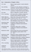
Box 1
Figure 1: Identification of studies of uterine artery Doppler ultrasonography used to predict pre-eclampsia and intrauterine growth restriction, for inclusion in the meta-analysis. *Includes 6 studies on pre-eclampsia and 8 on intrauterine growth restriction that were added after manual search of bibliographies of selected articles.
Figure 2: Plots of receiver operating characteristics showing pooled and single accuracy estimates, with 95% confidence intervals, for uterine artery Doppler indices to predict pre-eclampsia and intrauterine growth restriction in the second trimester according to patient risk. Note: the x axis shows reversed specificity. The closer the index values are to the upper left corner of each graph, the greater the accuracy of that index. The test index that best predicted the development of pre-eclampsia (highest positive likelihood ratio) in low-and high-risk patients was an increased pulsatility index with notching. This index was also the best predictor of intrauterine growth restriction in low-risk patients. For intrauterine growth restriction in high-risk patients, the Doppler indices showed low predictive value. (The thresholds for the Doppler indices reported in the studies we reviewed are provided in Appendices 3 and 4 [available online at
www.cmaj.ca/cgi/content/full/178/6/701/DC2
].)
Figure 3: Plots of receiver operating characteristics showing pooled and single accuracy estimates, with 95% confidence intervals, for uterine artery Doppler indices to predict severe pre-eclampsia and severe intrauterine growth restriction in the first and second trimester. Note: the x axis shows reversed specificity. The closer the index values are to the upper left corner of each graph, the greater the accuracy of that index. In low-risk patients, an increased pulsatility index was the test characteristic that best predicted the development of severe pre-eclampsia or severe intrauterine growth restriction (highest positive likelihood ratio). In high-risk patients, the best predictor of each outcome was an increased resistance index > 0.58. (The thresholds for the Doppler indices reported in the studies we reviewed are provided in Appendices 3 and 4 [available online at
www.cmaj.ca/cgi/content/full/178/6/701/DC2
].)
Comment in
- How useful is uterine artery Doppler ultrasonography in predicting pre-eclampsia and intrauterine growth restriction?
McLeod L. McLeod L. CMAJ. 2008 Mar 11;178(6):727-9. doi: 10.1503/cmaj.080242. CMAJ. 2008. PMID: 18332389 Free PMC article. No abstract available. - Use of Doppler ultrasonography to predict pre-eclampsia.
Conde-Agudelo A, Lindheimer M. Conde-Agudelo A, et al. CMAJ. 2008 Jul 1;179(1):53; author reply 53-4. doi: 10.1503/cmaj.1080039. CMAJ. 2008. PMID: 18591529 Free PMC article. No abstract available.
Similar articles
- First-trimester uterine artery Doppler and adverse pregnancy outcome: a meta-analysis involving 55,974 women.
Velauthar L, Plana MN, Kalidindi M, Zamora J, Thilaganathan B, Illanes SE, Khan KS, Aquilina J, Thangaratinam S. Velauthar L, et al. Ultrasound Obstet Gynecol. 2014 May;43(5):500-7. doi: 10.1002/uog.13275. Epub 2014 Apr 4. Ultrasound Obstet Gynecol. 2014. PMID: 24339044 Review. - Screening for pre-eclampsia and fetal growth restriction by uterine artery Doppler and PAPP-A at 11-14 weeks' gestation.
Pilalis A, Souka AP, Antsaklis P, Daskalakis G, Papantoniou N, Mesogitis S, Antsaklis A. Pilalis A, et al. Ultrasound Obstet Gynecol. 2007 Feb;29(2):135-40. doi: 10.1002/uog.3881. Ultrasound Obstet Gynecol. 2007. PMID: 17221926 - Multicenter screening for pre-eclampsia and fetal growth restriction by transvaginal uterine artery Doppler at 23 weeks of gestation.
Papageorghiou AT, Yu CK, Bindra R, Pandis G, Nicolaides KH; Fetal Medicine Foundation Second Trimester Screening Group. Papageorghiou AT, et al. Ultrasound Obstet Gynecol. 2001 Nov;18(5):441-9. doi: 10.1046/j.0960-7692.2001.00572.x. Ultrasound Obstet Gynecol. 2001. PMID: 11844162 - How useful is uterine artery Doppler flow velocimetry in the prediction of pre-eclampsia, intrauterine growth retardation and perinatal death? An overview.
Chien PF, Arnott N, Gordon A, Owen P, Khan KS. Chien PF, et al. BJOG. 2000 Feb;107(2):196-208. doi: 10.1111/j.1471-0528.2000.tb11690.x. BJOG. 2000. PMID: 10688503 Review.
Cited by
- Differences in uterine artery blood flow and fetal growth between the early and late onset of pregnancy-induced hypertension.
Mitsui T, Masuyama H, Maki J, Tamada S, Hirano Y, Eto E, Nobumoto E, Hayata K, Hiramatsu Y. Mitsui T, et al. J Med Ultrason (2001). 2016 Oct;43(4):509-17. doi: 10.1007/s10396-016-0729-6. Epub 2016 Jun 28. J Med Ultrason (2001). 2016. PMID: 27352079 - Mary Crosse project: systematic reviews and grading the value of neonatal tests in predicting long term outcomes.
Malin GL, Morris RK, Khan KS. Malin GL, et al. BMC Pregnancy Childbirth. 2009 Oct 29;9:49. doi: 10.1186/1471-2393-9-49. BMC Pregnancy Childbirth. 2009. PMID: 19874579 Free PMC article. - Defining normal and abnormal fetal growth: promises and challenges.
Zhang J, Merialdi M, Platt LD, Kramer MS. Zhang J, et al. Am J Obstet Gynecol. 2010 Jun;202(6):522-8. doi: 10.1016/j.ajog.2009.10.889. Epub 2010 Jan 13. Am J Obstet Gynecol. 2010. PMID: 20074690 Free PMC article. Review. - B-Flow imaging of the placenta: A feasibility study.
Dighe MK, Moshiri M, Jolley J, Thiel J, Hippe D. Dighe MK, et al. Ultrasound. 2018 Aug;26(3):160-167. doi: 10.1177/1742271X18768841. Epub 2018 Apr 6. Ultrasound. 2018. PMID: 30147740 Free PMC article. - Implementation of Uterine Artery Doppler Scanning: Improving the Care of Women and Babies High Risk for Fetal Growth Restriction.
Ekanem E, Karouni F, Katsanevakis E, Kapaya H. Ekanem E, et al. J Pregnancy. 2023 Jan 23;2023:1506447. doi: 10.1155/2023/1506447. eCollection 2023. J Pregnancy. 2023. PMID: 36726451 Free PMC article.
References
- Sibai B, Dekker G, Kupferminc M. Pre-eclampsia. Lancet 2005;365:785-99. - PubMed
- Khan KS, Wojdyla D, Say L, et al. WHO analysis of causes of maternal death: a systematic review. Lancet 2006;367:1066-74. - PubMed
- Montan S, Sjoberg NO, Svenningsen N. Hypertension in pregnancy — fetal and infant outcome: a cohort study. Clin Exp Hypertens — Part B Hypertens Pregnancy 1987;6:337-48.
- Rich-Edwards JW, Colditz GA, Stampfer MJ, et al. Birthweight and the risk for type 2 diabetes mellitus in adult women. Ann Intern Med 1999;130:278-84. - PubMed
- Barker DJ. The developmental origins of chronic adult disease. Acta Paediatr Suppl 2004;93:26-33. - PubMed
Publication types
MeSH terms
LinkOut - more resources
Full Text Sources
Other Literature Sources
Miscellaneous


