Interconversion between two unrelated protein folds in the lymphotactin native state - PubMed (original) (raw)
Interconversion between two unrelated protein folds in the lymphotactin native state
Robbyn L Tuinstra et al. Proc Natl Acad Sci U S A. 2008.
Abstract
Proteins often have multiple functional states, which might not always be accommodated by a single fold. Lymphotactin (Ltn) adopts two distinct structures in equilibrium, one corresponding to the canonical chemokine fold consisting of a monomeric three-stranded beta-sheet and carboxyl-terminal helix. The second Ltn structure solved by NMR reveals a dimeric all-beta-sheet arrangement with no similarity to other known proteins. In physiological solution conditions, both structures are significantly populated and interconvert rapidly. Interconversion replaces long-range interactions that stabilize the chemokine fold with an entirely new set of tertiary and quaternary contacts. The chemokine-like Ltn conformation is a functional XCR1 agonist, but fails to bind heparin. In contrast, the alternative structure binds glycosaminoglycans with high affinity but fails to activate XCR1. Because each structural species displays only one of the two functional properties essential for activity in vivo, the conformational equilibrium is likely to be essential for the biological activity of lymphotactin. These results demonstrate that the functional repertoire and regulation of a single naturally occurring amino acid sequence can be expanded by access to a set of highly dissimilar native-state structures.
Conflict of interest statement
The authors declare no conflict of interest.
Figures
Fig. 1.
Human Ltn exists in a two-state conformational equilibrium. (A) Members of the C (Ltn10), CX3C (fractalkine), CC (RANTES), and CXC (IL-8) chemokine families display a conserved tertiary structure. Distinct modes of dimerization are observed for the CC and CXC chemokines. (B) 1H-15N HSQC spectra of Ltn at 10°C, 200 mM NaCl (Left) and 37°C, 150 mM NaCl (11) (Center), and 40°C, 0 mM NaCl (Right). Ltn exchanges slowly on the NMR chemical shift time scale between the Ltn10 and Ltn40 conformations, which are equally populated at near-physiological solution conditions. Arrows connect resonances for the same residue in the two conformational states. (C) Exchange peaks between the Ltn10 and Ltn40 1H-15Nε1 resonances of Trp55 in a 2D 15N zz-exchange spectrum (39), acquired with a 0.4-s mixing period.
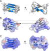
Fig. 2.
Structure of the Ltn40 dimer. (A) Ensemble of 20 NMR structures with backbone rmsd ≈0.5 Å. Disordered N- and C-terminal residues (residues 1–7 and 53–93) are not shown. (B) Hydrophobic and electrostatic stabilization of the Ltn40 dimer. Lys25 and a cluster of basic residues (blue) at one end of the dimer interface pair with Glu31 and other acidic side chains (red) at the other end of the opposing monomer. Aliphatic and aromatic residues in the center (white surface) form the hydrophobic core of the dimer. (C) The electrostatic potential surface of the Ltn40 dimer is highly positive, owing to the large number of basic residues.
Fig. 3.
Native Ltn exchanges between two unrelated structures. (A) Ltn10 ↔ Ltn40 interconversion alters all tertiary contacts. Val15 and Ala49 pack together in the Ltn10 hydrophobic core (Left) but are separated by 18 Å in Ltn40 (Right), whereas the converse is true for Leu14 and Leu45. (B) Ltn10 and Ltn40 exhibit markedly different patterns of long-range contacts. NOE distance constraints for Ltn10 (blue circles) and Ltn40 (orange circles, intramolecular; red circles, intermolecular) are plotted below the diagonal. Close contacts (<2.5 Å) observed in >80% of the NMR structure ensemble are plotted above the diagonal for residues of Ltn10 (blue squares) and Ltn40 (white squares, intramolecular; red squares, intermolecular). (C) Relative changes in solvent-accessible surface (SAS) calculated as SASLtn40 − SASLtn10, expressed as the percentage of total surface area for each side chain. Highlighted residues are more solvent-exposed in either Ltn10 (cyan) or Ltn40 (orange). Residues on the Ltn10 surface (orange) reside in the core of the Ltn40 structure, whereas Ltn40 surface residues (cyan) are located in the Ltn10 interior. (D) (Left) The odd-numbered residues of β1 (orange), β2 (cyan), and β3 (orange) contribute to the Ltn10 core. (Right) In the Ltn40 native-state conformer, the odd-numbered side chains of β1 and β3 pack in the dimer interface, whereas the odd-numbered residues of β2 are reoriented to the opposite face of the β-sheet and reside on the surface. (E) Rearrangement of hydrogen bonds defining the Ltn secondary structure. Each bar denotes a pair of backbone N–H···O = C hydrogen bonds connecting β1–β2 (cyan), β2–β3 (orange), and β0–β3 (green). Ltn10 ↔ Ltn40 interconversion shifts β2 by one residue relative to β1 and β3, which rotate 180° and establish a new hydrogen bond pattern with residues of β0 and β2.
Fig. 4.
Ltn10 and Ltn40 are functionally distinct. (A) An engineered disulfide (red) locks the CC3 variant into the Ltn10 fold (15), but replacement of Trp55 (blue) with Asp (W55D) yields exclusively the Ltn40 species. With the exception of signals adjacent to the amino acid substitution, the HSQC spectrum of W55D is identical to the Ltn40 spectrum obtained with WT Ltn. (B) Ca2+ flux response of XCR1 expressing HEK293 cells to WT (black), CC3 (orange), and W55D (blue) Ltn. Dashed line marks the addition of 200 nM chemokine. (C) Elution profiles for WT (black), CC3 (orange), and W55D (blue) Ltn from heparin-Sepharose. (D) Heparin selectively precipitates the Ltn40 conformation. (Left) Before the addition of heparin tetrasaccharide, HSQC signals are observed for both W55D (blue) and CC3 (orange). (Center and Right) Heparin addition results in the broadening (Center) and disappearance of W55D amide peaks (Right). (E) Titration of WT Ltn with semipurified heparin shifts the conformational equilibrium toward Ltn40 based on changes in the Trp55 emission wavelength (blue). CC3 emission is unaltered by heparin (orange). Emission maxima for Ltn10 and Ltn40 are indicated. (F) Ca2+ flux response of WT (black) and CC3 (orange) after incubation with an equimolar concentration of semipurified heparin. Heparin prevents WT Ltn-XCR1 association, as sequential addition of CC3 elicits XCR1 activation.
Similar articles
- An engineered second disulfide bond restricts lymphotactin/XCL1 to a chemokine-like conformation with XCR1 agonist activity.
Tuinstra RL, Peterson FC, Elgin ES, Pelzek AJ, Volkman BF. Tuinstra RL, et al. Biochemistry. 2007 Mar 13;46(10):2564-73. doi: 10.1021/bi602365d. Epub 2007 Feb 16. Biochemistry. 2007. PMID: 17302442 Free PMC article. - Lymphotactin: how a protein can adopt two folds.
Camilloni C, Sutto L. Camilloni C, et al. J Chem Phys. 2009 Dec 28;131(24):245105. doi: 10.1063/1.3276284. J Chem Phys. 2009. PMID: 20059117 - Engineering Metamorphic Chemokine Lymphotactin/XCL1 into the GAG-Binding, HIV-Inhibitory Dimer Conformation.
Fox JC, Tyler RC, Guzzo C, Tuinstra RL, Peterson FC, Lusso P, Volkman BF. Fox JC, et al. ACS Chem Biol. 2015 Nov 20;10(11):2580-8. doi: 10.1021/acschembio.5b00542. Epub 2015 Sep 2. ACS Chem Biol. 2015. PMID: 26302421 Free PMC article. - Lymphotactin.
Hedrick JA, Zlotnik A. Hedrick JA, et al. Clin Immunol Immunopathol. 1998 Jun;87(3):218-22. doi: 10.1006/clin.1998.4546. Clin Immunol Immunopathol. 1998. PMID: 9646830 Review. No abstract available. - A backbone-based theory of protein folding.
Rose GD, Fleming PJ, Banavar JR, Maritan A. Rose GD, et al. Proc Natl Acad Sci U S A. 2006 Nov 7;103(45):16623-33. doi: 10.1073/pnas.0606843103. Epub 2006 Oct 30. Proc Natl Acad Sci U S A. 2006. PMID: 17075053 Free PMC article. Review.
Cited by
- Structural perspectives on antimicrobial chemokines.
Nguyen LT, Vogel HJ. Nguyen LT, et al. Front Immunol. 2012 Dec 28;3:384. doi: 10.3389/fimmu.2012.00384. eCollection 2012. Front Immunol. 2012. PMID: 23293636 Free PMC article. - The "CPC clip motif": a conserved structural signature for heparin-binding proteins.
Torrent M, Nogués MV, Andreu D, Boix E. Torrent M, et al. PLoS One. 2012;7(8):e42692. doi: 10.1371/journal.pone.0042692. Epub 2012 Aug 6. PLoS One. 2012. PMID: 22880084 Free PMC article. - Evolutionary dynamics on protein bi-stability landscapes can potentially resolve adaptive conflicts.
Sikosek T, Bornberg-Bauer E, Chan HS. Sikosek T, et al. PLoS Comput Biol. 2012;8(9):e1002659. doi: 10.1371/journal.pcbi.1002659. Epub 2012 Sep 13. PLoS Comput Biol. 2012. PMID: 23028272 Free PMC article. - Structural insight into the evolution of a new chemokine family from zebrafish.
Rajasekaran D, Fan C, Meng W, Pflugrath JW, Lolis EJ. Rajasekaran D, et al. Proteins. 2014 May;82(5):708-16. doi: 10.1002/prot.24380. Epub 2013 Dec 14. Proteins. 2014. PMID: 23900850 Free PMC article. - Moonlighting Proteins in the Fuzzy Logic of Cellular Metabolism.
Liu H, Jeffery CJ. Liu H, et al. Molecules. 2020 Jul 29;25(15):3440. doi: 10.3390/molecules25153440. Molecules. 2020. PMID: 32751110 Free PMC article. Review.
References
- Anfinsen CB. Principles that govern the folding of protein chains. Science. 1973;181:223–230. - PubMed
- Chothia C. Proteins: One thousand families for the molecular biologist. Nature. 1992;357:543–544. - PubMed
- Cordes MH, Burton RE, Walsh NP, McKnight CJ, Sauer RT. An evolutionary bridge to a new protein fold. Nat Struct Biol. 2000;7:1129–1132. - PubMed
- Luo X, et al. The Mad2 spindle checkpoint protein has two distinct natively folded states. Nat Struct Mol Biol. 2004;11:338–345. - PubMed
Publication types
MeSH terms
Substances
Grants and funding
- UO1 AI053877/AI/NIAID NIH HHS/United States
- R01 AI045843/AI/NIAID NIH HHS/United States
- R01 AI45843/AI/NIAID NIH HHS/United States
- R01 AI063325/AI/NIAID NIH HHS/United States
- U01 AI053877/AI/NIAID NIH HHS/United States
LinkOut - more resources
Full Text Sources
Other Literature Sources
Molecular Biology Databases


