Selective death of newborn neurons in hippocampal dentate gyrus following moderate experimental traumatic brain injury - PubMed (original) (raw)
Selective death of newborn neurons in hippocampal dentate gyrus following moderate experimental traumatic brain injury
Xiang Gao et al. J Neurosci Res. 2008.
Abstract
Memory impairment is one of the most significant residual deficits following traumatic brain injury (TBI) and is among the most frequent complaints heard from patients and their relatives. It has been reported that the hippocampus is particularly vulnerable to TBI, which results in hippocampus-dependent cognitive impairment. There are different regions in the hippocampus, and each region is composed of different cell types, which might respond differently to TBI. However, regional and cell type-specific neuronal death following TBI is not well described. Here, we examined the distribution of degenerating neurons in the hippocampus of the mouse brain following controlled cortical impact (CCI) and found that the majority of degenerating neurons observed were in the dentate gyrus after moderate (0.5 mm cortical deformation) CCI-TBI. In contrast, there were only a few degenerating neurons observed in the hilus, and we did not observe any degenerating neurons in the CA3 or CA1 regions. Among those degenerating cells in the dentate gyrus, about 80% of them were found in the inner granular neuron layer. Analysis with cell type-specific markers showed that most of the degenerating neurons in the inner granular neuron layer are newborn immature neurons. Further quantitative analysis shows that the number of newborn immature neurons in the dentate gyrus is dramatically decreased in the ipsilateral hemisphere compared with the contralateral side. Collectively, our data demonstrate the selective death of newborn immature neurons in the hippocampal dentate gyrus following moderate injury with CCI in mice. This selective vulnerability of newborn immature dentate neurons may contribute to the persistent impairment of learning and memory post-TBI and provide an innovative target for neuroprotective treatment strategies.
Figures
Fig. 1
Secondary neuronal death in the hippocampus following CCI injury. Fluoro-Jade B (FJB) staining to show the degenerating neurons in the mouse brain 24 hr following controlled cortical impact. a: Degenerating neurons are stained by FJB in green. The section was stained with DAPI in blue. b: Enlarged image from panel a to show the degenerating neurons in the hippocampal dentate gyrus. c: High-magnification view to show the morphology of degenerating neurons with FJB staining in green in the dentate gyrus. d: DAPI staining to show the nucleus. e: Merged image of c and d. f: Enlarged image from c to show the degenerating neurons. g: DAPI staining in blue to show the apoptotic neurons with condensed or cleaved nuclear morphologies, marked with a white arrow. h: Merged imaged of f and g. [Color figure can be viewed in the online issue, which is available at
.]
Fig. 2
Distribution of degenerating neurons in hippocampal dentate gyrus following moderate traumatic brain injury. FJB staining to show the degenerating neurons in green in the mouse brain following controlled cortical impact. The section was stained with DAPI in blue. a–d: Sections rostral to epicenter. e: Section at epicenter. f–i: Sections caudal to epicenter. [Color figure can be viewed in the online issue, which is available at
.]
Fig. 3
Layer-specific distribution of secondary neuronal death in the hippocampal dentate gyrus following CCI injury. FJB staining to show the degenerating neurons in green in the mouse brain following controlled cortical impact. DAPI was used to stain with DAPI in blue to show the structure of the hilus and the dentate gyrus. Left: Degenerating neurons were observed in the hilus, SGZ, inner granular layer, outer granular layer, and molecular layer (ML). Right: Quantification of degenerating neurons in the hippocampal dentate gyrus. [Color figure can be viewed in the online issue, which is available at
.]
Fig. 4
Cell type specificity of secondary neuronal death in the hippocampal dentate gyrus following CCI injury. a: Nestin antibody was used to show the neural stem cells in yellow in the hippocampal dentate gyrus. b: Dcx antibody to show newborn neurons in pink in the hippocampal dentate gyrus. c: NeuN antibody to show mature neurons in the hippocampal dentate gyrus. d: Merged image of a–c to show the layer of neural stem cells, newborn neurons, and mature neurons in the hippocampal dentate gyrus. e: Degenerating neurons were stained with FJB in green. f: Merged image of a–e to show the location of FJB-positive neurons between the nestin-positive cell layer and the NeuN-positive cell layer. g: The section was counterstained with DAPI in blue to show the location of FJB-positive cells in the hippocampal dentate gyrus. h: Separately merged images of Dcx staining and FJB staining to show the position of newborn neurons and degenerating neurons in the hippocampal dentate gyrus. i: Enlarged view of h to show the cells doubly stained with Dcx and FJB, marked by a white arrow. j: Separately merged images of nestin staining and FJB staining to show the position of neural stem cells and degenerating neurons in the hippocampal dentate gyrus. k: Enlarged view of j to show that there is no colocalization of Dcx with FJB. l: Separately merged images of NeuN staining and FJB staining to show the position of mature neuron and degenerating neurons in the hippocampal dentate gyrus.
Fig. 5
Confocal microscopy to confirm that FJB stained degenerating newborn neurons in the hippocampal dentate gyrus following TBI. a: Confocal image of FJB-positive cell expressing Dcx in the hippocampal dentate gyrus using FJB staining combined with immunostaining with antibody against Dcx. The section was counterstained with DAPI in blue. Enlarged view of FJB- and Dcx-positive cell in the white box is shown in separated channels in b–d. e: Merged image of b–d. Focal sections through the FJB- and Dcx-positive cell are shown in f–i. f: Merged image of f1–f3. g: Merged image of g1–g3. h: Merged image of h1–h3. i: Merged image of i1–i3.
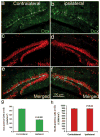
Fig. 6
The number of newborn neurons in the adult hippocampal dentate gyrus dramatically decreased following CCI injury. Double immunostaning to show the newborn neurons with Dcx antibody and mature neurons with NeuN antibody in the contralateral (a,c,e) and ipsilateral (b,d,f) sides of the hippocampal dentate gyrus 24 hr after moderate CCI injury. a: Dcx-positive newborn neurons in the contralateral side of adult hippocampal dentate gyrus. b: Dcx-positive newborn neurons in the ipsilateral side of adult hippocampal dentate gyrus. c: NeuN-positive mature neurons in the contralateral side of adult hippocampal dentate gyrus. d: NeuN-positive mature neurons in the ipsilateral side of adult hippocampal dentate gyrus. e: Merged image of a and c. f: Merged imaged of b and d. g: Quantification of Dcx-positive newborn neurons in the contralateral and ipsilateral sides of the adult hippocampal dentate gyrus. h: Quantification of NeuN-positive mature neurons in the contralateral and ipsilateral sides of the adult hippocampal dentate gyrus.
Similar articles
- Moderate traumatic brain injury causes acute dendritic and synaptic degeneration in the hippocampal dentate gyrus.
Gao X, Deng P, Xu ZC, Chen J. Gao X, et al. PLoS One. 2011;6(9):e24566. doi: 10.1371/journal.pone.0024566. Epub 2011 Sep 13. PLoS One. 2011. PMID: 21931758 Free PMC article. - Moderate traumatic brain injury triggers rapid necrotic death of immature neurons in the hippocampus.
Zhou H, Chen L, Gao X, Luo B, Chen J. Zhou H, et al. J Neuropathol Exp Neurol. 2012 Apr;71(4):348-59. doi: 10.1097/NEN.0b013e31824ea078. J Neuropathol Exp Neurol. 2012. PMID: 22437344 Free PMC article. - Post-traumatic hypoxia exacerbates neuronal cell death in the hippocampus.
Feng JF, Zhao X, Gurkoff GG, Van KC, Shahlaie K, Lyeth BG. Feng JF, et al. J Neurotrauma. 2012 Apr 10;29(6):1167-79. doi: 10.1089/neu.2011.1867. Epub 2012 Jan 30. J Neurotrauma. 2012. PMID: 22191636 Free PMC article. - Cell death mechanisms following traumatic brain injury.
Raghupathi R. Raghupathi R. Brain Pathol. 2004 Apr;14(2):215-22. doi: 10.1111/j.1750-3639.2004.tb00056.x. Brain Pathol. 2004. PMID: 15193035 Free PMC article. Review.
Cited by
- Moderate traumatic brain injury promotes neural precursor proliferation without increasing neurogenesis in the adult hippocampus.
Gao X, Chen J. Gao X, et al. Exp Neurol. 2013 Jan;239:38-48. doi: 10.1016/j.expneurol.2012.09.012. Epub 2012 Sep 26. Exp Neurol. 2013. PMID: 23022454 Free PMC article. - Delayed and progressive damages to juvenile mice after moderate traumatic brain injury.
Zhao S, Wang X, Gao X, Chen J. Zhao S, et al. Sci Rep. 2018 May 9;8(1):7339. doi: 10.1038/s41598-018-25475-9. Sci Rep. 2018. PMID: 29743575 Free PMC article. - The Small-Molecule TrkB Agonist 7, 8-Dihydroxyflavone Decreases Hippocampal Newborn Neuron Death After Traumatic Brain Injury.
Chen L, Gao X, Zhao S, Hu W, Chen J. Chen L, et al. J Neuropathol Exp Neurol. 2015 Jun;74(6):557-67. doi: 10.1097/NEN.0000000000000199. J Neuropathol Exp Neurol. 2015. PMID: 25933388 Free PMC article. - Hyperbaric Oxygenation Prevents Loss of Immature Neurons in the Adult Hippocampal Dentate Gyrus Following Brain Injury.
Jeremic R, Pekovic S, Lavrnja I, Bjelobaba I, Djelic M, Dacic S, Brkic P. Jeremic R, et al. Int J Mol Sci. 2023 Feb 21;24(5):4261. doi: 10.3390/ijms24054261. Int J Mol Sci. 2023. PMID: 36901691 Free PMC article. - Post-traumatic seizures exacerbate histopathological damage after fluid-percussion brain injury.
Bao YH, Bramlett HM, Atkins CM, Truettner JS, Lotocki G, Alonso OF, Dietrich WD. Bao YH, et al. J Neurotrauma. 2011 Jan;28(1):35-42. doi: 10.1089/neu.2010.1383. Epub 2010 Oct 12. J Neurotrauma. 2011. PMID: 20836615 Free PMC article.
References
- Altman J, Bayer SA. Migration and distribution of two populations of hippocampal granule cell precursors during the perinatal and postnatal periods. J Comp Neurol. 1990a;301:365–381. - PubMed
- Altman J, Bayer SA. Mosaic organization of the hippocampal neuroepithelium and the multiple germinal sources of dentate granule cells. J Comp Neurol. 1990b;301:325–342. - PubMed
- Altman J, Bayer SA. Prolonged sojourn of developing pyramidal cells in the intermediate zone of the hippocampus and their settling in the stratum pyramidale. J Comp Neurol. 1990c;301:343–364. - PubMed
- Amaral DG, Witter MP. The three-dimensional organization of the hippocampal formation: a review of anatomical data. Neuroscience. 1989;31:571–591. - PubMed
- Anderson KJ, Miller KM, Fugaccia I, Scheff SW. Regional distribution of fluoro-jade B staining in the hippocampus following traumatic brain injury. Exp Neurol. 2005;193:125–130. - PubMed
Publication types
MeSH terms
Grants and funding
- R01 NS046566/NS/NINDS NIH HHS/United States
- R21 NS072631/NS/NINDS NIH HHS/United States
- R21 NS075733/NS/NINDS NIH HHS/United States
- 1R01 NS046566/NS/NINDS NIH HHS/United States
LinkOut - more resources
Full Text Sources
Miscellaneous


