Antiangiogenic effect of inhibitors of cytochrome P450 on rats with glioblastoma multiforme - PubMed (original) (raw)
Antiangiogenic effect of inhibitors of cytochrome P450 on rats with glioblastoma multiforme
Drazen Zagorac et al. J Cereb Blood Flow Metab. 2008 Aug.
Abstract
Cytochrome P450 epoxygenase catalyzes 5,6-, 8,9-, 11,12-, and 14,15-epoxyeicosatrienoic acids (EETs) from arachidonic acid (AA). In 1996, our group identified the expression of the cytochrome P450 2C11 epoxygenase (CYP epoxygenase) gene in astrocytes. Because of our finding an array of physiological functions have been attributed to EETs in the brain, one of the actions of EETs involves a predominant role in brain angiogenesis. Blockade of EETs formation with different epoxygenase inhibitors decreases endothelial tube formation in cocultures of astrocytes and capillary endothelial cells. The intent of this investigation was to determine if pharmacologic inhibition of formation of EETs is effective in reducing capillary formation in glioblastoma multiforme with a concomitant reduction in tumor volume and increase in animal survival time. Two mechanistically different inhibitors of CYP epoxygenase, 17-octadecynoic acid (17-ODYA) and miconazole, significantly reduced capillary formation and tumor size in glial tumors formed by injection of rat glioma 2 (RG2) cells, also resulting in an increased animal survival time. However, we observed that 17-ODYA and miconazole did not inhibit the formation of EETs in tumor tissue. This implies that 17-ODYA and miconazole appear to exert their antitumorogenic function by a different mechanism that needs to be explored.
Figures
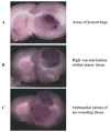
Figure 1
Pathological characteristics of tumors derived from RG2 cells (n = 6). (A) Depicts representative areas of hemorrhage, (B) Shows high vascularization within the tumor and (C) Substantial edema of the surrounding tissue.
Figure 2
(A) Inhibition of CYP epoxygenase activity with miconazole or 17-ODYA decreases tumor size in the Fischer rat. RG2 cells were implanted into the forebrain of male Fischer rats and 3 days later drug or vehicle was delivered using osmotic mini pumps. Animals were killed, perfusion fixed with formalin and frozen sections prepared for microscopy. (B) Depicts quantitative representation of tumor volume (n = 6 per group).
Figure 3
(A) Representative images of tumor vasculature captured following perfusion with FITC-conjugated dextran in the absence or presence of the CYP epoxygenase inhibitors. At the termination of the experiment, the animals were anesthetized, briefly perfused with FITC-conjugated dextran in a saline solution, the brains quickly removed and frozen sections prepared for fluorescence microscopy. The green spots represent blood vessels. (B) Bar graph representing the average number of blood vessels in 200 μm2 area (n=6 per group).
Figure 4
Shows the survival time of rats in days of different treatment groups with glioblastoma multiforme. Red line denotes the control group with the least survival time compared to blue and black lines that are miconazole and 17-ODYA treated groups, respectively. The statistical significance of survival analysis were computed using multiple comparison procedures of Holm-Sidak method, n = 15 per group P <0.05.
Figure 5
Differential expression of CYP2C11 epoxygenase protein in normal and tumor brain samples as determined by western blot analysis using male rat liver microsomes as positive control.
Figure 6
Representative LC-MS profile of CYPAA metabolites in normal and tumor rat brain tissue samples. Normal brain and tumor core tissue were removed from rat brain and frozen in liquid nitrogen. The samples were homogenized and assayed for CYP activity as described in the methods section. Epoxyeicosatrienoic acids and Di-HETEs (8, 9- 11, 12- and 14,15-EETs) and HETEs ( 5-, 8-, 12-,15- and 20-HETE) were detected in both normal and tumor tissues.
Figure 7
Effect of 17-ODYA and miconazole on CYP activity in normal and tumor samples. The brain tissue samples from the rats infused by implanted minipumps containing vehicle control, 17-ODYA (0.3 mmol/L) or miconazole (0.3 mmol/L) were removed and assayed for CYP activity by LC-MS as described in the methods section. There was no significant difference between the levels of CYP metabolites in control and treated tumor brain tissues.
Similar articles
- Molecular characterization of an arachidonic acid epoxygenase in rat brain astrocytes.
Alkayed NJ, Narayanan J, Gebremedhin D, Medhora M, Roman RJ, Harder DR. Alkayed NJ, et al. Stroke. 1996 May;27(5):971-9. doi: 10.1161/01.str.27.5.971. Stroke. 1996. PMID: 8623121 - Hypoxic preconditioning and tolerance via hypoxia inducible factor (HIF) 1alpha-linked induction of P450 2C11 epoxygenase in astrocytes.
Liu M, Alkayed NJ. Liu M, et al. J Cereb Blood Flow Metab. 2005 Aug;25(8):939-48. doi: 10.1038/sj.jcbfm.9600085. J Cereb Blood Flow Metab. 2005. PMID: 15729289 - Effects of epoxyeicosatrienoic acids on levels of eNOS phosphorylation and relevant signaling transduction pathways involved.
Chen R, Jiang J, Xiao X, Wang D. Chen R, et al. Sci China C Life Sci. 2005 Oct;48(5):495-505. doi: 10.1360/062004-36. Sci China C Life Sci. 2005. PMID: 16315601 - Functional hyperemia in the brain: hypothesis for astrocyte-derived vasodilator metabolites.
Harder DR, Alkayed NJ, Lange AR, Gebremedhin D, Roman RJ. Harder DR, et al. Stroke. 1998 Jan;29(1):229-34. doi: 10.1161/01.str.29.1.229. Stroke. 1998. PMID: 9445355 Review. - CYP epoxygenase derived EETs: from cardiovascular protection to human cancer therapy.
Chen C, Wang DW. Chen C, et al. Curr Top Med Chem. 2013;13(12):1454-69. doi: 10.2174/1568026611313120007. Curr Top Med Chem. 2013. PMID: 23688135 Review.
Cited by
- Epoxyeicosanoids stimulate multiorgan metastasis and tumor dormancy escape in mice.
Panigrahy D, Edin ML, Lee CR, Huang S, Bielenberg DR, Butterfield CE, Barnés CM, Mammoto A, Mammoto T, Luria A, Benny O, Chaponis DM, Dudley AC, Greene ER, Vergilio JA, Pietramaggiori G, Scherer-Pietramaggiori SS, Short SM, Seth M, Lih FB, Tomer KB, Yang J, Schwendener RA, Hammock BD, Falck JR, Manthati VL, Ingber DE, Kaipainen A, D'Amore PA, Kieran MW, Zeldin DC. Panigrahy D, et al. J Clin Invest. 2012 Jan;122(1):178-91. doi: 10.1172/JCI58128. Epub 2011 Dec 19. J Clin Invest. 2012. PMID: 22182838 Free PMC article. - Cytochrome P450-derived eicosanoids: the neglected pathway in cancer.
Panigrahy D, Kaipainen A, Greene ER, Huang S. Panigrahy D, et al. Cancer Metastasis Rev. 2010 Dec;29(4):723-35. doi: 10.1007/s10555-010-9264-x. Cancer Metastasis Rev. 2010. PMID: 20941528 Free PMC article. Review. - Epoxyeicosanoid signaling in CNS function and disease.
Iliff JJ, Jia J, Nelson J, Goyagi T, Klaus J, Alkayed NJ. Iliff JJ, et al. Prostaglandins Other Lipid Mediat. 2010 Apr;91(3-4):68-84. doi: 10.1016/j.prostaglandins.2009.06.004. Epub 2009 Jun 21. Prostaglandins Other Lipid Mediat. 2010. PMID: 19545642 Free PMC article. Review. - EET signaling in cancer.
Panigrahy D, Greene ER, Pozzi A, Wang DW, Zeldin DC. Panigrahy D, et al. Cancer Metastasis Rev. 2011 Dec;30(3-4):525-40. doi: 10.1007/s10555-011-9315-y. Cancer Metastasis Rev. 2011. PMID: 22009066 Free PMC article. Retracted. Review. - Preclinical Models and Technologies in Glioblastoma Research: Evolution, Current State, and Future Avenues.
Slika H, Karimov Z, Alimonti P, Abou-Mrad T, De Fazio E, Alomari S, Tyler B. Slika H, et al. Int J Mol Sci. 2023 Nov 14;24(22):16316. doi: 10.3390/ijms242216316. Int J Mol Sci. 2023. PMID: 38003507 Free PMC article. Review.
References
- Alkayed NJ, Birks EK, Narayanan J, Petrie KA, Kohler-Cabot AE, Harder DR. Role of P-450 arachidonic acid epoxygenase in the response of cerebral blood flow to glutamate in rats. Stroke. 1997;28:1066–1072. - PubMed
- Alkayed NJ, Narayanan J, Gebremedhin D, Medhora M, Roman RJ, Harder DR. Molecular characterization of an arachidonic acid epoxygenase in rat brain astrocytes. Stroke. 1996;27:971–979. - PubMed
- Barth RF. Rat brain tumor models in experimental neuro-oncology: the 9L, C6, T9, F98, RG2 (D74), RT-2 and CNS-1 gliomas. J Neurooncol. 1998;36:91–102. - PubMed
- Bhardwaj A, Northington FJ, Carhuapoma JR, Falck JR, Harder DR, Traystman RJ, Koehler RC. P-450 epoxygenase and NO synthase inhibitors reduce cerebral blood flow response to N-methyl-D-aspartate. Am J Physiol Heart Circ Physiol. 2000;279:H1616–1624. - PubMed
- Blackhall FH, Shepherd FA. Angiogenesis inhibitors in the treatment of small cell and non-small cell lung cancer. Hematol Oncol Clin North Am. 2004;18:1121–1141. ix. - PubMed
Publication types
MeSH terms
Substances
Grants and funding
- R37 HL033833/HL/NHLBI NIH HHS/United States
- P01 HL059996-06A1/HL/NHLBI NIH HHS/United States
- HL33833-21/HL/NHLBI NIH HHS/United States
- P01 HL068769-04/HL/NHLBI NIH HHS/United States
- 2P01 HL059996-06A1/HL/NHLBI NIH HHS/United States
- P01 HL059996/HL/NHLBI NIH HHS/United States
- P01 HL068769/HL/NHLBI NIH HHS/United States
- R01 HL033833/HL/NHLBI NIH HHS/United States
- 5P01 HL068769-04/HL/NHLBI NIH HHS/United States
- R01 HL033833-21/HL/NHLBI NIH HHS/United States
LinkOut - more resources
Full Text Sources


