In vivo natural killer cell depletion during primary simian immunodeficiency virus infection in rhesus monkeys - PubMed (original) (raw)
Comparative Study
. 2008 Jul;82(13):6758-61.
doi: 10.1128/JVI.02277-07. Epub 2008 Apr 23.
Affiliations
- PMID: 18434394
- PMCID: PMC2447079
- DOI: 10.1128/JVI.02277-07
Comparative Study
In vivo natural killer cell depletion during primary simian immunodeficiency virus infection in rhesus monkeys
Elisa I Choi et al. J Virol. 2008 Jul.
Abstract
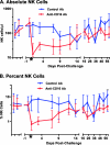
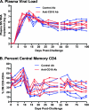
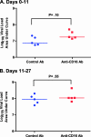
The contribution of natural killer (NK) cells to the immune containment of human immunodeficiency virus infection remains undefined. To directly assess the role of NK cells in an AIDS animal model, we depleted rhesus monkeys of >88% of CD3(-) CD16(+) CD159a(+) NK cells at the time of primary simian immunodeficiency virus (SIV) infection by using anti-CD16 antibody. During the first 11 days following SIV inoculation, when NK cell depletion was most profound, a trend toward higher levels of SIV replication was noted in NK cell-depleted monkeys compared to those in control monkeys. However, this treatment did not result in significant changes in the overall levels or kinetics of plasma viral RNA or affect the SIV-induced central memory CD4(+) T-lymphocyte loss. These findings are consistent with a limited role for cytotoxic CD16(+) NK cells in the control of primary SIV viremia.
Figures
FIG. 1.
Monoclonal anti-CD16 antibody (Ab) administration depleted NK cells from blood during primary SIV infection. Rhesus monkeys were administered either anti-CD16 (red) or a control monoclonal antibody (blue) and infected 1 day later with SIV. NK cell numbers (A) and percentages (B) were monitored by flow cytometric analysis of blood lymphocytes, with NK cells defined as CD3− CD16+ CD159A+. Data are shown as medians ± IQRs. An asterisk indicates a significant difference between groups (Kruskal-Wallis test/Dunn's multiple-comparison posttest, P < 0.05).
FIG. 2.
NK cell depletion during primary SIV infection had minimal effects on plasma viral RNA levels and virally induced loss of CD4 central memory cells. (A) Plasma viral RNA levels are shown for the anti-CD16 (red) and control (blue) monoclonal antibody-treated monkeys. (B) The peripheral blood central memory (CM) CD4+ T lymphocytes were defined as CD3+ CD4+ CD28+ CD95+. Significant differences were not detected between groups at any time point (Kruskal-Wallis test/Dunn's multiple-comparison posttest, P < 0.05).
FIG. 3.
Area-under-the-curve analysis of plasma viral load during primary SIV viremia. Plasma viral load was analyzed by integrating the area under the curve for the first 11 days of infection leading up to the peak of replication (A) and for the 15 days following the peak of viremia (days 11 to 27) as virus replication declined to the set point (B). Significant differences were not detected between groups (Mann-Whitney U test, P < 0.05).
Similar articles
- Use of an anti-CD16 antibody for in vivo depletion of natural killer cells in rhesus macaques.
Choi EI, Wang R, Peterson L, Letvin NL, Reimann KA. Choi EI, et al. Immunology. 2008 Jun;124(2):215-22. doi: 10.1111/j.1365-2567.2007.02757.x. Epub 2008 Jan 12. Immunology. 2008. PMID: 18201184 Free PMC article. - Natural Killer Cells Regulate Acute SIV Replication, Dissemination, and Inflammation, but Do Not Impact Independent Transmission Events.
Woolley G, Mosher M, Kroll K, Jones R, Hueber B, Sugawara S, Manickam C, Terry K, Varner V, Lifton M, Ram D, Fennessey CM, Keele BF, Reeves RK. Woolley G, et al. J Virol. 2023 Jan 31;97(1):e0151922. doi: 10.1128/jvi.01519-22. Epub 2022 Dec 13. J Virol. 2023. PMID: 36511699 Free PMC article. - Simian immunodeficiency virus (SIV)/immunoglobulin G immune complexes in SIV-infected macaques block detection of CD16 but not cytolytic activity of natural killer cells.
Wei Q, Stallworth JW, Vance PJ, Hoxie JA, Fultz PN. Wei Q, et al. Clin Vaccine Immunol. 2006 Jul;13(7):768-78. doi: 10.1128/CVI.00042-06. Clin Vaccine Immunol. 2006. PMID: 16829614 Free PMC article. - Cytotoxic T lymphocytes specific for the simian immunodeficiency virus.
Letvin NL, Schmitz JE, Jordan HL, Seth A, Hirsch VM, Reimann KA, Kuroda MJ. Letvin NL, et al. Immunol Rev. 1999 Aug;170:127-34. doi: 10.1111/j.1600-065x.1999.tb01334.x. Immunol Rev. 1999. PMID: 10566147 Review. - Monkeying Around: Using Non-human Primate Models to Study NK Cell Biology in HIV Infections.
Manickam C, Shah SV, Nohara J, Ferrari G, Reeves RK. Manickam C, et al. Front Immunol. 2019 May 22;10:1124. doi: 10.3389/fimmu.2019.01124. eCollection 2019. Front Immunol. 2019. PMID: 31191520 Free PMC article. Review.
Cited by
- No monkey business: why studying NK cells in non-human primates pays off.
Hong HS, Rajakumar PA, Billingsley JM, Reeves RK, Johnson RP. Hong HS, et al. Front Immunol. 2013 Feb 18;4:32. doi: 10.3389/fimmu.2013.00032. eCollection 2013. Front Immunol. 2013. PMID: 23423644 Free PMC article. - Characterization of circulating natural killer cells in neotropical primates.
Carville A, Evans TI, Reeves RK. Carville A, et al. PLoS One. 2013 Nov 11;8(11):e78793. doi: 10.1371/journal.pone.0078793. eCollection 2013. PLoS One. 2013. PMID: 24244365 Free PMC article. - Mathematical modeling of N-803 treatment in SIV-infected non-human primates.
Cody JW, Ellis-Connell AL, O'Connor SL, Pienaar E. Cody JW, et al. PLoS Comput Biol. 2021 Jul 28;17(7):e1009204. doi: 10.1371/journal.pcbi.1009204. eCollection 2021 Jul. PLoS Comput Biol. 2021. PMID: 34319980 Free PMC article. - Innate immune natural killer cells and their role in HIV and SIV infection.
Bostik P, Takahashi Y, Mayne AE, Ansari AA. Bostik P, et al. HIV Ther. 2010 Jul 1;4(4):483-504. doi: 10.2217/HIV.10.28. HIV Ther. 2010. PMID: 20730028 Free PMC article. - Fc-dependent functions are redundant to efficacy of anti-HIV antibody PGT121 in macaques.
Parsons MS, Lee WS, Kristensen AB, Amarasena T, Khoury G, Wheatley AK, Reynaldi A, Wines BD, Hogarth PM, Davenport MP, Kent SJ. Parsons MS, et al. J Clin Invest. 2019 Jan 2;129(1):182-191. doi: 10.1172/JCI122466. Epub 2018 Nov 26. J Clin Invest. 2019. PMID: 30475230 Free PMC article.
References
- Alter, G., and M. Altfeld. 2006. NK cell function in HIV-1 infection. Curr. Mol. Med. 6621-629. - PubMed
- Biron, C. A., K. S. Byron, and J. L. Sullivan. 1989. Severe herpesvirus infections in an adolescent without natural killer cells. N. Engl. J. Med. 3201731-1735. - PubMed
- Cooper, M. A., T. A. Fehniger, and M. A. Caligiuri. 2001. The biology of human natural killer-cell subsets. Trends Immunol. 22633-640. - PubMed
- Fehniger, T. A., G. Herbein, H. Yu, M. I. Para, Z. P. Bernstein, W. A. O'Brien, and M. A. Caligiuri. 1998. Natural killer cells from HIV-1+ patients produce C-C chemokines and inhibit HIV-1 infection. J. Immunol. 1616433-6438. - PubMed
Publication types
MeSH terms
Substances
Grants and funding
- U01 AI067854/AI/NIAID NIH HHS/United States
- RR000168/RR/NCRR NIH HHS/United States
- AI040101/AI/NIAID NIH HHS/United States
- R24 RR016001/RR/NCRR NIH HHS/United States
- AI067854/AI/NIAID NIH HHS/United States
- RR016001/RR/NCRR NIH HHS/United States
- P30 AI060354/AI/NIAID NIH HHS/United States
- R01 AI040101/AI/NIAID NIH HHS/United States
- P51 RR000168/RR/NCRR NIH HHS/United States
- K26 RR000168/RR/NCRR NIH HHS/United States
- AI060354/AI/NIAID NIH HHS/United States
- U19 AI067854/AI/NIAID NIH HHS/United States
LinkOut - more resources
Full Text Sources
Other Literature Sources
Research Materials


